Cell & Developmental Biology
Open Access
ISSN: 2168-9296
ISSN: 2168-9296
Research Article - (2016) Volume 5, Issue 1
This paper represents the numerical and analytical characterization of ion uptake of osteoblast cell under the influence of different electrical stimuli. The effects of pico electrical field, electrode configuration, micro channel dimension and property of suspension media on ion uptake were investigated. The electrodes were assumed to be embedded in the walls of the microchip & osteoblast cell was suspended between these electrodes. In this context it is observed that the efficient ion uptake is conducted in very short duration low amplitude DC impulse. It is found that the ion uptake is sinusoidal distributed over the surface of osteo cell membrane and it is minimum at pole θ=90 &θ=270 and maximum value is obtained at pole θ=180 which is independent of specification of electrical pulse, geometry of electrode, dimension of micro channel & property of suspension media. But their value depends on the above parameters. So the region near by the θ=90 & θ=270 are assigned as high permeable area of the osteoblast cell. The effect of neighbour cells on ion-uptake is considered to make the result more authentic and realistic.
<Keywords: Bi layer membrane; Ion uptake; Electrical stimuli; Osteoblast cell; Micro electrode
All biological cells have bi-layer selectively-permeable membrane made by phospholipids that controls the exchange of ions and different materials such as sodium or potassium [1]. Inspire of this Lipid and proteins on the cell membrane surface often have short carbohydrate chains protruding out from the cell surface, known as glycolipids and glycoproteins. They form hydrogen bonds with the water molecules surrounding the cell and thus help to stabilize membrane structure. However, more importantly, they are used as receptor molecules, binding with hormones or neurotransmitters to trigger a series of chemical reactions within the cell itself. Among them sodium ion is more use full to generate nerve impulse and maintain the electrolyte balance and fluid balance [2]. The distribution of sodium ions are mediated in all animals by Na+/K+-ATPase, which is an active transporter pumping ions against the gradient, and sodium/potassium channels [3]. Transition metals such as iron, copper, manganese, and zinc are essential nutrients. High-affinity systems are active in metal-limited cells, whereas lowaffinity systems play the predominant roles when the substrate is more abundant. Metal ion uptake systems of cells are tightly controlled, and both transcriptional and posttranscriptional regulatory mechanisms have been identified [4]. So ion uptake is an inherent property of biological cell. It is also applicable in rigid cell like osteolast [5]. But due to non-uniform distribution and concrete structure of osteoblast cell gives the challenges to researchers for its ion uptake.
A number of research works has been done considering the soft biological cell [6-8] but it is not applicable in osteoblast cell. We are focusing on that limitation and consider the original bi-layer structure of osteoblast cell. In this context a numbers of experimental study carried on at the outside of the micro or nano fluidic but all the reported studies on cell membrane surface pressure considered the cells in an infinitely large medium [9]. In micro fluidic devices, the cells are usually located in micro channels or micro chambers. The results of the current published studies may not reflect the boundary effects of micro fluid-based electroporative devices [10]. It should be focus out that all the reported studies considered that the cells contained single layer placed in an infinitely large medium and electrodes are placed at bottom or top layer of the micro fluidic devices [11]. As a result the results are influenced by reflect the boundary effects of micro fluid based electroporative devices & perfect optimization is to be needed [12]. As a result the original and realistic information about the developed ion uptake is limited for multi-layer or bi layer cell mainly in rigid cell like bone cell.
With references to the above limitations the bi-layer rigid bone cell like osteoblast is considered and ion uptake of bi-layer osteoblast cell is investigated, which is located in a micro channel in between two side wall electrodes. We also find out the effect of neighbour cells on it. The pulse shape was chosen as a square wave, which is widely used in the micro fluidic devices. This electric pulse shape can be easily generated by contracting the cross-sectional area of the micro channels or using a high-voltage Pulse generator [13]. Although we can also use the triangular or saw tooth electrical pulse but the results are not expected, where as in micro-fluidic channel the rectangular pulse is more convenient to apply.
In first part of the present study the analytical study of ion uptake for bi layer osteoblast which is placed in between two electrodes at the micro fluidic channel is addressed. In the remaining part of this paper the effects of the specification of rectangular electric pulse (duration, intensity, interval), electrode geometry (width, gap, and shape, material), dimension of micro fluidic channel as well as the nature of suspension media on ion uptake of osteoblast cell at radio frequency (10GHz) have been studied. As the cells are in contact together tightly so we also consider the effect of neighbour cells on membrane electroporation to make the result more important and realistic. The mathematical modelling and governing equations of cell membrane ion uptake are introduced in numerical simulation section and Result and Discussion deals with graphical and numerical approach of our current study. All the information of our study gives the new aid to the osteo clinical diagnosis and bone cancer treatment.
Effect of rectangular pulse specification on ion uptake
The following Schematic diagram of double -shelled spherical cell in suspension, which is used for theoretical explanation of membrane potential of a biological cell [14,15], which is related with ion uptake of the cell (Figure 1).
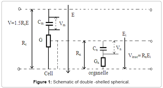
Figure 1: Schematic of double -shelled spherical.
According to the transfer functions defined by C. Yao the inner and outer membranes to a given rectangular pulse electric field E(s) can be obtained
 (1)
(1)
 (2)
(2)
Where,
After simplification of equation (1) & (2) we get the outer membrane potential  m (t) are as follows
 (3)
(3)
It is known from the bio chemistry that the ion uptake can be calculated by following mathematical calculation,
 (17)
(17)

 (4)
(4)

T=Temp in kelvin.
Kb=Boltz man constant
 = permitivity of water,
 = permitivity of cytoplasm. &
 = pulse duration.
 = radius of pore,
r=radius of initial pores,
 = membrane potential
By replacing from equation 3 into the equation 4, the dependency of pulse specification on ion uptake is addressed.
Effect of micro-electrode specification on ion uptake
As we know that 
where V=applied voltage &  = distances in between two electrode. We replace E(t)= v/d in equation (3) & (4) and get outer membrane potential is
 (5)
(5)
If the value of is replaced in equation (4) than the relation among the ion uptake occurred with intra-electrode gape is established.
In this context it is known from the theory of electrochemistry the value of produced voltage at metal electrode  expressed as
 (6)
(6)
Where K = Boltz man constant, T = absolute temperature, z = valancy of electrode material & e =charge of an electrode, Cmet = ionic concentration of electrode material. If the value of v is replaced in equation (5) and (4) than the relation among the ion uptake occurred with electrode material and shape of electrode is established.
From the knowledge of dielectric spectroscope of cell membrane it is find out that
 (7)
(7)
Where 
Replaces  in equation (3), it is obtained that
By replacing, and  in equation (3) and (4), then the dependency of electrode width, length, material and geometry on ion uptake of the osteo blast cell is expressed.
Effect of micro-channel specification on ion uptake
It is reported that if radius of cell is  and channel height H than relative resistances of cell placed in the micro channel is consider as
  If we replaces in equation ( 3) and (4) then the effect of channel height on ion uptake is established..
The basic concept of micro fluidic devices the channel resistances R ch is numerically defined as  and
and 
Whree k=2 for biological cell,  = Conductivity of medium and
Thickness of Membrane.
After simplification it is turned as
 (9)
(9)
On the other hand width of the channel is defined as
 (10)
(10)
Where, V 0 = Initial velocity of fluid Raqu = resistances of aqua medium. By replacing value of equation (9) and (10) in equation (3) and (4) then the effect of channel resistances on ion uptake is exposed.
Effect of micro-fluidics specification on ion uptake
In primary concept of fluids dynamic it reveals that flow rate  , As channel resistances has a great influences on membrane potential so flow rate has also a large influences on membrane ion uptake.
Numerical Analysis of Ion Uptake
Simulation tools
In this study, above Equations are solved numerically to find the electric potential developed in outer and inner membrane to investigate the creation of nano-pores on the cell membrane. The Mat lab 7.2 & Comsol-4.3a, commercial package was used in the numerical simulations.
Used simulation parameter
Table 1 Values for constants and parameters used in the simulations [15] (Table 1).
| Parameter | Cell parameters | Osteoblast |
|---|---|---|
| conductivity (S/m) |
Extracellular medium (σe) | 10×10(-3) |
| Cell membrane (σm) | 1.2×10(-7) | |
| Cell cytoplasm(σc) | 0.039s | |
| Nuclear membrane(σn) | 10×10(-1) | |
| Nuclear cytoplasm(σn) | 0.08s | |
| relative permittivity |
Extracellular medium(εe) | 80 |
| Cell membrane(εm) | 22 | |
| Cell cytoplasm(εc) | 93 | |
| Nuclear membrane(εn) | 22 | |
| Nuclear cytoplasm(εn) | 93 | |
| Geometry parameter (μm) |
Cell radius(rc) | 12μm |
| Cell membrane thickness(d) | 0.006μm | |
| Nuclear radius(rn) | 6μm | |
| Constant parameters | N0 | 1* 109 |
| D | 5*10(-14) | |
| K | 1.38065*10(-23) | |
| T | 300 | |
| β | 1.4* 10-19 | |
| γ | 1.8* 10-11 | |
| Fmax | 0.7* 10-9 | |
| σ | 1* 10-6 |
Table 1: Values for constants and parameters used in the simulations.
Electrical field analysis
Figure 2a and 2b depicts the potential distribution with the micro devices having the non uniform distribution of potential and intra cellular organelle also effect by this field. At pole  and
and  maximum potential are exposed which is similar as our numerical result. And complete electrical potential distribution within the 3D hybrid micro chip. Where cell is affected through the whole channel. Figure 3 shows the Graphical view of polarization and electric field with respect to height of the bi metallic electrode within the 3D hybrid micro chip which helps us to find out the maximum polarization area of Nono pores. It explore that the centre part (300-600) μm of the chip holds the maximum uniform potential where the nano pores are generated at the intra organelle. Maximum velocity excreted at the centre of the chip, it affects the ion uptake of intra cellular unit of the rigid cell. All the information exposed in COMSOL simulation is as similar as numerical and experimental values of intra organelle nano poration of multi-layer dense osteoblast cell (Figures 2 and 3).
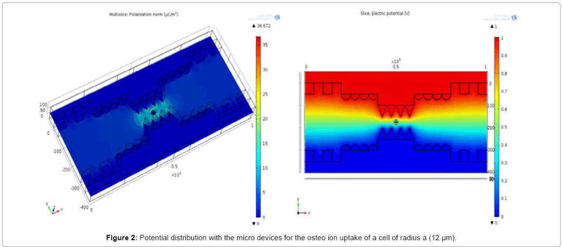
Figure 2: Potential distribution with the micro devices for the osteo ion uptake of a cell of radius a (12 μm).
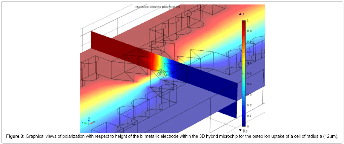
Figure 3: Graphical views of polarization with respect to height of the bi metallic electrode within the 3D hybrid microchip for the osteo ion uptake of a cell of radius a (12μm).
Numerical simulation results and discussion
In this study, above equations are solved numerically to find the surface pressure developed in outer and inner membrane to investigate the creation of nonporous on the cell membrane. The MATLAB7.2 & COMSOL 4.2a commercial package was used in the numerical simulations.
The quantitative information used in the simulations is provided in Table 1. A cell of radius a (12 μm) is considered in the microchannel of height (500μm). The necessary electric field (1v) was applied by the two electrodes of width (50 m) located on the walls of the micro channel. The electric pulse span was on the order of microseconds to pico second operated at radio frequency (10 GHz hertz).The results of the numerical simulations are as follows.
Evaluation of pulse: Figure 4(a) shows that the amount of ion uptake of osteoblast cell is sinusoidally varied with respect to azimuthal angle of the electric field. It is observed that if pulse interval is increased than the amount of molecules (ion uptake) taken by the cell is reduced and maximum ion uptake is occurred at pole  whereas minimum value at the pole
  . But Figure 4(b) shows that ion uptake is in dependent with pulse amplitude variation (Figure 4).
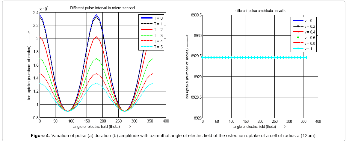
Figure 4: Variation of pulse (a) duration (b) amplitude with azimuthal angle of electric field of the osteo ion uptake of a cell of radius a (12μm).
Evaluation of micro-electrode: In Figure 5(a) shows that ion uptake depends on the shape of the micro electrode and if the shape is saw-tooth than maximum ion uptake is occurred but polar location is fixed and it is .
From the Figure 5(b), it has also been observed that the value of ion uptake is inversely proportional with inter electrode distances and it is maximum when electrode gape is 10 μm.
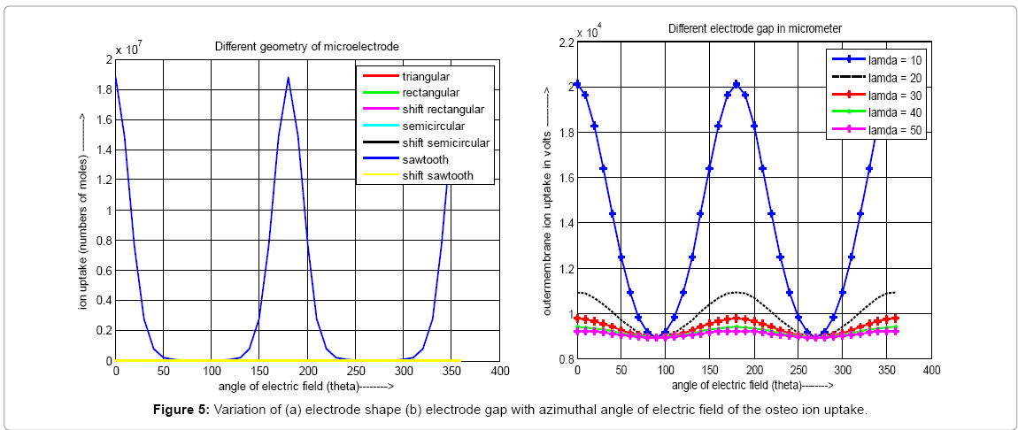
Figure 5: Variation of (a) electrode shape (b) electrode gap with azimuthal angle of electric field of the osteo ion uptake.
Evaluation of micro-channel: Figure 6(a) shows that micro channel height does not effect the ion uptake and it is uniform throughout the whole micro channel but channel resistances effects the molecule incursion which is depicted in Figure 6(b). It also observed that the ion uptake inversely proportional with channel resistances and maximum value is obtained when channel resistances is 10 MΩ (Figure 6).
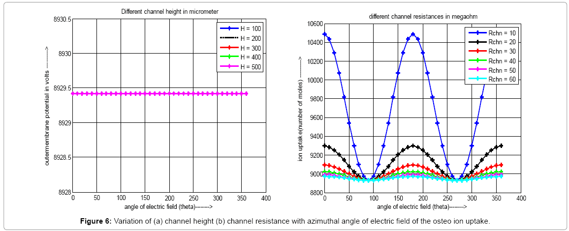
Figure 6: Variation of (a) channel height (b) channel resistance with azimuthal angle of electric field of the osteo ion uptake.
Evaluation of micro-fluidics: Figure 7 illustrates the effects of conductivity and flow rate of suspension media on ion uptake. Figure 7(a) shows that ion uptake of osteoblst cell is inversely proportional to conductivity of the micro flouds but flow rate does not effect on it. Finally it is concluded that the flow rate and conductivity of suspension medium have great influences on ion uptake. For efficient ion uptake the flow rate and conductivity should be as low as possible (Figure 7).
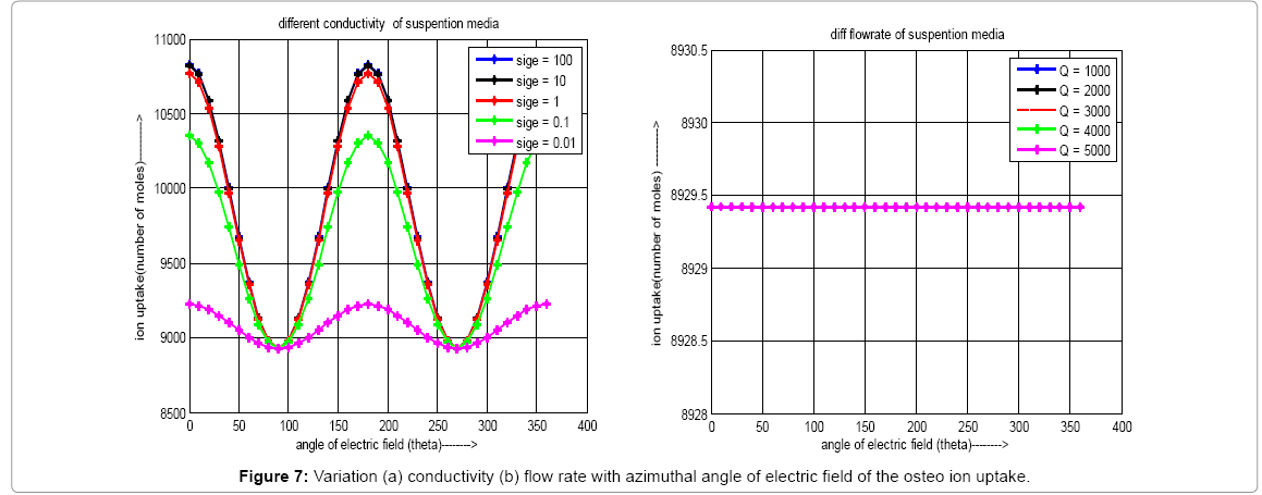
Figure 7: Variation (a) conductivity (b) flow rate with azimuthal angle of electric field of the osteo ion uptake.
After all discussion it is concluded that ion uptake is not same throughout the whole surface of the osteoblast cell and it is sinusoidally occurred. The maximum ion uptake is occurred at pole  and minimum at pole
  which is independent of pulse ,micro electrode, micro channel and micro floud specifications. Low amplitude (1V) and very short interval DC pulse is required for effective ion uptake. In addition, the saw tooth micro electrode having the electrode gap of 10 μm for effective ion uptake. It also observed that micro channel height does not effect the ion uptake and the ion uptake inversely proportional with channel resistances and maximum value is obtained when channel resistances is 10 MΩ. These analytical results are independent with nature of pulse, geometry of electrode and microchannel. In present study it is explored that the width of electrode in micro channel has no effect on cell ion uptake because they are placed in the sidewall of micro channel.
In brief it is conclude that osteo ion uptake can be externally controlled by short impulse electrical stimuli. In future the researcher could be think about the design and fabrication of 3D bi-layer micro bio chip for more effective in osteo ion uptake which would be widely used in drug delivery system.
All of we wish to thank Dr. Soumen Das (IIT, KGP) for assistance with analytical data processing throughout the whole work.