Journal of Hematology & Thromboembolic Diseases
Open Access
ISSN: 2329-8790
ISSN: 2329-8790
Review Article - (2016) Volume 4, Issue 5
Antiphospholipid antibody syndrome is a serious autoimmune disorder which can lead to multisystem manifestations from recurrent thrombosis to pregnancy loss to intrauterine death and other obstetric morbidities. In few cases it may lead to catastrophic syndrome. Antiphospholipid antibodies are circulating antibodies which bind with the plasma proteins which in turn bind to phospholipids and thus lead to pathogenesis. Despite of so many researches done in the field of APLA, the mechanisms leading to obstetric complications are still debatable. Antiphospholipid antibodies are detected on the basis of solid phase assays which comprised of anticardiolipin (acl) antibody and Anti β2 glycoprotein (aB2GP) and the other one is liquid phase assays which identifies lupus anticoagulants (LAC). While aB2GP and acl are being detected most commonly by ELISA, LAC is being detected by clot based test. Lupus anticoagulant is a double misnomer as most patients don't have systemic lupus erythematous and in vivo reacts as procoagulants.
Keywords: Antiphospholipid; Catastrophic syndrome; Anticoagulant; Thromboembolism
Antiphospholipid antibody syndrome is a serious autoimmune disorder which can lead to multisystem manifestations from recurrent thrombosis to pregnancy loss [1] to intrauterine death and other obstetric morbidities [2]. In few cases it may lead to catastrophic syndrome. Antiphospholipid antibodies are circulating antibodies which bind with the plasma proteins which in turn bind to phospholipids and thus lead to pathogenesis. Despite of so many researches done in the field of APLA, the mechanisms leading to obstetric complications are still debatable [3]. Antiphospholipid antibodies are detected on the basis of solid phase assays which comprised of anticardiolipin (acl) antibody and Anti β2 glycoprotein (aB2GP) and the other one is liquid phase assays which identifies lupus anticoagulants (LAC). While aB2GP and acl are being detected most commonly by ELISA, LAC are being detected by clot based test. Lupus anticoagulant is a double misnomer as most patients don’t have systemic lupus erythematous and in vivo reacts as procoagulants. Term anticoagulant was assigned to LAC because in liquid phase assay in vitro they prolong the tests and therefore acting as anticoagulants. Four LAC tests based upon clot based assays are being defined and recommended by various bodies and studies-Diluted Russel Viper Venom test (dRVVT), Activated Partial Prothrombin Time-Lupus Anticoagulant (APTT-LA), Kaolin clotting time (KCT), Dilute Prothrombin Time (dPT). The diagnostic utilities of of KCT and dPT has been discouraged because of mainly of their poor performance [4]. Therefore ISHTH recommends dRVVT and APTT-LA. But the debate of the diagnostic utilities of each test is on as various studies have varied results. Both the false positive and the false negative have their issues while the false negative issue is understood the false positive cases will lead to unnecessary exposure of patients to anticoagulants. This review article will concentrate on the diagnostic issues of LAC tests and its impact.
Defnition and Diagnostic Criteria
While the Antiphospholipid syndrome (APS) routed its major cornerstone [3,4] decades back but the defining criteria of Antiphospholipid syndrome (APS) opened its new chapter in a discussion on antiphospholipid antibodies in an International congress in Sydney in 2004 which got published in 2006. Serological criteria were one of the leading topics in the discussions mainly in the Journal Thrombosis and Hemostasis and chapter update on Antiphospholipid syndrome (APS). Pengo et al. and Tripodi updated the Lupus anticoagulants recommendations for its detection. While Gianacopulous B et al., Pengo V et al., Roubey RAS analysed the lab diagnostic work up for the Antiphospholipid Syndrome (APS).
Though ELISA based investigations anti β2 GP and anticardiolipin also have an important role in Antiphospholipid syndrome (APS) but the LAC was the first and even today the foremost runners among all anti-phospholipid antibodies. It is shown in the study that LAC has a stronger risk for thrombosis than anticardiolipin antibodies [5]. There are several reasons for supremacy of LAC over the detection of antiphospholipid antibodies. Firstly, inter laboratory interpretation for lupus anticoagulant detection is more consistent than solid phase.
This was shown in studies by Favaloro et al. [6,7] in which it was seen that interpretation for LA as ‘positive’ or ‘negative’ for the dRVVT ratios is fairly consistent between laboratories with mostly having similar cut off of 1.2 while with using cut off of 20 GPL units for aCL interpretation as ‘positive’ or ‘negative’ it was less consistent between laboratories. Secondly, there is a stronger association of LA than anticardiolipin antibodies with adverse events such as thrombosis [8]. Thirdly, only higher titer and more clinically relevant forms of aPL are detected by the clot-based test system [6]. LAC investigations are clot based tests. International Society on Thrombosis and Haemostasis (ISTH) has updated its recommendations in 2009 for the detection of the Lupus anticoagulants [4].
Importance of LAC tests lies on the fact that long term anticoagulation depends upon how accurately Antiphospholipid Syndrome (APS) has been diagnosed. Though in the earlier period of time LAC tests like kaolin clotting time, dilute prothrombin time were also given emphasis [9,10] but after the recommendations of International Society on Thrombosis and Haemostasis (ISTH) in favour of dilute Russell viper venom time (dRVVT) and a sensitive partial thromboplastin time (PTT-LA) the former investigations have been questioned (Figure 1). As per the guidelines of ISTH guidelines the basis of using 2 assays was that the risk for false-positive results is increased if more than 2 LAC assays are performed but one large retrospective study [11] data showed that even after using 4 panel assays (dRVVT, APTT_LA, dPT, KCT) there was low LAC-positive prevalence which thus indicates that a wider array of assays is not necessarily result in a spuriously high prevalence Antiphospholipid syndrome (APS). This study was unable to characterize a single particular assay that was associated with either stillbirth or a history of TEs. Therefore they highlighted the need of more than 2 assays for the detection of APS. In fact they found that the increased number of positive assay results per panel were associated with an increased occurrence of clinical events in from of thrombotic events, recurrent abortions which thus suggests to correlate with poorer prognosis, hence giving an evidence that if we limit the assessment of LAC to only 2 assays as per ISHTH guidelines it may not be the optimal choice.
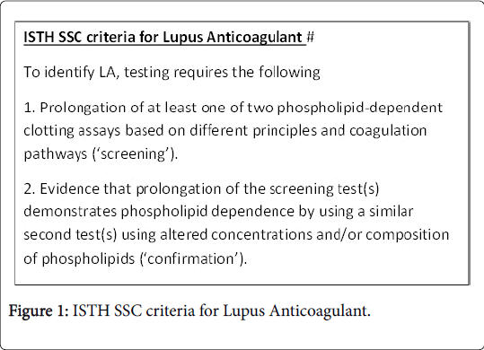
Figure 1: ISTH SSC criteria for Lupus Anticoagulant.
The diagnostic utility of any test is based upon the patient selection, timing of the tests, pre-analytical variables, choice of the tests, indications of the test, interfering variables, standardization of the tests and the interpretation of the tests therefore LAC is also not an exceptional to these factors. In the following sections we will discuss each of the above factors.
Patient Selection
Pengo et al. [4] recommended that testing for LA would be most appropriate in individuals with:
Unprovoked venous thromboembolism
Unexplained arterial thrombosis in young patients (<50 years of age)
Thrombosis at unusual sites
Late pregnancy loss
Any thrombotic events or pregnancy morbidity in patients with other autoimmune disorders like SLE
In other patients though not most appropriate but still in whom testing is reasonable include individuals.
With recurrent spontaneous early pregnancy loss and
Provoked venous thromboembolism in young patients.
While patients in whom testing for antiphospholipid antibodies are unlikely to be helpful include.
Elderly patients with venous or arterial thromboembolism.
There is another category of individuals that are asymptomatic but found to have an unexplained prolonged aPTT in whom work up for APLA is sometimes useful.
Timing of the Tests
Testing for LAC should be performed when a patient is not having an acute event and not on any anticoagulants. Except for Fondaparinux other anticoagulants have either variable effects or LACs are prolonged [12].
Pre-analytical Variables
It is now a very well-known and has been emphasised in various studies [11,13] that residual platelets affects the phospholipid based coagulation factors through the exposure from platelet membranes of anionic phospholipids that quench LA activity thus leading to shorter coagulation time. This effect is more pronounced in plasmas which has undergone freezing and thawing before analysis and with reagents whose phospholipid content is relatively low [14]. One of the best reasons given for the effect of freezing and thawing is that weak LAs may lose during freezing and thawing. It is recommended to have platelet free (<109/L) plasma for LA testing. Though filtration is considered to be effective means to have free platelet free plasma but filtration may cause loss of high molecular weight coagulation proteins (von Willebrand factor and consequently factor VIII) [15] which will lead to artifactual prolonged APTT.
Which Test to Choose for the Screening of APLA?
It’s a debatable question till now since every test has its pros and cons and the various studies have suggested one or other test based on their studies.
APTT-LA
Its disadvantage lies on the fact that it has varied sensitivity because of the class and concentration of phospholipids [16]. For example use of reagents rich in phosphatidyl serine abolishes the LA effect on the APTT. Besides conformation of phospholipids also play a role in the detection of LA. As shown in one study [17] that Lupus anticoagulants from patients with SLE specifically recognize hexagonal (II) phase instead of bilayer phosphatidylethanolamine [18].
KCT
KCT is considered to be the most sensitive screening test as it has low content of phospholipids. However things are not that good for it also as it has some drawbacks. Firstly, Kaolin is a particulate substance and therefore interferes with the optical clot detection systems. Secondly, it forms sediment within the coagulometer dispensing systems and cuvettes. Thirdly, it has been seen that it has long coagulation time even in normal individuals therefore loses its reproducibility. In view of it there was a long search of replacement of kaolin by micronized silica (SCT) which has the same sensitivity as kaolin but does not form sediment.
dRVVT
It has been considered to be the most important test for investigations of APLA and therefore no doubt has been incorporated into recommendations to be the foremost test. Studies by Galli et al. [10] and Pengo et al. [19] proved that dRVVT was more sensitive and predictor of thrombosis than the KCT.
Dilute prothrombin time test
Though not considered to better test than the above tests but can improve on using recombinant thromboplastin. As it has been quoted that every test has some or other issues and therefore no single test is 100% sensitive to LA. Therefore it was recommended that at least 2 tests are required and that to with different assay principles [20]. Many studies have suggested that 1 of the 2 should be the dRVVT and the other may be based on the intrinsic pathway of coagulation i.e. APTT, KCT, or SCT. Some have suggested using 3 assays one drVVT and resting two ones based upon the coagulation based.
Mixing test
Next part ascertaining the screening test for APLA we have to rule out the coagulation factor deficiency. The role of the mixing test is to differentiate between the factor deficiency and the inhibitor. It is quite understood that if we mix the test plasma with normal pooled plasma (prepared from at least 20 normal samples for ensuring normal concentrations of all coagulation factors), if it is due to deficiency coagulation factors it will correct and if it doesn’t than it’s an inhibitor. There are 3 ways to interpret inhibitors. Firstly if when the coagulation time of the mixture doesn’t falls within the reference interval (Note each lab has its own normal reference value). Secondly the mixture of test and normal plasma falls >2 SDs from the mean than that of a normal plasma mixed with various non-LA plasmas (i.e., plasmas from patients with coagulation factor deficiencies). Thirdly, though the calculation of Rosner index circulating anticoagulant (ICA) i.e. [21] defined as ICA=[(Coagulation Time mix- Coagulation Time Normal Pooled Plasma)/Coagulation time T patient)] × 100. If ICA is prolonged then it’s an indication that the inhibitor is there
It is not simple to interpret as it seems to be especially when patient coagulation time is only slightly prolonged and secondly there are high chances of false-negative results for weak LA because of the dilution effect. Therefore there is a debate on the ground that whether mixing should be done or not. Though the current ISTH SSC guidelines added recommended but not mandated requirement for mixing studies which was later clarified to be the strongly recommended by the authors of SSC guidelines. Even the new CLSI guidelines seem to have reduced the relative importance of the mixing studies. The foremost reason coming out for the doubt in conducting mixing studies is that mixing tends to dilute the weak Lupus anticoagulants and therefore could lead to false negative results. Further it will just lead to more testing and thus more complex issues and the cost for the patients or the hospital.
Confirmatory tests
After ruling out the factor deficiencies confirmation is required for involvement of lupus anticoagulants by adding phospholipid in LAC tests. As we know that the lupus anticoagulants acts upon the phospholipids or the factors dependent upon the phospholipid and if we increases the concentration of phospholipids in the test system which it will neutralizes the effect of LA and therefore shortens the coagulation time which was prolonged in the screening tests if it is because of Lupus anticoagulants while it will remain prolonged if it is due to factor inhibitors. The agents used as the source of phospholipids is aged platelets or the commercial prepared phospholipids, either bilayer or hexagonal (II) phase. But again the confirmatory tests too have some drawbacks. Firstly heparin may behave like LA. Therefore it is advisable to thrombin time test/Factor Xa assays to rule out heparin. To counter the effects of heparin some commercial reagents include anti-heparin substances like polybrene are mixed in the reagents. Secondly sometimes antifactor antibody may behave like LA [22,23]. This again can be done by scrutinising through detailed clinical and family history.
Currently most of the laboratories perform LA testing using activated partial thromboplastin time (APTT) and dilute Russell viper venom time (dRVVT) methods, and few also employ the kaolin clotting time (KCT). Rest other methods like silica clotting time (SCT) and the platelet neutralization procedure (PNP) are only used by <5% of laboratories. These tests are shown to have lesser inter-laboratory CVs in comparison to solid-phase assays such as aCL and anti 2GPI. These CVs are increased slightly with increasing LA positivity. Though most of the laboratories correctly interpreted test findings for LA but some of the laboratories still found interpretation to be challenging especially for samples having weak LA (which was reported as normal by around 50% of laboratories). Though currently we are having three assays namely LA, aCL, and anti 2GPI but they all have some drawbacks. Firstly, these assays lack standardization. Secondly, these assays may not detect the autoantibody which is responsible for the clinical manifestations and thirdly, none of these assays predict the risk of recurrence. Therefore, there is an essential need for novel assays which also has prognostic information that can be used clinically in the management of APLA.
While considering the diagnosis utility of LAC one should be aware of the choice of patients and tests besides the pre and post analytical factors of the LACs. LACs are no doubt the better options in comparison to solid phase tests like 2GPI and aPL antibodies detection. While the detailed history is required for the selection of patients based upon his clinical features and his drug history, in depth knowledge is required among the medical fraternity involved in the diagnostic field to be aware of in and out of the technical aspects of the tests done in APLA. While weak LACs can be missed there are scenarios when false positive cases are also included. Therefore ISTH recommendations it’s to be followed strictly in order to avoid these false negatives and false positives. Simultaneously it is required to repeat the tests after 12 weeks to rule out the transient positive LACs because of some infections. However if the test comes positive even after the repetition after 12 weeks then there is no need of follow up. Beside these selection of the LAC test is again a very important factor. ISTH recommends two tests dRVVT and one test based upon APTT (Kaolin clotting test, Silica clotting tests, APTT- Lupus sensitive). Besides there are studies which are against the conducting two tests and recommend more than two tests. In future the assay is required to develop which can be prognostically useful and can be used up for follow up of the disease and treatment.