Journal of Plant Biochemistry & Physiology
Open Access
ISSN: 2329-9029
ISSN: 2329-9029
Research Article - (2018) Volume 6, Issue 1
Octacosanol, a primary alcohol, was isolated from rice bran wax and characterized by GCMS and XRD techniques, which confirmed the identity and purity of the isolated octacosanol. Five different concentrations of the compound ranging from 0.01 mg/ml, 0.05 mg/ml, 0.1 mg/ml, 0.5 mg/ml to 1.0 mg/ml were prepared in isopropanol. The antioxidant activities of octacosanol were studied at these concentrations for four in vitro assay systems including DPPH radical scavenging activity, reducing activity, metal chelation activity and inhibition of lipid peroxidation. Maximum antioxidant potency was displayed at 1.0 mg/ml for all the assays except the metal-chelation assay which demonstrated highest activity at 0.5 mg/ml. Octacosanol also showed anti-bacterial activities against Escherichia coli, Pseudomonas aeruginosa and Bacillus subtilis as observed by disc assay against concentrations of 1 mg/ml and 2 mg/ml. At 2 mg/ml, octacosanol was observed to inhibit microbial activity by gram negative bacteria Escherichia coli and Pseudomonas aeruginosa.
Keywords: Octacosanol; Rice bran wax; Fatty alcohol; Nutraceutical; Antioxidant assay; Anti-microbial activities
FTIR: Fourier Transform Infrared Spectroscopy; GC-MS: Gas Chromatography and Mass Spectrometry; XRD: X-ray Diffraction; DPPH: 2,2-diphenyl-1-picrylhydrazyl.
Vegetable oils are the principal sources for waxes of nonhydrocarbon origin. They are basically esters of fatty alcohols and fatty acids [1]. However other lipid components, such as hydrocarbons, ketones, mono-, di-, tri-acylglycerols, sterol esters etc. are also coextracted with waxes when they are isolated from oils [2]. Waxes obtained from Rice Bran Oil (RBO) are mostly composed of C16-18 fatty acids and C24-34 fatty alcohols. Octacosanol, a long chain 28-carbon primary fatty alcohol present in rice bran wax [3], shows several healthpromoting effects like anti-inflammatory effects, lowering of low density lipoprotein, preventing coronary heart diseases etc. [4]. Furthermore, due to the amphiphilic character of such fatty alcohols they also find immense applications in the surfactant and cosmetic industries [5].
Extensive study regarding the antioxidant and anti-bacterial activity of octacosanol is still lacking. A complete knowledge about these potencies would enable the pharmaceutical, nutraceutical, surfactant, and cosmetic industries to utilize octacosanol and other such fatty alcohols in several all-round applications. Under normal conditions of temperature and pressure octacosanol is highly resistant to oxidation because of its typical structural make-up. Moreover, due to the presence of the hydroxyl group it can be converted to aldehydes and carboxylic acids only in the presence of very strong oxidants [6]. It was previously observed that 1-octacosanol showed protective effects against Parkinson’s disease when tested on a rat model and showed quick recovery from liver injury by reducing the effect of reactive oxygen species [7,8]. Till date octacosanol has been isolated from crops like wheat, Euphorbia species, sugarcane etc. [9-11]. A major challenge is the isolation of pure octacosanol from the mixture of different lipid components and polycosanols like triacontanol, hexacosanol etc. Previously 1-octacosanol was isolated from sugarcane rind by saponification process. However, in the cited work apart from octacosanol, other products like triacontanol, campesterol, stigmasterol etc. were also co-extracted with octacosanol [12]. Furthermore, rice bran wax has not been very much explored till date as a resource for extraction of octacosanol. Rice bran wax is an important by-product of the edible oil refining industries which is mostly disposed-of later. A number of polycosanols have been co-extracted from rice bran wax and their nanoemulsions are prepared to enhance stability [13]. Previous reports have pointed out that the anti-bacterial activity of fatty alcohols was dependant on the number of carbon atoms in the aliphatic chain, with clear indication of the importance of the polar end group [14-16]. The fatty alcohols thus are the ideal compounds to display anti-bacterial properties according to this study.
However, in none of the previous studies 1-octacosanol has been isolated uniquely, in a pure form, from rice bran wax. A possible reason could be the minimal concentration of octacosanol in rice bran wax. None of the strategies or developed methods completely justified the isolation of octacosanol from rice bran wax cost-effectively. However, recovery of octacosanol from rice bran wax would ensure isolation of ‘wealth’ from ‘waste’. Hence in the present study the technology to isolate pure 1-octacosanol from rice bran wax has been developed, with proper characterisations to prove the authenticity of the developed method. Furthermore, the importance of the isolated octacosanol in acting as an antioxidant and inhibiting bacterial infestations has also been explored subsequently with encouraging results. Additionally, if the product is from a natural source it will prevent any untoward sideeffects associated with synthetic products.
Materials
Rice bran wax was supplied by Sethia Oils Limited, West Bengal, India. The wax was de-oiled with acetone, then dried and the crude wax obtained was preserved at 4°C for further use.1-octacosanol, tocopherol, beta-sitosterol and squalene standards, 2,2-diphenyl-1-picryl-hydrazyl (DPPH) was acquired from Sigma-Aldrich ChemieGmbh (Munich, Germany). The micro-organisms used in this study, viz., Escherichia coli, Bacillus subtilis and Staphylococcus aureus, were procured from IMTECH, Chandigarh, India. All other reagents were of analytical grade and obtained from Merck Ltd., Mumbai, India.
Extraction of unsaponifiable matter
About 20 g of the de-oiled wax taken in a 250-ml round-bottomed flask, attached to an air condenser. It was saponified with 150 ml of 0.2 (M) sodium hydroxide in 95% ethyl alcohol (base hydrolysis) at 80°C following the method of Ishaka et al. [13] with suitable modifications. The mixture was boiled on a water bath for two hours. The strong base expedites cleavage of the ester bond and releases the fatty acid as its sodium salt along with glycerol as the by-product.
The unsaponifiable material, which was a hard-yellow portion, was collected with hexane under hot condition (60°C). The hexane portion was made completely soap-free by repeated aqueous washing (at least 10 times). The complete removal of soap from the final hexane layer collected was confirmed in the presence of phenolphthalein indicator which did not change color. The hexane layer was then evaporated to dryness. The unsaponifiable matter consisted of chiefly fatty alcohols along with small amounts of sterols, tocopherol and hydrocarbon.
Isolation of 1-octacosanol from the unsaponifiable matter
The soap-free unsaponifiable matter was subjected to fractional distillation. It is the process of separation of a mixture into its component fractions based on their boiling points. It was done using the Claisen-Vigreax flask. The product was distilled from 200°C to 230°C (Fraction 1) and between 230°C to 260°C (Fraction 2). Each fraction was collected, weighed and their gas chromatographic analyses were performed, against standard octacosanol, tocopherol, betasitosterol and squalene (hydrocarbon). It was observed that the fraction 1 (200°C – 230°C) was rich in 1-octacosanol and hexacosanol (Figure 1). This temperature range corresponds to the boiling point of these fatty alcohols (1-octacosanol: 225°C and 1-hexacosanol: 220°C at 100 Pa pressure). The second fraction of higher temperature range was rich in the sterols, tocopherol and hydrocarbon.
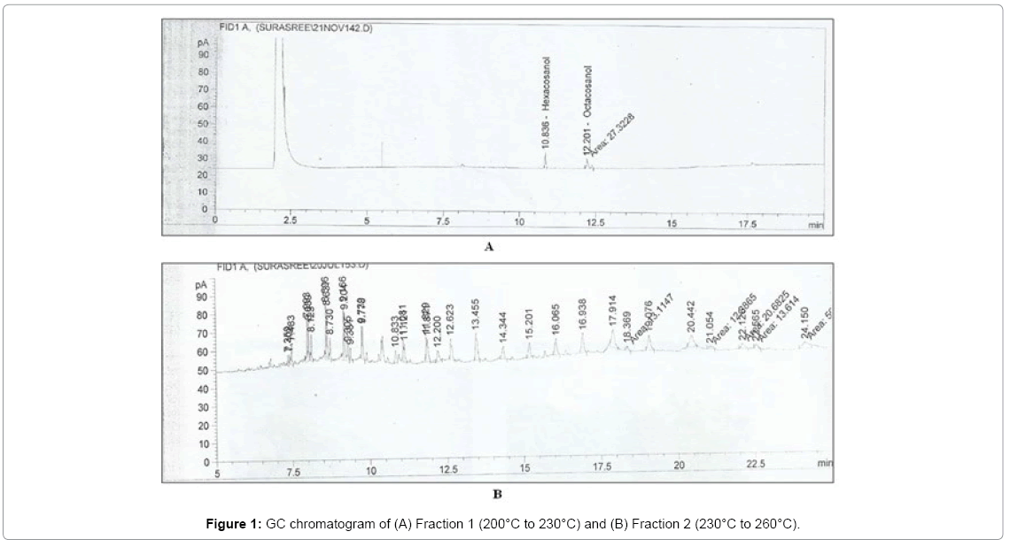
Figure 1: GC chromatogram of (A) Fraction 1 (200°C to 230°C) and (B) Fraction 2 (230°C to 260°C).
Crystallization with isopropanol
Fraction 1 was collected and dissolved in minimum volume of isopropanol heated to 90°C. It was then gradually cooled to room temperature. At 84°C the first appearance of precipitate was observed. This precipitate corresponded to pure 1-octacosanol as this particular temperature of crystallization is that of 1-octacosanol. GC-MS analysis of this product was performed to confirm its identity. X-ray crystallographic analysis was also performed to determine its bond length and crystallinity.
Characterization of isolated compound
GC-MS analysis: Gas chromatography and mass spectrometry was the coupled instrument used for identification purpose. The instrument used was manufactured by Thermofisher Scientific with model no. 5890 model series II. Here non-polar, low bleed, polysiloxane DB-5 capillary columns (30 mm × 0.25 mm × 0.25 μm) was used which was manufactured by Agilent Technologies. The analysis was done in a split mode with the split ratio of 1:12 and split flow (ml/min) was 10. Manual injection was done for the analysis with 1 μl sample.
Carrier gas was Helium used at a flow rate of 0.8 ml/minute and the flow mode was constant. The column temperature was programmed in the following steps starting from 50°C to 150°C (held for 1 minute at each temperature) at a rate of 10°C/minute, then up to 205°C with 5°C/minute, increasing the temperature 10°C/minute and 5°C/minute with 1 minute holding up to 310°C where it was held for 10 minutes. ION Trap mass analyzer of the mass spectrometry was within the vacuum pressure of 50 – 60 millitorr. The temperature of GC to MS transfer line was 300°C. Electron induced ionization (EI) detector was the main source of detection which was 70 (–eV). Mass spectrum of the compound was produced by mass/charge (m/z) ratio which was 50 to 500 m/z. These were recorded by the data processing software XCALIBUR.
Identification of the compound was done by NIST (National Institute of Standards and Technologies) Library. They were matched with the arithmetic index derived from primary alcohol series and authentic standards. Other related authentic, published articles were also considered for detection of the compound.
XRD analysis: Crystallinity was analyzed by X-ray diffraction (XRD) [17]. A diffractometer (XPERT-PRO from Panalytical Diffractometer) using Cuα (λ=1.5406) as X-ray source. Kα1α2β radiation from copper was used at 40 kV and 30 mA. Kα2 /Kα1 ratio was 0.50000. A scanning velocity of 1°/minute from 2° to 80° was maintained. Experiments were performed at ambient temperature (25°C).
Antioxidant assays: The antioxidant activities of 1-octacosanol were examined by five different in vitro assay systems by preparing different concentrations of 0.01 mg/ml, 0.05 mg/ml, 0.1 mg/ml, 0.5 mg/ml and 1.0 mg/ml in isopropanol. A control of only isopropanol was also studied in each case. The scavenging activity of 1-octacosanol for the stable DPPH free radical was studied following the method of Katerere et al. [18]. Thereafter the reductive potential of 1-octacosanol was determined according to the method of Dorman et al. [19]. The Fe2+ chelating ability of 1-octacosanol with ferrozine was estimated by the method of Dinis et al. [20]. Prevention of lipid oxidation in a linoleic acid emulsion model system by 1-octacosanol using ammonium thiocyanate was performed following the procedure of Gulcin et al. [21].
Microorganisms and culture media: The microorganisms used in this study included three types namely Escherichia coli, Bacillus subtilis and Staphylococcus aureus. Cultures were grown in an agar slant consisting of glucose (1%), peptone (0.5%), beef extract (0.3%), yeast extract (0.3%) and NaCl (0.9%) in distilled water where pH was adjusted to 6.8 to 7.0 with 1(N) NaOH. The agar (2%) was dissolved in it by steaming the mixture in an autoclave for 30 minutes. The molten agar was quickly transferred in test tubes and then plugged with cotton. They were sterilized in an autoclave for 15 minutes and then incubated at 28-30°C for 24 hours. The nutrient agar was then streaked with the 48-hour old cultures. The cultures were incubated for 24 hours at 28- 30°C [22].
Discassay
The first validation method involved the preparation of a sterile test solution consisting of 1 mg/ml and 2 mg/ml of octacosanol in isopropanol. Concentrations lower than 1 mg/ml did not display sufficient anti-bacterial activity on a trial assay. Hence 1 mg/ml and subsequent higher concentration of 2 mg/ml were chosen for disc assay. The assay plates (100 mm in diameter), covered with a thin layer of agar medium were initially sterilized and incubated for 24 hours. Next day they were streaked with a sterile swab consisting ofa 0.85% saline solution, spiked with a minimal concentration of the bacterial species.
Assay plates were prepared as described above. After properly demarcating the plates according to the sample to be analyzed in each, they were streaked by each of the three bacterial solutions. Filter paper discs (Whatman, no. 3) of a uniform diameter of 5 mm were loaded with 1 mg/ml and 2 mg/ml sample solutions in isopropanol, 1 μg/ ml antibiotic solution (tetracycline 500) and isopropanol was used as control. The diluent solvents from the discs were evaporated and then they were placed on the surface of the agar plates. After incubation at 28-30°C for 24 hours the bacterial growths were observed, and their zones of inhibition were calculated [23]. Tests were performed in triplicate.
Statistical analysis
Statistical analysis was performed using one-way analysis of variance (ANOVA). When significant differences between mean values were detected, means were compared using Tukey’s test. Statistical studies were performed using OriginLab software (OriginLab Corporation, Northampton, UK) was used. Statistical significance was measured at P<0.05. Values were expressed as Mean ± SEM.
Isolation of pure octacosanol
The unsaponifiable fraction was at first subjected to fractional distillation where the first fraction (200°C-230°C) consisted of the desired fatty alcohols and the second fraction (230°C-260°C) consisted of other unsaponifiable matters present in rice bran wax like tocopherols, sterols, hydrocarbons etc. (Figure 1). On GC analysis it was observed that the first fraction consisted of fatty alcohols hexacosanol and octacosanol which were obtained by comparison with authentic standards and reliable published data.
The component parts of the first fraction were separated by crystallization technique using isopropanol. The crystals precipitating at 82°C-83°C corresponded to octacosanol. The amount of octacosanol crystals collected was estimated to be about 1.3% octacosanol with respect to the amount of rice bran wax taken. Octacosanol crystals were then subjected to several identification and characterization techniques.
Characterization of octacosanol
Octacosanol was characterized using different analytical techniques like GCMS and XRD.
GC-MS analysis: The GC-MS chromatogram of the isolated octacosanol is shown in Figure 2. Octacosanol was identified initially by subjecting it to gas chromatography followed by its mass fragmentation pattern. The ionization of high molecular weight primary alcohols, like octacosanol, was an extremely difficult job. However, the high temperatures characteristic of the typical instrumental conditions, led to the formation of fragments due to the loss of a water molecule. 1-octacosanol eluted at 22.56 minutes under the specified GC conditions. The retention time of 1-octacosanol in GC chromatogram corresponded with that of the standard 1-octacosanol.
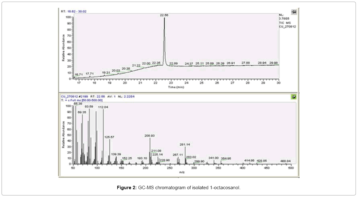
Figure 2: GC-MS chromatogram of isolated 1-octacosanol.
In the mass fragmentation the spectra corresponding to the larger molecules were indicated by less conspicuous peaks. This was due to the fragmentation of 1-octacosanol during the electron ionization process. The following peaks were formed due to the several alkyl radicals, generated (Figure 2). Fragments having m/z ratio of 283.02 with 10% relative abundance corresponded to nonadecane (C20H42); m/z ratio 341.00 with 6% relative abundance corresponded to tricosanol (C23H48O) and finally m/z ratio of 354.95 with 5% relative abundance corresponded to tetracosanol (C24H50O) (Table 1). The chemical composition of the actual target compound was determined from the individual fragment characteristics, comparison with standard and consulting the NIST library. The compound belonged to the polycosanol group, with long hydrocarbon chain, as elaborated in Table 2. The characteristics of the target molecule were determined on the basis of the relative abundance of each fragment formed along with their mass-to-charge ratios. In addition, the single peak obtained from the gas chromatographic data was corroborated by comparison with the standard. The chemical formulae can be represented as shown in Figure 3.
| Hit | m/z ratio | Relative abundance (%) |
|---|---|---|
| 1 | 55.38 | 100.0 |
| 2 | 69.35 | 86.0 |
| 3 | 83.59 | 96.0 |
| 4 | 112.04 | 96.0 |
| 5 | 125.57 | 42.0 |
| 6 | 139.39 | 12.0 |
| 7 | 208.93 | 44.5 |
| 8 | 211.08 | 20.0 |
| 9 | 225.14 | 12.0 |
| 10 | 267.11 | 11.0 |
| 11 | 281.14 | 31.5 |
| 12 | 283.02 | 10.0 |
| 13 | 341.00 | 6.0 |
| 14 | 354.95 | 5.0 |
Table 1: Mass fragmentation of 1-octacosanol.
| GC Retention time (minutes) | Molecular formulae | Molecular weight (g.mol-1) | Nature of compound | Name of compound |
|---|---|---|---|---|
| 22.56 | C28H58O | 410.77 | Aliphatic alcohol | Octacosanol-1-ol or 1-Octacosanol |
Table 2: Characteristics of the target molecule.

Figure 3: Molecular formulae of 1-octacosanol.
X-ray diffraction patterns: Very sharp and high intensity peaks were observed on the x-ray chromatogram of 1-octacosanol. The distinct crystalline nature of the isolated compound was evident. Table 3 shows the diffraction data for 1-octacosanol, measured using filtered CuKα radiation at λ=1.5406, along a 0.1 slit system at 1° (2θ)/ minute. Kα2/Kα1 ratio was 0.50000. Reflections were recorded at half of the maximum intensity. Also, 2θ values were evaluated. The two large Bragg peaks indicated the presence of well-ordered domains within the alcohol crystals (Figure 4). However, at small 2θ angles the lattice spacing could not be accurately correlated with the Bragg equation [17]. The lattice structure and lattice planes played important roles in determining the d-spacing corresponding to the reflection angles. In consequence it was evident from Table 3 that as the 2θ value increased subsequently the d-spacings were diminished.
| Pos. [°2Th.] | Height [cts] | FWHM [°2Th.] | d-spacing [Å] | Rel. Int. [%] |
|---|---|---|---|---|
| 19.8623 | 111.95 | 111.95 | 4.47012 | 9.45 |
| 22.1097 | 1185.25 | 1185.25 | 4.02057 | 100.00 |
| 24.6098 | 482.40 | 482.40 | 3.61749 | 40.70 |
| 27.3385 | 81.74 | 81.74 | 3.26230 | 6.90 |
| 36.5649 | 48.12 | 48.12 | 2.45755 | 4.06 |
| 41.3705 | 62.91 | 62.91 | 2.18071 | 5.31 |
Table 3: X-ray Diffraction analysis of 1-octacosanol.
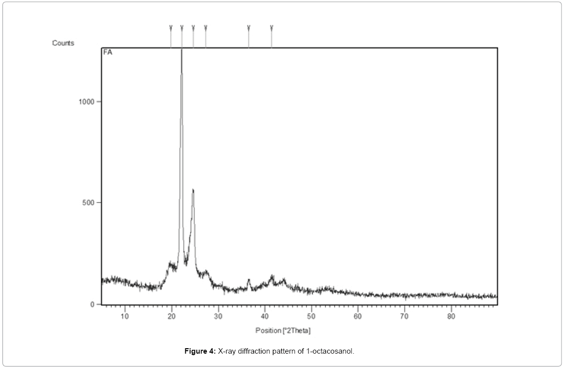
Figure 4: X-ray diffraction pattern of 1-octacosanol.
Antioxidant activities: Antioxidant activities of 1-octacosanol displayed unique behaviors. Five different concentrations were formulated, viz., 0.01 mg/ml, 0.05 mg/ml, 0.1 mg/ml, 0.5 mg/ml and 1.0 mg/ml. It was observed that initially with increasing concentration of octacosanol from 0.01 mg/ml to 0.1 mg/ml, the DPPH radical scavenging activity increased very slowly. However, at 0.5 mg/ml concentration the activity rapidly and significantly escalated to a value of 13.36% from an initial value of 8.05%. But at 1.0 mg/ml concentration, the hike was slow again from 13.36% to 14.38% activities (Table 4). Similar behavior was also observed for the reducing activity. The highest DPPH radical scavenging and reducing activities were observed at 1.0 mg/ml octacosanol concentration. Efficacy of octacosanol in acting as a metal chelator was similar to its radical scavenging activities. However here maximum metal-chelation activity was observed at 0.5 mg/ml beyond which the activity gradually diminished. This could be due to its prooxidant effect at very high concentrations, which barred its metalchelating property. Again, in the present study, low concentrations of octacosanol were not very effective in inhibiting the peroxidation process. But at the highest concentration of 1.0 mg/ml maximal inhibition was observed (Figure 5).
| Concentration (mg/ml) | DPPH radical scavenging activity (%) | Reducing activity (Å) | Metal chelation activity (%) | Inhibition of Lipid peroxidation activity (%) | |
|---|---|---|---|---|---|
| After 1 day | After 5 days | ||||
| 0.01 | 6.07±0.45a | 0.025±0.005a | 14.62±0.95a | - | 1.02±0.56a |
| 0.05 | 7.70±0.23a | 0.026±0.008a | 14.71±0.83a | - | 5.25±0.71b |
| 0.1 | 8.05±0.15b | 0.027±0.003a | 22.22±0.77b | - | 5.39±0.83b |
| 0.5 | 13.36±0.62c | 0.034±0.007b | 22.36±0.68b | 2.90±0.988a | 10.30±1.11c |
| 1.0 | 14.38±0.59c | 0.040±0.006c | 16.15±0.52a | 13.15±1.21b | 19.68±1.23d |
Values are expressed as mean ± S.E.M, n=3. Means were compared using Tukey’s test. At 0.05 level (p < 0.05), the difference in superscripts (a, b, c, d) along a column indicates statistically significant difference in mean values.
Table 4: Antioxidant activities of 5 different concentrations of 1-octacosanol.
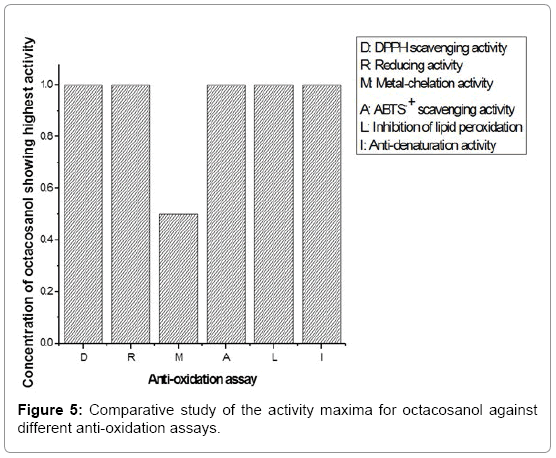
Figure 5: Comparative study of the activity maxima for octacosanol against different anti-oxidation assays.
Anti-bacterial activity: The anti-bacterial activities of 1-octacosanol were studied at two concentrations viz., 1 mg/ml and 2 mg/ml in isopropanol by agar disc-diffusion assay. They were compared with the activity of a standard antibiotic, tetracycline 500 using an isopropanol control. According to the results the zones of inhibition for both the concentrations were recorded. Among the two concentrations used, the higher concentration of 2 mg/ml exhibited good antibacterial activity against the three enteric bacterial strains of Pseudomonas aeruginosa and Escherichia coli with Gram negative origin and Bacillus subtilis of Gram positive origin.
The disc assays against the three strains of bacteria are shown in Figure 6 (for octacosanol concentration of 1 mg/ml) and Figure 7 (for octacosanol concentration of 2 mg/ml). The bacterial strains were plated out as a lawn and filter paper discs (about 5 mm in diameter), soaked in appropriate solution/solvent, were placed on top. The halolike appearance surrounding the filter paper discs indicated the degree of inhibition displayed by the corresponding compound/solvent in which the disc was soaked. The zones of inhibition for B. subtilis were found to be 0.1 cm for 1 mg/ml octacosanol concentration and 0.2 cm for 2 mg/ml octacosanol concentration against antibiotic with 1.0 cm and isopropanol control with only 0.03 cm. In case of E. coli, it was found to be 0.15 cm for 1 mg/ml octacosanol concentration and 0.7 cm for 2 mg/ml octacosanol concentration against antibiotic which was 1.5 cm and isopropanol control with only 0.05 cm only. Finally, best results were obtained for P. aeruginosa where the zones of inhibition were found to be 0.1 cm for 1 mg/ml octacosanol concentration and 0.25 cm for 2 mg/ml octacosanol concentration where for antibiotic it was 1.5 cm and for isopropanol control it was 0.01 cm (Table 5).
| Strain of bacteria | Concentrations | Zones of inhibition (cm)* | |
|---|---|---|---|
| B. subtilis | Concentration of octacosanol | 1 mg/ml | 0.1±0.011a |
| 2 mg/ml | 0.2±0.087b | ||
| Isopropanol control | - | 0.03±0.001c | |
| Concentration of antibiotic | 1 μg/ml | 1.0±0.12d | |
| E. coli | Concentration of octacosanol | 1 mg/ml | 0.15±0.062m |
| 2 mg/ml | 0.70±0.095n | ||
| Isopropanol control | - | 0.05±0.004o | |
| Concentration of antibiotic | 1 μg/ml | 1.5±0.083p | |
| P. aeruginosa | Concentration of octacosanol | 1 mg/ml | 0.1±0.011w |
| 2 mg/ml | 0.25±0.037x | ||
| Concentration of antibiotic | 1 μg/ml | 1.5±0.13y | |
| Isopropanol control | - | 0.01±0.003z | |
Table 5: Zones of inhibition for 1 mg/ml and 2 mg/ml concentrations of octacosanol, isopropanol control and 1 μg/ml concentration of antibiotic.
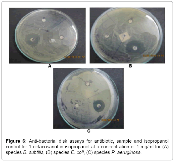
Figure 6: Anti-bacterial disk assays for antibiotic, sample and isopropanol control for 1-octacosanol in isopropanol at a concentration of 1 mg/ml for (A) species B. subtilis, (B) species E. coli, (C) species P. aeruginosa.
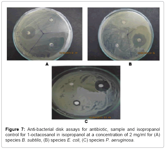
Figure 7: Anti-bacterial disk assays for antibiotic, sample and isopropanol control for 1-octacosanol in isopropanol at a concentration of 2 mg/ml for (A) species B. subtilis, (B) species E. coli, (C) species P. aeruginosa.
The initial stage of isolation of octacosanol involved de-oiling of the wax with acetone. This ensured removal of major oil components like tri-, di-, mono-glycerides and free fatty acids from wax. The wax was then saponified with ethanol and alkali. As a result, the wax ester bonds were split, and fatty acids and fatty alcohols were released into the reaction medium. The fatty acids react with the alkali to form soap which was removed by water-washing. The fatty alcohols were collected with hexane as the unsaponifiable fraction. Previously when 1-octacosanol was isolated from sugarcane rind [12] the products obtained from the unsaponifiable fraction were analysed by GC-MS technique. But there pure 1-octacosanol was not isolated. Further process treatments need to be done to obtain pure products. This was exploited in the present study. In the present work, the unsaponifiable fractions were further treated by fractional distillation followed by crystallization, to isolate 1-octacosanol in pure form.
Octacosanol was identified by the gas chromatography-mass spectrometry method where octacosanol was bombarded with high energy electron beam resulting in generation of low molecular weight fragments. No molecular ion peak or parent peak corresponding to octacosanol had appeared there, clearly emphasizing the fact that complete ionization of the compound had occurred. The instability of the alcohol molecular ion was a major reason for its not being detected as a parent peak. The base peak (100% relative abundance) corresponded to an oxonium ion (CH3CH2CH=O+H). This was the result of the cleavage at the hydrocarbon tail of the fatty alcohol. The isolated compound was an even carbon numbered alcohol of 28 carbons. It contained one hydroxyl group which determined most of its reactivity.
The result of GC-MS analysis was corroborated by the diffraction studies where the Kα1 and Kα2 peak doublets appeared at 22.1097° and 24.6098° respectively, and the relative intensity of Kα1 was more than double that of Kα2. At larger angles they would have appeared further apart from each other. The sharp and large peak at 22.1097° indicated a uniform, homogeneous composition of the fatty alcohol crystals, free from defects. The inter-atomic distance, correlated from the d-spacing data, was a clear indication of the unit cell dimension. The uniformity of the inter-atomic distances also corroborated by the integral breadth (FWHM) at 2θ and was clear of any changes in the crystals brought about by temperature, pressure, etc. It was reflected from the uniformity of the peak positions, crystalline structure and narrow peak width that the compound was highly stable to any kind of environmental stresses. This could be attributed to the long hydrophobic chain that provided immense stability to the compound. The characterizations were in correlation with previous studies on isolation of 1-octacosanol from sugarcane rind [12].
Among the different antioxidant studies DPPH radical scavenging and reducing activitiesare the most popular assays to evaluate the antioxidant potency of natural products. Again, chelation with prooxidant metals is an established method for prevention of oxidation. Several d-block elements exhibit accentuated oxidation behavior by acting as a catalyst in free-radical initiated reactions. Metal-chelators either cause steric hindrance or reduce redox potential, thus inhibiting such oxidation reactions [24]. Furthermore, lipid peroxidation originates due to the presence of free radicals and reactive oxygen species. These are unstable in nature and attack lipids thus degrading them by generation of peroxides [25]. A radical or reactive oxygen species quencher can inhibit such deteriorating effects. In all types of antioxidant assays, concentration of octacosanol plays a pivotal role [4,7,8]. The hydroxyl group was the only functional unit effective in mediating the antioxidant properties of octacosanol. This molecule consisted of a single hydroxyl group. The antioxidant activity could be related to the assembly of the hydroxyl groups [26]. Hence with increasing concentration of octacosanol, the hydroxyl content of the solutions also increased. The free radical scavenging activity was directly correlated to the reducing nature of 1-octacosanol. The reducing property assisted in adsorbing/quenching and then neutralizing the free radicals, which are the primary cause of several degenerative and lethal diseases. Octacosanol effectively scavenged the free radicals as it showed potent reducing activities generating from the hydroxyl group, and would be thus effective against free-radical induced diseases. By comparing the different concentrations of octacosanol for their anti-oxidation behavior, it was observed that the concentration of 1 mg/ml showed the best result for most of the assays followed by the concentration 0.5 mg/ml. Thus, higher concentrations of 1-octacosanol were more effective in preventing oxidation. Our studies were limited to the highest concentration of 1 mg/ml. However probably at higher concentrations greater quenching properties will be observed, leading to further enhancement of nutritional and therapeutic values of the compound. Hence the present study indicates the possibility of future studies at higher octacosanol concentrations.
The antibacterial studies of octacosanol also provided promising results. E. coli normally resides in the lower intestine of organisms and may be the cause of food poisoning, diarrhea, severe anemia and sometimes kidney failure [27]. P. aeruginosa is a harmful, pathogenic drug resistant bacterium associated with serious illnesses like pneumonia, sepsis, urinary tract infection, gastro-intestinal infections, etc. [28-30]. B. subtilis resides in the upper layers of soil and the gastrointestinal tract of humans and is highly tolerant to drastic environmental conditions. It has no known harmful effects, instead contributes towards the health and well-being of humans. It is the most widely studied bacterium today [31]. In the present work, disc assay was performed to study the antibacterial activity of octacosanol. Octacosanol diffuses from the disc to the agar and then prevents the growth of the test microorganisms up to a certain distance. This distance or zone of inhibition can be measured in terms of the diameter of the halo-like appearances. In each case along with the zones of inhibition of the sample solutions, a corresponding antibiotic (tetracycline 500) solution with a concentration of 1 μg/ml and an isopropanol control were also studied. From the zones of inhibition values, it was evident that 1-octacosanol was not as strong an antibacterial agent as the antibiotic tetracycline, however, displayed enough encouraging results for industrial application as a weak antibacterial agent. Furthermore, the fact that it was a primary alcohol isolated from oil industry waste products, surplused as an added advantage as the raw material cost was almost negligible.
The studies have suggested that 1-octacosanol possessed a broad spectrum of activities against both gram positive and gram negative bacterial strains. In fact, its activity was higher against gram negative bacteria E. coli and P. aeruginosa, indicating its possible medicinal application at higher concentrations. This fact justifies the arduous extraction technologies leading to its isolation in pure form. This study was also in correlation to a previous study on anti-microbial activities of plant extracts, where one of the compounds identified was octacosanol [32]. This implied that 1-octacosanol can be useful as antiseptic formulations in the pharmaceutical, food and nutrition, neutraceutical, or health and hygiene sectors.
Our study showcased that 1-octacosanol can be isolated in a considerable pure form from rice bran wax by adopting suitable technologies. Additionally, 1-octacosanol displayed good anti oxidant activity in the range of 0.5 mg/ml to 1.0 mg/ml. Furthermore, the presence of 28 carbon atoms led to the demonstration of antibacterial activity by 1-octacosanol against three different types of bacterial strains and the hydroxyl group was the genesis of the antioxidant properties. The isolation of 1-octacosanol in a pure form was a convoluted process, entailing intricate separation of the pure compound from other naturally occurring products in wax. Thereafter its identification and characterization involved choice of suitable technologies to pin-point its structural make-up as was done using FTIR and GCMS. Its stress resistance, derived from diffraction studies, indicated its structural stability. Finally, the anti-oxidation and anti-bacterial properties defined its beneficial aspects in relation to the upheaval of human health. Thus, our studies distinctly justified the isolation of 1-octacosanol and implicated the fields in which it can find application. The study also encouraged further researches using higher concentrations of 1-octacosanol. Other long chain fatty alcohols should also be studied in this regard.
The authors would like to acknowledge the ‘Council of Scientific and Industrial Research’ (CSIR) for their financial support. Furthermore, the Centre for Research in Nanoscience and Nanotechnology (CRNN), University of Calcutta, is acknowledged for extending their FTIR facilities. Bose Institute, Centenary Campus, Kolkata, West Bengal, is also acknowledged for providing their GCMS facilities.
The authors have declared no conflict of interest.