Journal of Psychology & Psychotherapy
Open Access
ISSN: 2161-0487
ISSN: 2161-0487
Research Article - (2015) Volume 5, Issue 3
Objectives: Meditation is described in
traditional yoga
texts as three stages, which follow each other in sequence: (i)
Focused attention
(FA), (ii) Focused attention on the object of meditation (MF), and (iii) Meditation with one-pointed focused attention without effort (ME). When not in meditation the mind is considered to be in a state of normal consciousness characterized by random thinking (RT). The objective of the present study was to determine the brain areas activated during the three stages of meditation compared to the control state using fMRI. Methods:
Functional magnetic resonance images
were acquired from twenty-six right handed meditators during MF, ME and random thinking (RT) for comparison. Ten of them were experienced (average age ± SD, 37.7 ± 13.4 years; 9 males) with 6048 hours of meditation, whereas 16 (group average age ± SD, 23.5 ± 2.3 years; all males) were less experienced, with 288 hours of meditation. During the fMRI recordings the participants practiced RT, non-meditative focused thinking (FA), MF and ME, each lasting for 2 minutes. Brain areas activated during the intervention were scanned using a 3.0-Tesla Philips-MRI scanner. Results: During the third phase of meditation (ME) the experienced meditators alone showed significant activation in the
right middle temporal cortex
(rMTC), right inferior frontal cortex (rIFC) and left lateral orbital gyrus (LOG) (p < 0.05), Bonferroni adjusted t-tests for unpaired data, comparing ME and random thinking. Conclusions: These changes suggest that ME is associated with sustained attention, memory, semantic cognition, creativity and an increased
ability to detach mentally
<Keywords: Meditation; Yoga; Traditional texts; Random thinking; fMRI; Focused attention; Effortless focused attention
Meditation can be considered to be a training in awareness which produces definite changes in perception, attention, and cognition [1]. Meditation is also recognized as a specific consciousness state in which deep relaxation and increased internalized attention co-exist [2]. Perhaps related to this is the concept that directing and regulating attention are considered an inherent part of different meditation techniques [3].
Multiple neuroimaging studies on meditation have attempted to describe the cognitive processes involved. The most common examples are of focused attention and open monitoring meditation [4,5]. There appears to be no neuroimaging study which has categorized the process of meditation based on traditional texts whether Buddhist, Yoga, Chinese or any others. The present study aimed to compare three stages of yoga meditation described in Indian yoga texts with the mental state that is described to exist when not in meditation. This non-meditative state is characterized by both mind-wandering and switching of attention at random. It has been described in traditional texts as the characteristic mental state when the mind is not directed or instructed (Cancalata in Sanskrit; Bhagavad Gita, Circa 500 B.C.; Chapter 6, Verse 34; simplified here as random thinking or RT) [6]. This was considered as the control state against which the stages of meditation were compared. In an attempt to describe this mental state with contemporary descriptions it can be considered as normal consciousness [7].
¨Traditionally it is mentioned that in order to reach a meditative state attention should be focused and maintained. In order to do this different meditation techniques use varied objects, mantras as well as interoception [8].
As a practitioner attempts to meditate there are three successive stages, these are: (i) Focused attention (FA), (ii) Meditative focusing (MF) and (iii) Pure meditation (ME). In FA the practitioner attempts to return to focus when the mind wanders and attention is directed to several thoughts about the same subject (in the present study the thoughts were on the concepts of meditation). During Meditative focusing (MF) focusing of attention is directed to a single thought (in the present case the Sanskrit syllable ‘Om’), with the exclusion of all distractions, which requires effort. Pure meditation (ME) occurs as the practitioner continues with the second stage the stage of pure meditation is spontaneously reached, where attention is on a single thought (in this case the syllable ‘Om) but there is no effort involved.
The descriptions of each stage of meditation in the traditional texts give greater clarity about the processes involved. The first stage (FA) is called ekagrata in Sanskrit (Bhagavad Gita, Chapter 6, Verse 12), during which attention is directed to a series of associated thoughts. As mentioned above if the thoughts are related to meditation, the person would then be able to progress to the next two stages, dharana (MF) and dhyana (ME). Dharana (or focusing with effort), is described as ‘confining the mind within a limited mental area’ (‘deshabandhashchittasya dharana’, Patanjali’s Yoga Sutras, the sage Patanjali Circa 900 B.C.; Chapter 3, Verse 1) [9]. The next state is dhyana or effortless expansion called pure meditation This state is described as ‘the uninterrupted flow of the mind towards the object chosen for meditation’(‘tatra pratyayaikatanata dhyanam’, Patanjali’s Yoga Sutras, the sage Patanjali Circa 900 B.C.; Chapter 3, Verse 2).
The difference between dharana and dhyana in using effort to direct attention is supported by data which show a shift towards vagal dominance during dhyana [10]. Apart from the autonomic variables there have been electrophysiological recordings of short [11], middle [12] and long latency [13] auditory evoked potentials during FA, MF, ME as well as during the control state (RT) of random thinking. The changes were both in the time taken for information transmission (i.e., the latency) as well as in the number of neurons recruited (indicated by the amplitude). However auditory evoked potentials were specific for the auditory pathway and the neural generators were correspondingly specific to that pathway. Also evoked potential recordings do not give spatial and temporal resolution which fMRI provides to localize changes in brain functions.
Hence the present study was designed to compare the parts of the brain involved in three successive stages of traditionally described yoga meditation (i.e., FA, MF and ME) each with the mind wandering state (RT) using fMRI.
Participants
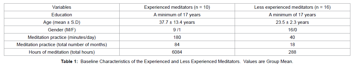
The participants were twenty-six right handed trained meditators. Ten of them (9 males; group average age ± SD; 37.7 ± 13.4 years) had 7 years of experience of meditation {(7 years × 12 months × 24 days × 180 minutes)/60} = 6048 hours), practiced as the two stages, meditative focusing (ME) leading to pure meditation (ME). The other sixteen meditators had 18 months experience of the same meditation. They were all males and had an average age of 23.5 ± 2.3 years with experience of 288 hours {(18 month × 24 days × 40 minutes)/60} = 288 hours). The two groups significantly differed with respect to age (t = 4.11 ; df = 24 ; p = 0.0003). Baseline characteristics of the experienced and less experienced meditators are given in Table 1. Participants were recruited for the trial by notices on the notice boards of the institution, the Indian Council of Medical Research Center for Advanced Research (ICMR-CAR), located in Bangalore, south India. This center is attached to a residential yoga training center where meditators receive training in meditation and come for advanced retreats. There was no incentive to take part in the study and while the study design was explained to the participants, the research question was not. To be included in the trial participants had to meet the following criteria (i) normal health based on a routine physical and mental health examination, (ii) right hand dominance based on a routine hand dominance inventory [14], and (iii) regularity in their practice of meditation, where regularity meant practicing for at least 40 minutes a day for six days in a week. The experienced meditators practiced for 180 minutes in a day while the inexperienced meditators practiced for 40 minutes each day. Pre-determined exclusion criteria were: (i) if they were not able to be scanned due to claustrophobia, metal implants, a pacemaker, or pregnancy, and (ii) inability to meditate in the scanner environment. None of the participants had to be excluded for these reasons. The study was approved by the Institutions’ ethics committees of the (i) Indian Council of Medical Research Center for Advanced Research (ICMR-CAR), and (ii) the National Institute of Mental Health and Neurosciences (NIMHANS), both located in Bangalore in south India. Signed informed consent was obtained from all the participants following the guidelines of the Indian Council of Medical Research.
Intervention
During the fMRI recordings, the participants were asked to practice the control and the three meditation sessions in the following order i.e., random thinking, non-meditative focused thinking, meditative focusing, and effortless meditation or pure meditation, each lasting for 2 min. The oral instructions were given from the control room through noise-canceling electrostatic headphones.
Random thinking
Participants were asked to keep their eyes closed and allow their thoughts to wander freely as they listened to a compiled audio CD consisting of brief periods of conversation, announcements, advertisements and talks on diverse topics recorded from a local radio station transmission. These conversations were not connected and hence it was thought that listening to them could induce a state of random thinking.
Non-meditative focused thinking (FT)
Participants were asked to keep their eyes closed and listened to a pre-recorded lecture on concepts of meditation. This was intended to induce a state of non-meditative focusing.
Meditative focusing (MF)
During training participants were asked to open their eyes and gaze at the Sanskrit syllable ‘Om’ as it is written in Sanskrit. However in the scanner they were asked to keep their eyes closed. During this time guided instructions through a pre-recorded audio tape required them to direct their thoughts to physical attributes of the syllable, i.e., the shape, the size and the color. The main emphasis during meditative focusing was that thoughts are consciously brought back if they wander to the single thought of ‘Om’.
Effortless meditation or pure meditation (ME)
During this session participants were instructed to keep their eyes closed and dwell on thoughts of ‘Om’, particularly on the subtle (rather than physical) attributes and connotations of the syllable. This would gradually allow the participants to experience brief periods of silence, which they reported after the session.
A block design was used. The paradigm consisted of two repeat sessions of 8 minutes duration. The session was repeated on another day at the same time of the day. Each session had 4 blocks corresponding to Random Thinking (RT), Focusing (FC), Meditative Focusing (MF) and ‘pure’ Meditation (ME) in a fixed sequence, for 120 seconds per block, 20 dynamic scans per block (20 × 4 = 80 dynamic scans in one session); hence in total 160 dynamic scans from the 2 sessions were used for analysis. Participants had been informed that a simple instruction to change their mental state would be given using the intercom to avoid their getting startled.
The sequence (i.e., RT-FC-MF-ME) was fixed. The fact that it was not randomized is a disadvantage of the study. However (i) this sequence is pre-determined in the traditional descriptions [9], and (ii) participants had been trained to follow a fixed sequence during familiarization sessions in the scanner environment.
For one month prior to the experiment the participants were trained to meditate in a fabricated ‘simulated scanner’ which was a cylinder of comparable dimensions. During this time the participants were required to listen to pre-recorded ‘scanner noise’ which was recorded during actual acquisition. These familiarization sessions were of the same duration as the actual recording sessions. The practice session included two trials : that is 2 minute sessions for each of the 4 states, practiced in 16 minute sessions, 5 days a week during the month.
Functional image data acquisition and reduction
MRI scanning was conducted using a 3.0-Tesla Phillips-MRI head scanner with an 8 channel head frequency coil. To minimize motion artifact the participants’ head was padded with foam coil. Functional images were acquired in 160 slices rotated about 30o above the anteriorposterior commissure (AC-PC) using a T2*-weighted EPI pulse sequence (repetition time, TR=3000; echo time, TE=35;flip angle, FA=90°;field of view, FOV=230×230×128 mm; slice thickness = 8mm, with 0mm slice gap). The 30oline offset was intended to reduce signal loss due to susceptibility artifact in the orbito-frontal cortex [15]. Scan acquisition was time-locked to the onset of each trial. Before functional scanning, a T1-weighted MP-RAGE high resolution 3D anatomical image was acquired. There were 160 slices, 1 mm thick; TR=8.1 ms; TE=3.7 ms; FA=90°; FOV=240×240×160 mm. The purpose was to evaluate structural abnormalities (there were none) and to allow for transformation of functional data into standard reporting space for spatial normalization [16]. With the block design paradigm used, which is detailed above and in Figure 1,160 dynamic scans from the 2 sessions were obtained.

Figure 1: Experienced meditators (n = 10): Areas showing supra threshold activation in right middle temporal cortex, right inferior frontal cortex and left orbital gyrus in meditation (p < 0.05, t-tests for unpaired data Bonferroni adjusted following one-way ANOVA).
Imaging data were processed using Brain Voyager (BVQX 2.1; Brain Innovation, Maastricht, The Netherlands). Preprocessing included (i) 3-D motion correction using trilinear interpolation, (ii) slice-scan time correction to temporally realign the slices, (iii) spatial smoothing using a 3D 6mm full width at half maximum (FWHM) Gaussian filter, (iv) voxel-wise linear detrending, and (v) temporal filtering of frequencies below 3 cycles per time course to remove low frequency non-linear drifts. Registration of the functional images to anatomical volumes was completed with standard BVQX methods. For group-wise analysis, spatial normalization of functional images was carried out by scaling the functional images into standard Talairach space.
Self–Report of Meditation on Visual Analog Scales (VAS)
At the end of each session participants were asked to rate the extent that they felt they were able to follow instructions on a liner continuous scale from 0 to 10, where 0 meant ‘not being able to at all’ and 10 meant ‘being able to do so perfectly’.
Data Analysis
Imaging data were analyzed using whole brain voxel-wise statistical tests (Brain Innovation Version 2.1, The Netherlands). The Talairach Client (Version 2.4.3) was used to assign Talairach atlas 3D coordinates and overlay statistical maps onto the reference anatomical image, transformed as standard reporting co-ordinates.
A General Linear Model was applied for group whole-brain analysis. Following separate one-factor ANOVAs for each of the two groups, separate t-tests were carried out to compare overlay values of (i) Focused attention (FA), (ii) Meditative Focusing (MF), and (iii) Pure Meditation (ME), where each of them were compared with Random Thinking (RT) for comparison, and for any change. The level of significance was p < 0.01 with a cluster threshold of 10. Comparisons were made with t-tests which were Bonferroni adjusted or FDR corrected to reduce Type I errors
(i) Self-rated ability to switch between states on the VAS: All participants rated their ability to switch between states as 7 or more on the 10 point scale, where 0 meant ‘not able to switch at all’ and 10 meant ‘able to switch perfectly without any difficulty at all’ [17]. There was no further analysis performed on the selfreports,
(ii) The imaging data of the two sets of participants, (a) experienced meditators with 6048 hours of meditation practice, and (b) the less experienced meditators with 288 hours of experience of meditation practice:
Experienced meditators
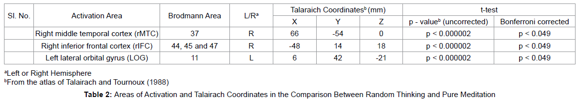
The 10 experienced meditators showed a significant change in the comparison between pure meditation (ME) and random thinking (RT) (p = 0.049, one tailed); One Factor ANOVA followed by Bonferroni adjusted t tests). Areas showing supra-threshold activation are mentioned in Table 2 and shown in Figure 1.
Less experienced meditators
There were no significant areas of activation for the three comparisons, which is (i) RT with FA, and (ii) RT with MF and (iii) RT with ME (p > 0.05); One Factor ANOVA after Bonferroni adjusted t tests
Meditators with a total of 6048 (7 years) of experience of meditation on the Sanskrit syllable “Om’ showed significant activation in the right medial temporal cortex (rMTG), right inferior frontal cortex (rIFG), and left orbital gyrus (LOG) during the stage of effortless or “pure” meditation. The comparison was with a period of random thinking. There were no changes during meditation with focusing or during focusing alone compared to random thinking.
In the present study the activation was observed in the right middle temporal cortex and right inferior frontal cortex which has been observed in earlier studies on meditation [18]. The medial temporal cortex is known to be involved in cognition and specifically in memory processing [19,20]. Other aspects of cognition required for memory such as attentional control are regulated by the inferior frontal gyrus [21,22]. Multichannel EEG of an advanced meditator during four different meditations using Low Resolution Electromagnetic Tomography (LORETA) was carried out. Functional images showed activation in the right fronto-temporal region along with other areas. The right fronto-temporal areas are considered to be involved in self-induced meditational dissolution and reconstitution of the experience of the self. Hence the results of the present and the earlier study [18] suggest that meditation activates brain areas concerned with self-representation. While the LORETA study [18] demonstrated activity in the right fronto-temporal region, the present study showed activity specifically in the right inferior frontal cortex. These results are comparable with an eLORETA study. Here eLORETA was used to compare differences in cortical source activity in intermediate (average experience 4 years) and advanced (average experience 30 years) Australian meditators of the Satyananda Yoga tradition [23]. Assessments were made during a body steadiness meditation, mantra meditation and non meditation mental calculation. Across all conditions differences were greatest in the same regions as the present study which included the right inferior frontal gyrus, and right anterior temporal lobe.
The above studies [18,23] demonstrated changes in the right inferior frontal gyrus and temporal region. The activation of the rMTG reported in the present study is in contrast to the findings of a report [24] which measured the performance of participants during an fMRI adapted Stroop word-color task. The comparison was between meditators and non-meditators. The Stroop task performance was comparable for the two groups. The MTG among other regions showed greater activity in the non-meditators than meditators during the incongruent task condition. The absence of activity during meditation in these areas was considered to suggest that meditation improves efficiency possibly through sustained attention and impulse control. The fact that the middle temporal gyrus was activated during pure meditation (ME) in the present study could be related to the fact that in this state attention was maintained on the object of focus without effort. The findings of the present fMRI study may be correlated with a morphometry assessment of cortical thickness in Brain Wave Vibration (BWV) meditation [25], a practice intended to increase awareness. Among other areas the meditators showed greater cortical thickness in the temporal cortex [25]. The regions with greater thickness were considered to be involved in internal mentation or attention that is detached from the external world [26]. While the present study demonstrated significantly greater activation in the right middle temporal cortex based on functional neuroimaging, structural cortical thickness mapping and diffusion tensor imaging showed greater cortical thickness in 46 experienced meditators compared with 46 matched meditation naïve volunteers in several brain areas including the middle temporal cortex [25].
The increased activation in the inferior frontal cortex in the present study has been reported in another neuroimaging study on meditation [27]. When two meditation techniques, a ‘focused based’ practice and a ‘breath based’ practice were studied, a strong correlation was found between the depth of meditation and activation in several areas of the brain including the inferior frontal cortex and temporal pole [28].
In the present study the increased activation of the lateral orbital gyrus during meditation may be associated with certain changes in mental attitude. The LOG is associated with specific personality traits including Machiavellian scores [29,30]. The Machiavelli personality is described as unemotional and detached from social morality for personal benefits. During meditation there is a possibility of attaining a mental state detached from all thoughts unrelated to meditation [31]. The activation of the LOG during ME suggests detachment which is ideal in meditation provided it co-exists with empathy, social consciousness and compassion. Also the orbital gyrus is considered to have a role in processing changes in reward related information [32]. Meditation could possibly influence factors involved in reward gratification with a detached attitude.
In meditation the ability to voluntarily shift from normal consciousness to meditation is enhanced. Thirty one meditators with meditation experience between 1.5 and 25 years were assessed using a block on-off design with 45 seconds alternating epochs. During the onset of meditation and normal relaxation SPM and ICA analysis showed activation in multiple regions in the frontal, temporal, parietal and limbic areas which was presumed to constitute a combination of fronto-parietal and cingulo-oppicular activation [33]. The block design in the present study which required practitioners to switch between random thinking and the three stages of meditation within a short period suggests that experienced meditators were able to change from non-meditation to meditation even though this was assessed subjectively without any biological marker.
It was also found by the study of Thomas et al. [23] that the networks greatly expanded during meditation practice to include homologous regions of the left hemisphere. It may be speculated that this may be true for the present study as well. Hence the apparent restriction of activation to the right hemisphere may be a partial result with the actual activation involving an extended network within the brain.
The absence of changes in the less experienced meditators is possibly related to their shorter duration of meditation experience, rather than to other differences between the groups such as the age. This is supported partly by a single study [34] which did not find any difference in self-focused attention between two groups whose mean age differed by 10 years. However the contribution of the difference in ages cannot be entirely ruled out.
The present study has certain unique features, particularly the attempt to study changes in the brain during meditation as described in traditional texts. This description does not specify a particular object or mantra, but describes a process to direct attention which can be used across different meditation techniques. The findings are limited by factors such as (i) the fixed sequence in the block design even though the stages of meditation are sequential, (ii) the absence of a group of non-meditators, (iii) the experienced meditators’ ages varied considerably, though their experience and intensity of meditation experience was comparable and (iv) the self-reports of efficacy to shift from state to state could have been influenced by subjectivity and the short time intervals of each block (2 minutes) made it all the more necessary to check this.
In conclusion, the present results showed that there are differences during effortless or ‘pure’ meditation as described by traditional yoga texts compared to random thinking, involving activation of areas involved in semantic cognition, memory, sustained attention, creativity and the ability to detach mentally.
The authors gratefully acknowledge the funding from the Indian Council of Medical Research (ICMR), Government of India, as part of a grant for a Center for Advanced Research in Yoga and Neurophysiology (CAR-Y&N), (Project No. 2001-05010).