Annals and Essences of Dentistry
Open Access
ISSN: 0975-8798, 0976-156X
ISSN: 0975-8798, 0976-156X
Research Article - (2024)Volume 16, Issue 1
Studies have been directed towards the production of new titanium alloys, aiming replacement of TiAlV alloy in the future. Therefore, many mechanisms related to biocompatibility and chemical characteristics have been studied in the field of implantology, but until now, enzyme defenses against oxidative stress have been underexplored. The objective of this study was to evaluate the activity of the antioxidant enzymes Superoxide Dismutase (SOD) and Catalase (CAT) in porous scaffolds of Ti-6 Aluminium-4 Vanadium, Ti-35 Niobium and Ti-35 Niobium-7 Zirconium-5 Tantalum. Porous titanium alloy scaffolds were prepared by powder metallurgy. Mesenchymal stem cells from mouse femur were used. After 24 hours the cells plated under the scaffolds were analyzed by Scanning Electron Microscopy (SEM). The antioxidant enzymes activity was measured after 72 hours of cell plating and after 3,7 and 14-days, cells were collected to evaluate RUNX2 expression by real-time PCR. The SEM images showed the presence of interconnected pores and growth, adhesion and cell spread in the 3 scaffolds. Our results for SOD and CAT were shown to be better for the TiNbZrTa and TiAlV scaffolds. The TiNb scaffold presented less activity for both enzymes. Despite differences between the scaffolds, there were not any statistical differences (p>0.05). RUNX2 presented high expression for TiNbZrTa and TiAlV at day 7 and presented statistical differences just for TiNbZrTa from the control group (TiAlV day 3). At day 14, all scaffolds showed expression above the baseline and exhibited statistical differences from the control group. It was possible to conclude that, as regards antioxidant enzymes, we are not able to confirm any differences which would justify the replacement of TiAlV, but gene expression showed a better capacity for mesenchymal stem cell differentiation in TiNbZrTa, making it a good choice for biomedical applications.
Titanium alloy; Porosity; Antioxidant enzyme; RUNX2
For a biomaterial to be considered excellent for bone replacement, it must show characteristics that are compatible with its use in the long term, that there are no adverse tissue reactions, that it has excellent corrosion resistance in the body fluid medium, exhibits high mechanical strength and fatigue resistance, low modulus of elasticity and good wear resistance. Titanium and TiAlV alloy, until now, has been indicated as the best for bone substitution, because it presents excellent physical and biocompatibility properties. However, one of its main disadvantages is the large difference in modulus of elasticity between biomaterial and bone. This fact is particularly important, because it can lead to long-term bone loss and consequent failure of implant function. TiAl alloy, which is widely used in the field of orthopedics, exhibits a singularly extra disadvantage: Its Al (Aluminum) and V (Vanadium) components have cytotoxic potential and can be released in the long term, causing adverse biological effects [1].
Therefore, studies have also been directed towards the production of new titanium alloys, with the insertion of other metals, aiming to improve their characteristics, especially with respect to the modulus of elasticity and long-term cytotoxicity. There are several studies showing the effectiveness of titanium bonding to metals such as Nb (Niobium), Zr (Zirconium) and Ta (Tantalum) resulting in non-cytotoxic alloys, with good biocompatibility and modulus of elasticity closer to the bone.
Porosity is another feature of titanium alloys that has been extensively studied in order to improve stress shielding in the bone-implant interface due to mismatching of elastic modulus between implant and bone. Studies show that interconnected pores increase bone implant contact and can be more favorable for transport and exchange of substances necessary for cell growth, allowing bone growth within the biomaterial, leading to a good biological fixation. The creation of interconnected pores may also help to decrease the modulus of elasticity, which would reduce the shielding stress, thus extending the implant life [2]. Porous titanium alloys have been studied for their physical and chemical characteristics, biocompatibility and osteogenesis potential, but there are no studies aiming to evaluate the oxidative stress on these biomaterials when they are in contact with cells, and what the role of oxidative stress is in the failure of biomaterials in the long term.
Oxidative stress occurs when there is a difficulty or lack of elimination of reactive oxygen species, which in high doses can cause damage to the cells and consequently to the tissues. Ozmen believes that this situation could play an important role in the development of complications in the implant, since at high concentrations, irreversible injury to cells and tissues is caused. There are defense mechanisms against oxidative stress, which are so-called primary antioxidant enzymes such as Catalase (CAT) and Superoxide Dismutase (SOD). These are able to neutralize Reactive Oxygen Species (ROS), making them harmless to the cellular environment [3].
We believe that a failure in the activity of these enzymes may be closely linked to implant loss. For this reason, we are interested in studying the activity of SOD and CAT on porous scaffolds of Ti6Al4V, Ti35Nb and Ti35Nb7Zr5Ta alloys, aiming with our results to contribute to a possible substitution of TiAlV alloy, which is one of the major challenges in current implantology. In addition, we also evaluated the expression of the RUNX2 gene to analyze whether the porosity associated to the chemical composition of the samples has a direct action in the cell differentiation process [4].
Porous scaffold preparation
The porous scaffolds of Ti6Al4V (control group), Ti35Nb (study group) and Ti35Nb7Zr5Ta (study group) used in this study were previously prepared by our research group using a special powder metallurgy process described in great detail. The powder of pure Ti grade II (CpTi) (purity 99.5%, <8 μm) was obtained by the Hydrogenation and Dehydrogenation process (HDDH) at the department of science and aerospace technology (DCTA) materials division of the institute of aeronautics and space (AMR/IAE) to be mixed with the powders aluminium (purity 99.5%, <5 μm), vanadium (99.9%, <325 mesh), zirconium (purity 99.5%, <325 mesh), tantalum (purity 99.9%, <325 mesh) and niobium (purity 99.8%, <45 μm), which were purchased (Sigma ± Aldrich, St. Louis, MO, USA). In order to create porosity, the alloy powders were mixed with urea (purity >98.0%, powder) (Sigma ± Aldrich, St. Louis, MO, USA), which acted as a spacer. Urea and the powders of each alloy were combined in a planetary grinding machine in order to obtain a completely uniform mixture. To remove the urea, the samples were incubated in a vacuum oven (Marconi, Piracicaba, Sao Paulo, Brazil), at 200°C for 2 hours. The samples obtained were disc-shaped scaffolds, fabricated using a steel mold: 12 mm in diameter and 5 mm in height and which were totally porous. The porosity of the samples was 40% and the pores showed a mean diameter of 300 μm. The samples were carefully grouped, because it is impossible to see differences macroscopically [5].
Cell culture
The project was approved by the Institutional Animal Care and Use Committee (IACUC) at the university of Michigan and is in conformity with the arrive guidelines. Bone marrow from the femurs of male mice Tg (Sp7/mCherry)2Pmay/J were collected. Two male mice (average body weight of 30 grams and 7-8 weeks old) were used for this propose. The animals were housed in individual cages, with freely available water and food and an artificial day/night cycle of 12 hrs/12 hrs in an air-conditioned room. Euthanasia occurred by carbon dioxide inhalation and the segments of femurs were removed to be processed for the experiments. The femurs were dissected and the proximal epiphysis were cut off so that the bone marrow could be flushed out. The femurs were placed in a 200 μl pipette tip with cut ends, placed in a 1.5 ml micro centrifuge tube and were centrifuged at 2,000 rpm for 5 minutes in order to obtain the cells. They were resuspended in 1 ml of MEM-Alfa modified with Earle's salts (MEM-α) (Gibco-Life, Grand island, NY, USA) supplemented with 10% fetal bovine serum (Gibco) and antibiotic/antimycotic (penicillin/streptomycin/amphotericin B) (Sigma chemical Co., St. Louis, MO, USA). Red blood cells were removed and total cells were counted. 1.5 × 10⁶ cells were plated onto prepared titanium scaffolds alloys in 250 μl of growth media [6].
Sample characterization
One sample of each scaffold was examined under a highresolution scanning electron microscope (Philips XL30 FEG, SEM, Philips, Eindhoven, Netherlands) after 24 hours of cell culture. Observations were made at three randomly selected points on the titanium alloy, at four different magnifications.
Superoxide dismutase assay
After 72 hours of growth in the scaffolds, cells were collected and the activity of the antioxidant enzyme Superoxide Dismutase (SOD) was measured using a Superoxide dismutase kit (Cayman chemicals, Ann Arbor-MI, USA). The media was removed and 250 μl of trypsin was placed on scaffolds. This was necessary because the cells had grown and adhered inside to the scaffolds. After one minute, cells were centrifuged at 2,000 rpm for 10 mins at 4°C in order to remove the trypsin. The cell pellets were homogenized in cold 20 mM Heppes buffer, pH 7.2, containing 1 mM EGTA, 210 mM manitol and 70 mM sucrose. This mixture was centrifuged at 1,500 rpm for 5 mins at 4°C. The supernatant was removed and placed on ice for analysis. The experiments were performed in triplicate following the manufacturer’s instructions. The assay used measured total SOD activity [7].
Catalase assay
After 72 hours growth under scaffolds samples, the cells were collected and the activity of the antioxidant enzymes Catalase (CAT) was measured using a catalase assay kit (Cayman chemicals, Ann Arbor-MI, USA). The media was removed and 250 μl of trypsin was placed on the scaffolds. After one minute, the cells were centrifuged at 2,000 rpm for 10 mins at 4°C in order to remove the media. The cell pellets were homogenized on ice in 1 ml-2 ml of cold buffer 50 mM potassium phosphate, pH 7.0, containing 1 mM EDTA. The mixture was centrifuged at 10,000 rpm for 15 mins at 4°C. The supernatant was removed and placed on ice for analysis. The experiments were performed in triplicate following the manufacturer’s instructions [8].
RNA isolation and analysis
Cells were collected and 14 days after plating. The media was removed and the scaffolds were rinsed twice with cold phosphate-buffered saline. For evaluation of mRNA expression on the titanium alloy scaffolds, adherent cells in each sample were lysed using Trizol (Invitrogen, Carlsbad, CA). The cell lysates were collected by pipetting and centrifugation. The samples were kept frozen at -80°C for at least 24 hours. Total RNA in the cell lysates was isolated according to the manufacturer’s protocol and collected by ethanol precipitation. Total RNA was quantified using a spectrophotometer (PowerWave HT-BioTek instruments) and the Gen5™ program (BioTek instruments). From each total RNA sample, cDNA was generated using SuperScript VILO cDNA synthesis-invitrogen) by a standard 20 μl reaction using 50 ng of the total RNA. Subsequently, equal volumes of cDNA were used to program real-time PCR reactions specific for mRNAs encoding the early osteogenic marker: RUNX2. The primer was obtained from Qiagen (Qiagen, Germantown, MD) and a qPCR reaction was performed in an ABI 7900HT fast real-time (Applied biosystems, Foster city, CA). Relative mRNA abundance was determined by the 2-ΔΔCt method and reported as a fold induction. GAPDH abundance was used for normalization. TiAlV scaffold at day 3 was used as the control group [9].
Statistical analysis
The GraphPad Prism 8 (GraphPad, San Diego, CA) was used to perform the statistical analyses. Data was analyzed using one-way ANOVA, followed by the Tukey test when necessary. The real time PCR result was shown as a fold change by the 2-ΔΔCt method, in baseline 2, with Ti6A4lV scaffold 9 (day 3) being used as the control. The test-t was used as a statistical test for comparison between the TiAlV control group and the other groups. For all statistical analyses, the level of significance was set at p<0.05 [10].
Surface analysis
At low magnification (500 ×) SEM images showed all titanium alloys scaffolds with the presence of interconnected pores. At this resolution it was possible to see the presence of cells interacting with the porous created in the scaffolds. At resolutions of 1000 × and 2000 ×, SEMs revealed growth and spreading of the cells in all scaffolds. At a high resolution of 4000 × it was possible to see better the interaction between the cells, with excellent adhesion, spreading of the cells and presence of filopodia and lamepodia (Figures 1-3) [11].
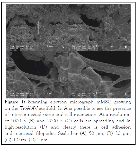
Figure 1: Scanning electron micrograph mMSC growing on the Ti6Al4V scaffold. In A is possible to see the presence of interconnected pores and cell interaction. At a resolution of 1000 × (B) and 2000 × (C) cells are spreading and in high resolution (D) and clearly there is cell adhesion and increased filopodia. Scale bar (A) 50 μm, (B) 20 μm, (C) 10 μm, (D) 5 μm.
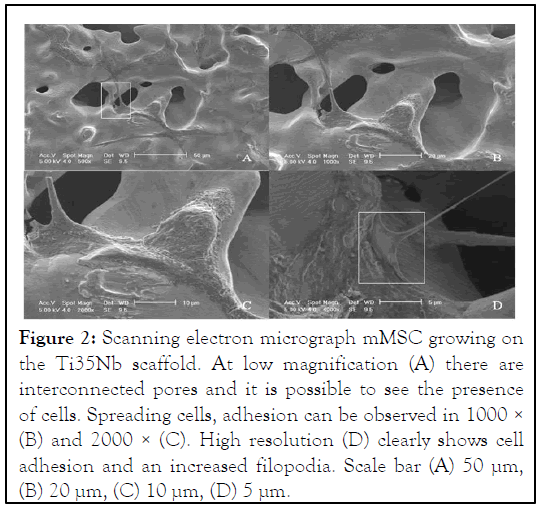
Figure 2: Scanning electron micrograph mMSC growing on the Ti35Nb scaffold. At low magnification (A) there are interconnected pores and it is possible to see the presence of cells. Spreading cells, adhesion can be observed in 1000 × (B) and 2000 × (C). High resolution (D) clearly shows cell adhesion and an increased filopodia. Scale bar (A) 50 μm, (B) 20 μm, (C) 10 μm, (D) 5 μm.
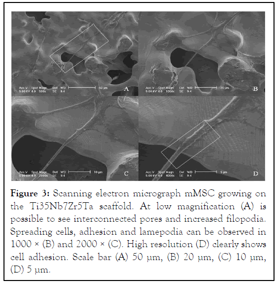
Figure 3: Scanning electron micrograph mMSC growing on the Ti35Nb7Zr5Ta scaffold. At low magnification (A) is possible to see interconnected pores and increased filopodia. Spreading cells, adhesion and lamepodia can be observed in 1000 × (B) and 2000 × (C). High resolution (D) clearly shows cell adhesion. Scale bar (A) 50 μm, (B) 20 μm, (C) 10 μm, (D) 5 μm.
Superoxide dismutase assay
In this study, it was possible to observe an increase of SOD activity in the Ti35Nb7Zr5Ta scaffold. However, for Ti6Al4V and Ti35Nb scaffolds the SOD activity was similar, but TiNb showed less activity of this antioxidant enzyme. Although Ti35Nb7Zr5Ta scaffold showed better results when compared to the other two scaffolds, no statistical difference was observed (Figure 4) [12].
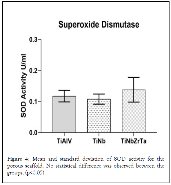
Figure 4: Mean and standard deviation of SOD activity for the porous scaffold. No statistical difference was observed between the groups, (p<0.05).
Catalase assay
Similar results of CAT activity were observed in Ti6Al4V and Ti35Nb7Zr5Ta, but Ti6Al4V showed better results. The Ti35Nb scaffold showed the lowest activity of antioxidant enzyme between the samples. No statistical difference was observed between the groups (p>0.05) (Figure 5).
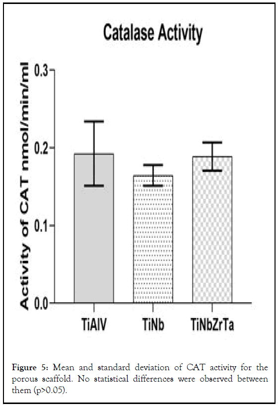
Figure 5: Mean and standard deviation of CAT activity for the porous scaffold. No statistical differences were observed between them (p>0.05).
Gene expression analysis (qPCR)
The results show the relative levels of expression in relation to the Ti6Al4V scaffold as the control group (Method 2-ΔΔCt), at days 3, 7 and 14 after cell culture. Our results showed high expression of RUNX2 gene after day 7 (Figure 6). At 3 days, the better result was for the Ti35Nb7Zr5Ta scaffold with a statistical difference from the control group (Ti6Al4V day 3). The Ti6Al4V scaffold was upregulated at day 7 but did not show any statistical difference from the control group. The Ti35Nb scaffold just showed expression at day 14, with values above Ti35Nb7Zr5Ta, when the levels of expression decline at this point in time but were above baseline. Ti6Al4V showed an increase of expression at this point in time. At day 14 all scaffolds showed a statistical difference from the control group, with p<0.05 [13].
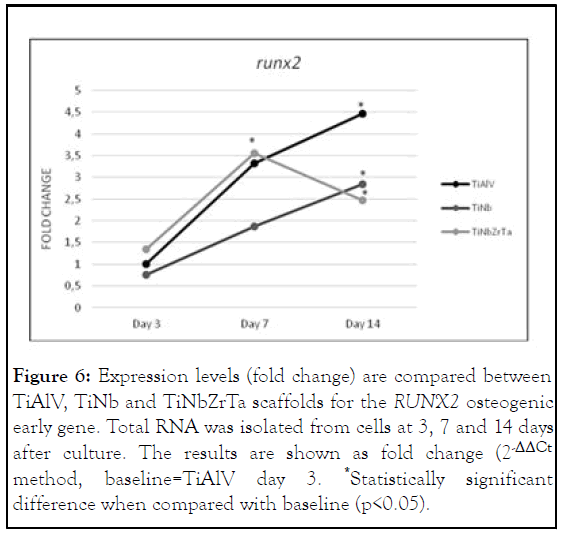
Figure 6: Expression levels (fold change) are compared between TiAlV, TiNb and TiNbZrTa scaffolds for the RUNX2 osteogenic early gene. Total RNA was isolated from cells at 3, 7 and 14 days after culture. The results are shown as fold change (2-ΔΔCt method, baseline=TiAlV day 3. *Statistically significant difference when compared with baseline (p<0.05).
The chemical composition of the titanium alloys, as well as their topographic structural characteristics, has been the focus of studies aiming to improve not only the modulus of elasticity of these implants, but also to promote good osseointegration and avoid future problems, such as aseptic loss of implant, which is a complication of unknown etiology. Therefore, there is the necessity for investigation of these new titanium alloys, not only with tests that show its biocompatibility and absence of cytotoxicity, as well as to evaluate other cellular mechanisms that may be linked to implant loss, since it is believed that the cellular response of the osseointegration process may be closely related to possible long-term failure [14].
The SEM images analysis showed some evidence that the cells could be in the process of cell differentiation, because the cells presented scattering and shape change. The images from the electron scanning microscopy also showed the topographic characteristics of the samples, with the presence of interconnected pores that mimic the porous structure of bone. Some authors suggest that surfaces that mimic the innate characteristics of bone could improve bone cell response by mimicking the bone cellular environment. Modification of the topography of the material is a most important criterion for the production of biomaterials, since it is believed to be decisive for cell-material interaction and for the integration of the material with the surrounding tissue. According to Horwitz, interconnected pores in three-dimensions serve as the temporary ECM for cell adhesion, growth and differentiation. This was exactly what we observed in all porous scaffolds, once there was a large number of cells growing and adhering to the scaffolds only 24 hours after the culture. This fact is extremely important and desirable in the osseointegration, since clinical success of the implants is related to rapid osseointegration, therefore decreasing the chances of failure due to the action of bacteria and other etiological agents that may be related to implant failure. We attribute the success of adhesion and spreading of the topographical characteristics of the implant to the creation of interconnected pores allowing greater anchoring of the cells.
An under-researched mechanism that is closely linked to tissue damage is oxidative stress and the enzyme defenses against it. Oxidative stress is faced by all aerobic organisms after antioxidant enzymes fail to eliminate Reactive Oxygen Species (ROS). Low levels of ROS are necessary for the regulation of different physiological mechanisms, including cell differentiation, apoptosis, cell proliferation and regulation of the redox-sentient signaling pathway. However, when its production exceeds the capacity of antioxidative activity, stress is unavoidable, causing serious damage to vital molecules such as proteins, enzymes or genetic material. In this regard, two important primary antioxidant enzymes, Superoxide Dismutase (SOD) and Catalase (CAT), play a significant role in the elimination of these oxygen reactive species [15].
We propose to analyze the activity of superoxide dismutase and catalase enzymes on a titanium alloy scaffold, since Tsark affirmed that extra evidence for permanent oxidative stress in the cells is based on the enzymatic activity of these two enzymes. SOD is the first and more important line of antioxidant defense enzymes against ROS damage, it being responsible for the dismutation of the superoxide anion in hydrogen peroxide and oxygen, which is then decomposed by the enzyme CAT in water and oxygen, which are innocuous to the cells. According to Chaudere, the activity of the antioxidant enzymes are synergistic; that is, these enzymes work simultaneously for the elimination of the oxygen reactive species. Thus, SOD activity protects against CAT inactivity and vice versa. Our study showed that both enzymes were active in all scaffolds, with quantitative differences in their expression. This fact is important because some studies suggest that SOD has its activity inhibited by hydrogen peroxide excess which would also generate a failure of CAT activity. Therefore, our results suggest that, although we did not perform a specific test to evaluate the accumulation of hydrogen peroxide in the cells, this substance is not present, because it is catalyzed by the activity of the enzymes.
In our results, SOD activity was higher in the TiNbZrTa scaffold and lower in the TiNb scaffold. Analyses of CAT activity showed very similar results in the TiAlVe TiNbZrTa scaffolds and TiNb scaffold presented lower activity. There are some studies that have indicated that TiNb alloy is a material with potential for use in biomedical implants and is a good possibility for the substitution of TiAlV alloy, based on biological and mechanical tests. Xu affirmed that TiNb alloy is not toxic, has good machinability and mechanical strength. In addition, it has an elastic modulus similar to that of human bone and has superior resistance to corrosion than titanium cp. TiNbZrTa alloy has also been studied and indicated as a good substitute for TiAlV alloy. According to Niommi, TiNbZrTa alloy is a newly developed alloy for biomedical use, due to its ideal modulus of elasticity close to human bone and its high biocompatibility. Do Padro14 showed interesting results regarding this alloy, mainly related to cell viability, which exhibited a higher number of viable cells when compared with other alloys, including TiNb and TiAlV, the latter exhibiting inferior results. Differing from these authors, we are not able to confirm (based only on enzymatic defenses), that TiNbZrTa and TiNb alloys are good or not good for replacement of TiAlV, because although they differed in their activities in both analyses, the difference between them failed to demonstrate any statistical significance.
This study used mesenchymal stem cells from mouse femurs, since our objective was also to evaluate the cell differentiation process, i.e., how much the chemical composition associated with surface topography would induce this differentiation. Thus, we evaluated the expression of RUNX2, a particularly important gene in the process of cell differentiation, since its presence indicates that mesenchymal stem cells are differentiating into pre-osteoblasts and osteoblasts which, when mature, will secrete bone matrix. This gene is called the key transcription factor, controlling osteoblast differentiation and are considered key transcriptional regulators that are expressed in osteoblasts and play multiple roles in osteogenesis. Our results showed lower and later expression of RUNX2 for the TiNb scaffolds. In the TiNrZrTa samples, there was a faster and greater expression in the adherent cells for this gene showing statistical differences, which indicates rapid osteoblastic differentiation in this sample. Due to the fact that RUNX2 was expressed at the beginning of the differentiation process, small changes in the level of expression were already sufficient to trigger the osteogenesis process. It is also important to note that RUNX2 elevations are related to an increase in the expression of other bone-related genes such as: Alkaline phosphatase, collagen type I, osteocalcin and osteopontin, genes closely linked to the process of bone neoformation. Therefore, even though we have not analyzed any of the aforementioned genes, we believe that TiNbZrTa porous scaffold can improve the osseointegration process.
In our study, we were able to observe excellent features for the presence of growth, adhesion and cellular spreading in all scaffold, concluding that these scaffolds presented good chemical and topographic characteristics for promoting osseointegration. Furthermore, we were able to conclude that by analyzing only the SOD and CAT activity, we were not able to confirm that TiNb and TiNbZrTa porous scaffolds are remarkable materials for the replacement of TiAlV alloy in the future, but the expression of RUNX2 in the quaternary alloy of TiNbZrTa demonstrated a better capacity for mesenchymal steam cell differentiation and can improve the osseointegration process, making it a good choice for biomedical applications. We believe that the porosity associated with the chemical composition of this alloy were key factors for the high RUNX2 gene expression.
[Crossref] [Google Scholar] [PubMed]
[Crossref] [Google Scholar] [PubMed]
[Crossref] [Google Scholar] [PubMed]
[Crossref] [Google Scholar] [PubMed]
[Crossref] [Google Scholar] [PubMed]
[Crossref] [Google Scholar] [PubMed]
[Crossref] [Google Scholar] [PubMed]
[Crossref] [Google Scholar] [PubMed]
[Crossref] [Google Scholar] [PubMed]
[Crossref] [Google Scholar] [PubMed]
[Crossref] [Google Scholar] [PubMed]
Citation: Carvalho LM, de Vasconcellos LMR, Zutin EAL, Sartori EM, Mendonca DBS, Mendonca G, et al. (2023) Activity of the Antioxidant Enzymes and RUNX2 Expression on Porous Titanium Alloy Scaffolds. Ann Essence Dent. 16:281.
Received: 06-Sep-2019, Manuscript No. AEDJ-23-2189; Editor assigned: 11-Sep-2019, Pre QC No. AEDJ-23-2189 (PQ); Reviewed: 25-Sep-2019, QC No. AEDJ-23-2189; Revised: 01-Sep-2023, Manuscript No. AEDJ-23-2189 (R); Published: 26-Mar-2024 , DOI: 10.35248/0976-156X.24.16.281
Copyright: © 2024 Carvalho LM, et al. This is an open-access article distributed under the terms of the Creative Commons Attribution License, which permits unrestricted use, distribution and reproduction in any medium, provided the original author and source are credited.