Medical & Surgical Urology
Open Access
ISSN: 2168-9857
ISSN: 2168-9857
Case Report - (2020)Volume 9, Issue 1
Tuberculosis remains a priority public health problem for the World Health Organization (WHO), The difficulty of often late diagnosis and the seriousness of lesions caused by BK even after anti-bacillary treatment, explains the seriousness of the disease with a high number of nephrectomies.
The clinical pictures can be misleading, hence the advantage of making this diagnosis before any recurring urological symptom.
In our observation, the diagnosis was mentioned in the face of the appearance of the "mastic kidney" indicates an already advanced stage of the pathology, after the anti bacillary treatment, the ablative surgical treatment takes place for the treatment sometimes serious consequences.
Tuberculosis; World Health Organization; Nephrectomies; Mastic kidney; Human immunodeficiency virus
With almost 10 million new cases per year worldwide, tuberculosis remains a priority public health issue for the World Health Organization (WHO) [1]. It is closely linked to the pandemic of infection with the human immunodeficiency virus (HIV) and to the emergence of multidrug-resistant strains of Mycobacterium tuberculosis [2], TUG is the 5th place in Morocco after pulmonary tuberculosis localizations, lymph node, osteoarticular and digestive [3]. It represents 15 to 30% of all extra pulmonary forms of tuberculosis worldwide [3].
The difficulty of the often late diagnosis and the seriousness of the lesions caused by BK even after anti bacillary treatment, explain the seriousness of the disease with a high number of nephrectomies and renal insufficiency [4,5]. Imaging is essential for the diagnosis of urogenital tuberculosis. Intravenous urography and computed tomography are the two basic tests. Tuberculosis generally digs the parenchyma and stenoses the excretory tract [4]. We report the case of a young patient of 45 years who consulted for bilateral low back pain, the Urinary Tree couple without preparation and CT allowing to objectify lesions characteristic of renal putty very suggestive of urogenital tuberculosis, a search for BK in the diagnostic.
Mrs. LA, 46 years old, having as a pathological antecedent a pulmonary tuberculosis treated 10 years ago, no other pathological antecedent having consulted for bilateral low back pain evolving within the framework of a deterioration of the general state, anorexia, quantified weight loss to 10 kg in 4 months without any notion of hematuria or emission of calculation, on clinical examination, apyretic patient, no lumbar contact, slight sensitivity of the lumbar fossa, biological renal failure with creatinine at 45 mg/l, clearance according to MDRD at 13.55 ml/min/1.72 m2, inflammatory syndrome biological with high SV and CRP, leukocytosis 15,000, cytobacteriological examination negative income.
A radiological examination was considered in the first place an AUSP in search of a lithiasic obstacle this last objective an opacity of the calcium tone that projects on the left renal region appearing as a complex coralliform calculus drawing the chalices (Figure 1) uro scanner a been achieved objective Extensive parenchymal calcifications on a non-functional kidney realizing the appearance of a pathognomonic mastic kidney of a late stage of renal tuberculosis (Figure 2) with a left kidney dilated on several staged ureteral stenosesThe patient underwent a Bk urine test 3 days in a row and a positive income confirmed the diagnosis of genitourinary tuberculosis. The patient urgently benefited from drainage of the right kidney by a double JJ probe which slightly improves renal function to 30 mg/l of creatinine; a cystoscopy was also performed with biopsy, the latter was not objectified with anomalies with a biopsy without anomalies. Therapeutically, the patient benefited from an anti-bacillary treatment combining rifampicin, isoniazid and pyrazinamide for a period of 9 months, the control creatinine after the treatment remained stationary at 30 mg/l and the radiological control still shows a non-functional putty kidney, the patient then underwent a nephrectomy for a destroyed kidney, the macroscopic examination found a kidney with destroyed pyelonephretic aspects. Histologically, it is a mononuclear inflammatory infiltrate of the renal parenchyma with an inflammatory lymphoplasmocytic filtrate associated with histiocytes in epitheloid inflections With a granulomatous inflammatory reaction and giant cells typical of urogenital tuberculosis. (Figures 3-5).
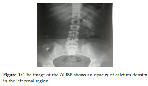
Figure 1: The image of the AUSP shows an opacity of calcium density in the left renal region.
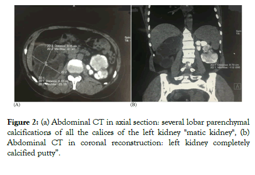
Figure 2: (a) Abdominal CT in axial section: several lobar parenchymal calcifications of all the calices of the left kidney "matic kidney", (b) Abdominal CT in coronal reconstruction: left kidney completely calcified putty”.
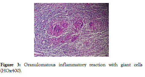
Figure 3: Granulomatous inflammatory reaction with giant cells (HGx400).
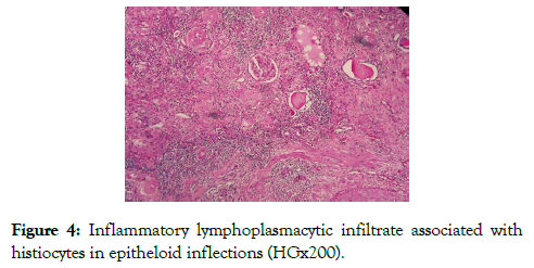
Figure 4: Inflammatory lymphoplasmacytic infiltrate associated with histiocytes in epitheloid inflections (HGx200).
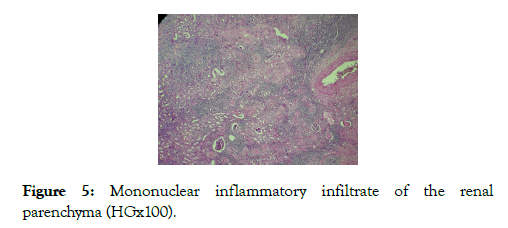
Figure 5: Mononuclear inflammatory infiltrate of the renal parenchyma (HGx100).
The operative follow-ups were simple with a stationary renal function at 30 mg/L of creatinine. And satisfactory morphological checks.
The epidemiology of renal tuberculosis is characterized by a significant variation in its frequency and its geographic distribution. It is difficult to know its exact prevalence, screening depending on the techniques used. However, renal (genitourinary) involvement represents around 30% of extrapulmonary forms [6]. Pulmonary localization is the most frequent, followed by lymph nodes and renal involvement. Renal involvement of tuberculosis, which is carried out by the hematogenous route, is most often bilateral and results in the formation of granulomas. These lesions heal in most cases and have not suffered from kidney disease. Granulomas can, however, become caseous and then rupture in the tubular lumen several years after primary pulmonary infection. In general, there is no impact on renal function, except in rare cases where the disease manifests itself as postrenal acute renal failure (ARF) on granulomatous ureteric stenosis [7]. Kidney damage is hematogenous with the formation of granulomas in the glomeruli. Macroscopically, white nodules, especially cortical, are observed, measuring 1 to 3 mm in diameter. Histologically, these are epithelioid granulomas, often containing giant Langhans type cells, with or without caseous necrosis [8]. The radiological translations of these lesions are renal calcifications which represent a classic manifestation of tuberculosis, found in 24 to 44% of cases [5,9]. Extensive parenchymal calcifications on a non-functioning kidney producing the appearance of a mastic kidney are pathognomonic of a terminal stage of urogenital tuberculosis. These calcifications visualized on the urinary tree without preparation are better studied on the computed tomography. The morphological evaluation requires the realization adapted imagery. The uroscanner is today considered as a reference for the diagnosis of the urinary tracts [10], very sensitive in particular for the analysis of whole perirenal and urinary trees [4,5]. this is the case of our patient whose orientation was first radiological and then confirmed by bacteriological examination after BK test in the urine.
The pathological lesions of renal tuberculosis are suggestive but can be non-specific [11], they vary depending on the location of the lesion, the route of spread, the virulence of the germ and the immune defenses. The initial lesions are similar to those of nonspecific chronic interstitial nephritis. In the state phase, hematogenous renal involvement is characterized macroscopically, by white nodules, mainly cortical, measuring between 1 and 3 mm in diameter [12]. They correspond to epithelioid and gigantocellular granulomas (giant Langhans type cells) with or without caseous necrosis. These lesions lead to the destruction of the renal parenchyma with areas of scar fibrosis. Cortical granulomas can rupture in the tubular and/or pyelic lumen, producing tuberculous pyelonephritis Kidney damage in tuberculosis can take other forms, such as cavitation or pyo nephrosis, which is called "mastic kidney". Calcifications are often observed [13].
Therapeutically as in the pulmonary and urinary forms, the quadritherapy anti-tuberculosis, the bitherapy must then be continued for a total duration of six months [14]. The caseous lesion of a partially functional kidney remains controversial.On the other hand, it is recommended to remove a ureteral obstruction by a JJ catheter or a subcutaneous nephrostomy if the kidney is functional with a correct cortical thickness and a creatinine clearance greater than 15 ml/min [15,16]. In our case, we performed an emergency drainage of the kidney on the right side with a double J probe, then a left nephrectomy after the persistence of radiological calcifications and in front of a nonfunctioning kidney and source of infection or secondary arterial hypertension.
The main common point of the urinary and genital forms of tuberculosis is an often insidious and destructive development, which can lead to irreversible sequelae, the main of which is an impairment of renal function. The diagnosis is often late due to the non-specificity of the clinical signs. Tuberculosis should therefore be considered in the face of any atypical and recurrent symptomatology. The treatment is first medical then surgical for the sequelae, the medical treatment is the same as that used for pulmonary tuberculosis but the renal insufficiency can be corrected in an incomplete way see the heavy sequelae like dialysis.
The authors declare no conflict of interest regarding the publication of this article.
Citation: Ziani I, Lakssir J, Derqaoui S, Deragamoun H, Ibrahimi A, Zouidia F, et al. (2020) Typical Aspects of the "Mastic Kidney" Suggestive of Renal Tuberculosis. Med Sur Urol 9:228. doi: 10.24105/2168-9857.9.228
Received: 30-Mar-2020 Accepted: 24-Apr-2020 Published: 02-May-2020 , DOI: 10.35248/2168-9857.20.9.228
Copyright: © 2020 Ziani I, et al. This is an open-access article distributed under the terms of the Creative Commons Attribution License, which permits unrestricted use, distribution, and reproduction in any medium, provided the original author and source are credited.