Medical & Surgical Urology
Open Access
ISSN: 2168-9857
ISSN: 2168-9857
Research - (2019)Volume 8, Issue 2
Modern literature data related to the erectile dysfunction was stated in this article. The issues of systemic and local endothelial dysfunction are discussed in this Research Work. Also the issues of the interaction of terahertz infrared radiation with biological objects of varying complexity of implementation are discussed. The implementation of the effect of infrared waves of the terahertz range in bio system is possible at the molecular, cellular and systemic levels of regulation.
Erectile dysfunction; Pathogenesis; Endothelial dysfunction; Terahertz range infra-red radiation
Erectile function in a healthy man is determined by the following factors:
-The function of the endothelium producing Nitric Oxide (NO);
-Inflow of arterial blood, providing adequate partial oxygen pressure, "breathing", nutrition and adequate filling of cavernous tissue; state, the degree of fibrotic changes;
-Function of the veno-occlusive erection mechanism and the ability to redistribute the pelvic blood flow during sexual arousal [1].
The cause of Erectile Dysfunction (ED) is endothelial dysfunction in 80% of patients. The average human body contains about 1.8 kg of endotheliocytes or one trillion cells [2]. According to modern concepts, endothelium is not just a semipermeable membrane that ensures the vessel’s non-wettability, but an active endocrine organ, the largest in the body, diffusely dispersing throughout all tissues [3]. One of the main functions of the endothelium is a balanced selection of regulatory substances that determine the integrated work of the circulatory system.
There are three main factors that stimulate endothelial secretion activity:
1. Change in blood flow velocity - for example, increase in blood pressure.
2. Circulating and/or "intraparietal" neurohormones (catecholamines, vasopressin, acetylcholine, bradykinin, adenosine, histamine, etc.) [3,4].
3. Factors released from platelets when they are activated (serotonin, ADP, thrombin).
Consequently, like any endocrine organ - The endothelium is under the influence of the center of regulation of homeostasis (hypothalamus).
The hypothalamus is the main coordinating and regulating center of the human autonomic nervous system, i.e. it regulates the neuroendocrine activity of the brain and the ability to maintain constancy of homeostasis.
This means that all three factors that stimulate the secretory activity of the endothelium need to be considered as conformity of the hypothalamus functions, and hence the pituitary gland, the autonomic nervous system, etc.
This part of the brain is very sensitive to various disorders: intoxication, infections, disorders of blood and cerebrospinal fluid circulation, pathological impulses from other parts of the Central Nervous System (CNS).
Mismatch of chemical reactions in the nuclei (over 30) of the hypothalamus (toxemia, aggravation by xenobiotics), and even worse damage to the nuclei (injuries, tumors) lead to unidirectional disorders.
Changes in the functional activity of the hypothalamus may be with increased or decreased level of activity of neurohormonal systems, i.e. with a reduced or increased tone of the autonomic nervous system. The increased tone of the neurohormonal systems is characterized by the predominance of the sympathetic orientation of the vegetative reactions. Lowering the tone, the predominance of parasympathetic autonomic reactions.
Hypothalamic dysfunction leads to impaired hemodynamics, which leads to tissue and cell hypoxia, which leads to tissue and cell hypoxia. Hence, there is a transition of energy-material cell exchange to the anaerobic (glycolytic) type. The cells use glucose from glycogen in the liver, muscles, which lead to hyperglycemia and hyperinsulinemia. Hyperinsulinemia leads to increased microcirculatory hemodynamic disorders. There is a "vicious circle." This is a pattern of systemic Endothelial Dysfunction (EnD). The local EnD of the cavernous arteries can be schematically represented as follows.
The imbalance between vasadilators and vasoconstrictors leads to impaired hemodynamics, which leads to tissue and cell hypoxia (endotheliocytes, smooth muscle cells). Anaerobic (glycolytic) type of energy exchange in endothelial cells is activated and dominates, leading to hyperglycemia and hyperinsulinemia. Microcirculatory hemodynamics worsens, which increases the hypoxia of tissues and cells, leading to fibrous changes in the cavernous bodies.
Currently known, the following mechanisms EnD:
- Impaired expression or structure of endothelial NO Synthase (eNOS);
- Impaired eNOS function;
-Increasing NO destruction;
-Decreased sensitivity of cells to NO.
All these mechanisms can be explained by one reason for the violation of the energy-material exchange of endothelial and smooth muscle cells [5].
The enzyme NO synthase is a protein that a certain amount of energy is spent on its formation by the cell; energy costs occur during the synthesis of NO from L-arginine, and increased destruction of NO, and a decrease in the sensitivity of cells to it (NO), all of this involves the use of energy. Consequently, the violation of energy and material metabolism in endothelial and smooth muscle cells is the basis of all the above EnD mechanisms.
Any cell of a tissue or organ uses the energy of Adenosine Triphosphate (ATP). ATP in the destruction of phosphate groups loses one phosphate, this forms Adenosine Diphosphoric acid (ADP) and energy is released. In contrast, in order to reattach the phosphate to ADP, it is necessary to expend energy.
ATP stocks in the cell are relatively small, so they should always to replenish. The energy for this is drawn by the oxidation of "combustible" substances- carbohydrates, fats and proteins. When they are split, the potential chemical energy contained in them is converted into other forms of energy.
ATP recovery goes in two ways:
1) Anaerobic - without oxygen;
2) Aerobic - with the participation of oxygen.
The most effective way is to restore APT through aerobic oxidation of carbohydrates.
Strengthening anaerobic synthesis of ATP is one of the most important mechanisms for regulating the energy metabolism of cells under various physiological conditions and adapting to changes in environmental conditions. Activation of glycolysis in many cells of the body occurs under conditions accompanied by ATP deficiency, which may occur as a result of: high speed of ATP consumption and/or a decrease in the level of aerobic ATP synthesis [6].
Changes in environmental conditions and the adverse effects of anthropogenic factors leads to increased anaerobic glycolysis in human cells and organs. This is a compensatory reaction that maintains the level of energy metabolism in the face of a decrease in the intensity of the process of oxidative phosphorylation in the mitochondria of cells. Reducing the level of aerobic synthesis of ATP in the cells of various organs and tissues is possible under the action of various factors. For example: while reducing the supply of oxygen to the cells due to various reasons. This is hypoxia, and violation of the structure and functions of mitochondria.
When exposed to pathogenic factors for preservation of homeostasis is necessary to expend some additional energy. The aerobic process, as already noted, is the most economical; however, it is quite slow and cannot provide enough energy. In such cases, the role of carbohydrates in the body's energy supply increases.
For faster energy, the body enhances the glycolytic type of energy exchange, as it is faster than oxygen and significantly longer than creatine phosphate.
Prolonged exposure to pathological factors (infection, toxins and/or stress) and not eliminating them, leads to impaired hemodynamic of organs, tissues, cells which makes aerobic type of energy exchange difficult, therefore the anaerobic type begins to predominate, which leads to local metabolic disorders (lactic acidosis). To eliminate these disorders, the body needs additional energy, so the role of carbohydrates (glycogen of the liver, muscles) increases, leading to hyperglycemia (increased glucose uptake by the tissues, for energy production), therefore the function of the pancreas is enhanced (insulin is producing impulsively), which is characterized by the appearance of hyperinsulinemia. This condition can lead to relative insulin deficiency; if the pathological factor is not eliminated, hence the depletion of β-cells occurs with the further development of absolute insulin deficiency. This leads to disruption of glucose uptake in insulin-dependent tissues, a decrease in oxidative phosphorylation and the formation of glucose-6-phosphate; the glycolytic oxidation of glucose, the Krebs cycle and the hexosomonophosphate (pentose) cycle are subsequently violated, glycogen synthesis is inhibited and glycogenolysis is enhanced. Very soon, glycogen stores are depleted, glycolysis is slowed down due to intracellular acidosis and the energy supply of cells and tissues is disturbed, which leads to dysfunction of the organ or system.
Thus, the violation of the energy supply of endothelial cells, due to the predominance of glycolytic (anaerobic) type of energy exchange, leading to metabolic disorders of the cell is the basis of endothelial dysfunctions.
Endothelial dysfunction is a pathological condition that occurs in the cells as a result of a disruption in the normal course of biochemical energy supply. This leads to an imbalance between the production of vasodilating, angioprotective, angioproliferative factors (nitric oxide, prostacyclin, tissue plasminogen activator, C-type natriuretic peptide, endothelial hyperpolarizing factor, and, on the one hand, vasoconstrictor, prothrombotic, proliferative, factors (endotelin, superoxideanion, thromboxane A2, tissue plasminogen activator inhibitor) on the other hand.
Cells, so to speak, begin to work off-board. There is a violation of energy and material exchange..
Energy-material exchange is a set of mechanisms that determine the vital processes at the cellular level [7].
The main stages of this multistep process of exchange transformations occur in mitochondria and lead to the accumulation of high-energy compounds, which ensures the vital activity of the cell and the whole organism. Today, the role of cellular energy metabolism disorders in the pathogenesis of diseases of various organs and systems has been proven, and mitochondria, identified in most tissues of the body, are responsible for implementing this process [8].
The main consequence of pathological changes in energy and material metabolism is a violation of ATP production and inhibition of β-oxidation of lipids. According to the literature, there is a certain threshold level for reducing the ATP content inside the cell, upon the achievement of which, the suppression of any energy-dependent processes begins [9].
It is known that the violation of electron transfer between the components of the respiratory chain is accompanied by the production of a superoxide radical by the mitochondria, which contributes to the activation of lipid peroxidation processes, oxidative damage to proteins and nucleic acids. Increased production of reactive oxygen species can have a damaging and dysregulatory effect on the cell. Elements that are particularly exposed to free radicals are lipids, proteins, redox enzymes and mitochondrial desoxyribonucleic acid (mtDNA). Direct damage to mitochondrial proteins reduces their affinity for substrates or coenzymes and impairs their function [10-14].
Mitochondrial dysfunction is a “vicious circle” in which damage to the cellular structure responsible for the production of ATP entails additional energy costs [15].
Following some tenets of information medicine: each element of the system (cell) has its own energy and material resources, sufficient to ensure information homeostasis; energymaterial and information exchange is carried out between the elements (cells) of the system; information exchange is carried out using signals in the millimeter wavelength range; impaired energy-material homeostasis of the cell leads to changes in the informational homeostasis of the cell, cell assembly in the structure of the organ and system [16].
According to numerous theoretical and practical researches in the field of medicine and biology, there is an electromagnetic connection between the elements of the system (any cells, organs). Consequently, the elements of the system exchange information electromagnetic signals [17,18].
It is known that in the process of vital activity the cell produces electromagnetic oscillations of a very wide range. However, the predominantly narrow millimeter and submillimeter range is used by cells for the exchange of information necessary for the regulation of intracellular functions and cell–cell interactions.
In recent years, a new direction of information therapy has emerged - terahertz therapy [19].
The terahertz frequency range of electromagnetic waves is located on the scale of electromagnetic waves between Extremely High Frequencies (EHF) and Optical Infrared (IR) ranges (Figure 1) and is interesting, first of all, because it contains the Molecular Specter of Emission and Absorption (MSEA) various cellular metabolites (NO, CO, reactive oxygen species, etc.).
Figure 1. Far-range infrared emitters.
It has been established that the reactivity of molecules excited by a terahertz quantum will be an order of magnitude higher than when excited by an SWF quantum [20].
The considered range of electromagnetic waves is used by living organisms for communication and control, while living organisms themselves emit millimeter-wave oscillations. Waves excited in the body when exposed to infrared radiation of the terahertz range, to a certain extent imitate the signals of internal communication and control (information communication) of biological objects. As a result, the radiation normal in the spectrum and power characteristic of a healthy organism is restored. Thus, the presented frequency range does not qualitatively change the organism, but can regulate and normalize its functional state within the limits inherent in this biological species [21,22].
Therefore, it is advisable for successful treatment and diagnosis to use narrow spectra of the far infrared range. Such narrowspectrum emitters are developed on the basis of oxide ceramics at the Institute of Materials Science (Tashkent, Republic of Uzbekistan). The spectrum of their radiation lies in the range from 8 to 50 cm. This means that the quantum energy of radiation transformed by ceramics is within or below the quantum energy of a person's own radiation, and, accordingly, cannot have a negative effect on the physiological processes of the human body [23,24].
We examined 61 men (main group) aged 32–74 years with CVD and Cardiovascular Risk Factors (CVRF), as well as 12 men (control group) aged 28–66 years without CVD and CVRF. All men underwent a comprehensive examination, which included the collection of general medical and sexological history, general examination, the study of hormonal status, lipid profile and blood glucose. All patients were surveyed by ICEF-5 (International Index of Erectile Function).
In addition, all men completed a study of the endothelial function of the cavernous arteries, using the ultrasound study of changes (ultrasound) diameter after exposure to narrowspectrum (long-range) IR emitters (Figure 1)
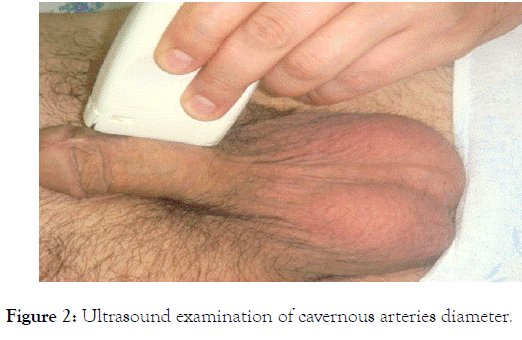
The study of the endothelial function of the cavernous arteries of the penis was performed according to the original method. In the position of the patient lying on his back, the linear sensor LA 523 10-5 was placed longitudinally along the ventral surface of the penis at a distance of 2-3 cm from the root (Figure 2).

Figure 2. Ultrasound examination of cavernous arteries diameter.
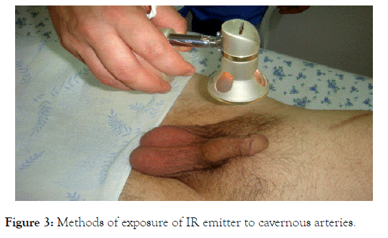
The diameter of the cavernous arteries was evaluated at least two times, measuring the distance between the opposite walls of each vessel, and mean values were used for the calculations. In addition, the following indicators were determined-Peak Systolic Velocity (PSS), Peak Diastolic Velocity (PDS), Reactivity Index (PI), Pulse Index (PI). Then an infrared emitter was applied to the ventral surface of the penis at a distance of 10-12 cm from the root with an exposure of 5 minutes (Figure 3). After exposure, re-ultrasound was performed with measurement of the diameter of the cavernous arteries in the same place. For calculations, we used the largest values of the diameter obtained during the repeated study.

Figure 3. Methods of exposure of IR emitter to cavernous arteries.
The Percentage of Increasing the Diameter of the Cavernous Arteries (PIDCA) was calculated by the formula:
PIDCA = 100% х (D2 –D1)\D1
Where,
D1-The average diameter of both cavernous arteries before irradiation with an infrared emitter;
D2-The average diameter of both cavernous arteries after irradiation.
All studies were conducted in the morning and one ultrasound specialist. Prior to the study, patients were asked to refrain from taking drugs acting on the cardiovascular system. The threshold value of the PIDCA to distinguish endothelial dysfunction from the norm was 30%.
Manifestations of Erectile Dysfunction (ED) were detected in 58 (95.1%) patients. According to ICEF-5: mild erectile disorders were detected in 15 (24.6%) men; moderate degree of impairment in 38 (62.3%) men; severe - in 5 (8.2%); signs of ED were absent in 3 (4.9%).
In the study of the endothelial function of the cavernous arteries, endothelial dysfunction was detected in 59 (96.7%) patients. Paradoxical vasoconstriction was determined in 44 (72.1%) patients (PIDCA<0%). In 15 (24.6%) patients, PIDCA was determined at a level of less than 30% and averaged 16.5%, which indicated endothelial dysfunction of the cavernous arteries.
A comparative analysis of the mean values of the PIDCA in patients of the main and control groups revealed that this indicator is statistically significantly (p<0.001) less in the group of patients with CVD and CVRF compared with the control group (Table 1).
| No | Group | Mean value PIDCA (%) |
|---|---|---|
| 1 | Control ( n=12) | 62.2 ± 12.4 |
| 2 | Main (n=61) | 12.5 ± 8.4* |
*p<0.001 compared to the control group
Table 1: Percent diameter increases of cavernous arteries in different groups.
Thus, the available reasoned data on the use of terahertz modulation of infrared radiation can be used to diagnose and treat endothelial dysfunction in erectile disorders.
Citation: Mirsagatovich AA (2019) The Disturbance of Energy and Material Exchange in Pathogenesis of Erectile Dysfunction. Med Sur Urol. 8:220.
Received: 02-Mar-2019 Accepted: 02-Apr-2019 Published: 09-Apr-2019 , DOI: 10.35248/2168-9857.19.8.220
Copyright: © 2019 Mirsagatovich AA. This is an open-access article distributed under the terms of the Creative Commons Attribution License, which permits unrestricted use, distribution, and reproduction in any medium, provided the original author and source are credited.