Cell & Developmental Biology
Open Access
ISSN: 2168-9296
ISSN: 2168-9296
Research Article - (2016) Volume 5, Issue 1
The aim of our study was the determination of prevalence of the sarcosporidiosis in the bovine carcasses in the slaughterhouses of El Harrach and the identification of involved species of Sarcocystis. Samples of oesophagi and diaphragms of 200 cattles brought down in the slaughterhouse of El Harrach were analyzed by the histo-pathological technique and the technique of digestion pepsique. We did not note macroscopic cyst in the carcasses inspection. The enzymatic digestion and the histo-pathological analysis revealed high rates of infestations (95%) and (80%) respectively. Cysts with thin wall of S. cruzi were dominant in diaphragms (94.2%) and in oesophagi (100%). A low rate of cysts with thick wall was detected in diaphragms (4.4%) and a rate of 0% in oesophagi. A single cyst of S. hominis was able to be diagnosed by photonique microscope in a diaphragm.
<Keywords: Slaughterhouses of El harrach; Bovine carcasses; Diaphragms; Oesophagi; Sarcocystis species
In Algeria, our cattle herd is very frequently affected by many muscular protozoan (Toxoplasma gondii, Neospora caninum and Sarcocystis sp.). The National Institute of the Veterinary (Ministry of Agriculture and the Algerian rural development) revealed during inquiries at national level, high prevalence of toxoplasmosis and bovine neosporose. For the sarcosporidiosis, it is less frequent in our slaughterhouses. No cases of éosinophilic myositis were observed till the current hour and the visible cysts are exceptionally observed. However, some studies on prevalence of the sarcosporidiosis were carried out, in particular those of Nedjari et al. [1] and of Harhoura et al. [2] who reported a very high prevalence at slaughterhouses of Hussein Dey and Rouiba respectively. But the sarcosporidiosis is underestimated and its search in our slaughterhouse (Algeria) is not obligatory. For this, a study on bovine sarcosporidiosis was conducted at slaughterhouses in the north of Algeria, in particular those of El Harrach. The study included 200 cattle slaughtered to determine the prevalence of sarcosporidiosis by two diagnostic techniques, enzyme digestion and histopathological study.
Method of sampling
At the slaughterhouse, a post-mortem examination was performed on bovine carcasses for research macroscopic cysts. Samples of oesophagi of 8 to 12 cm length and diaphragms were collected from each carcass and placed in identified freezer bags (sampling date, number, sex and the age of sampled cattle).
Preparation of samples in the laboratory: Samples are taken on the same day at the laboratory of parasitology and mycology ENSVAlgiers. They are cleaned and washed in water to remove blood and the food contents to the esophagus. The samples are cut into two parts a part suffered enzymatic digestion and the other is fixed in 10% formol for histological study.
Technique of enzymatic digestion: The principle of this technique is to reconstruct an artificial gastric juice (water, HCl, NaCl and pepsin) called digestion solution. This last combines the factors promoting digestion of the cysts and the release of bradyzoites, creating an optimum pH (pH 1-3) which allows the activation of pepsin (proteolytic enzyme) and denaturation proteins. For the pepsic digestion of 350 samples including 200 diaphragms and 150 oesophagi, we used the modified method of Seneviratna et al. [3,4]. The samples are hashed with a robot which is washed after each use and then weighed in an electronic balance. 10 g of diaphragm and 10 g of esophagi from each carcass are collected in a conical plastic tube, graduated, wherein 50 ml of digestion solution is added, the whole is homogenized using a spatula and incubated at 40°C in a constant agitation for 30 minutes. The digest is then filtered with a sieve on which two gauze layers are deposited, to remove muscle debris. The obtained filtrate was centrifuged at 3000 rpm for 5 minutes, the pellet is taken up in PBS (pH 7.2); the same operation is performed two times, in order to stop the digestion. A few drops of the pellet are aspirated and deposited on a slide for the preparation of smears and then dried in the incubator at 37°C. Dried smears are colored using the technique of May Grunwald Giemsa [5]. Stained smears are then observed with an optical microscope (Leica DMLS®) magnification (X400, X1000). A sample is considered positive when Sarcocystis typical bradyzoites are observed (a banana shape).
Histological technique: Histological technique was applied on 350 samples (200 and diaphragms 150 oesophagi). The method used is that cited by hould [6] with a hematoxylin and eosin. All steps of the technique was realised at the laboratory of anatomy and cytopathology of the Universitary Hospital of Bab El Oued (ex. hospital Maillot). The reading of the blades is performed with an optical microscope at a magnification (100X, 400X, 1000X). Positive slides reveal the presence of Sarcocystis cysts inside muscle fiber having a wall with variable thickness depending on the species involved (Gr. 1000).
Statistical analysis: For statistical analysis, we used the software program Microsoft Excel 2010. The comparison of the distribution of different populations were analyzed using Chi Square test with degree of significance as P<0.05.
Sarcocystis search by microscopic examination
On the 200 cattle that we have inspected at slaughterhouses of El Harrach, we did not find macroscopic cyst in the diaphragm or in the oesophagi. No cases of seizure for sarcosporidiosis were recorded and also there were no cases of eosinophilic myositis were observed in the inspected carcasses.
Research Sarcocystis by microscopic examination
Prevalence of Sarcocystis bradyzoites by enzymatic digestion: Enzymatic digestion revealed the presence of Sarcocystis bradyzoites in muscles of 190 cattle on the 200 analyzed, 95% of the study cattle are infested by Sarcocystis. Bradyzoites banana shaped, released after enzymatic digestion of the diaphragm and esophagi were observed under an optical microscope (Figure 1).
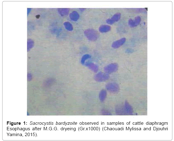
Figure 1: Sacrocystis bardyzoite observed in samples of cattle diaphragm Esophagus after M.G.G. dryeing (Gr.x1000) (Chaouadi Mylissa and Djouhri Yamina, 2015).
Prevalence of Sarcocystis according to sex: The prevalence of Sarcocystis in muscles (diaphragm and esophagus) was 94% in males and 96% in females. Parasite prevalence in both sexes is almost similar. The result is that sex has no influence on the prevalence of Sarcocystis studied in 200 cattle (Figure 2).
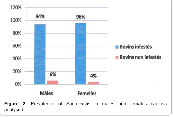
Figure 2: Prevalence of Sacrocystis in males and females carcass analysed.
Prevalence of Sarcocystis according to the age: The prevalence of Sarcocystis in our samples, in two age category, is 94.7% among cattle aged less than 2 years and 95.1% for cattle older than two years. The prevalence of Sarcocystis is very close in 2 age category, so age does not influence the prevalence of Sarcocystis in the 200 cattle studied (Figure 3).
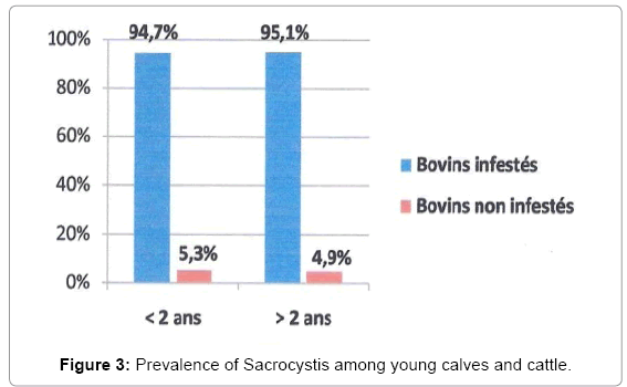
Figure 3: Prevalence of Sacrocystis among young calves and cattle.
Search for Sarcocystis cysts histologically
The histological test allowed to observe two types of cysts inside the muscle fibers. The first type is with a thin wall (Figure 4), characteristic of Sarcocystis cruzi. The second type is with a thick wall (Figure 5), it could be either Sarcocystis hominis or Sarcocystis hirsuta. Of the 350 samples analyzed, we observed unusually on a diaphragm, the cytophanères of a thick wall cyst. These were long, cylindrical (in the form of fingers) and almost perpendicular to the surface of the wall (finger-like) (Figure 6). It might therefore be S. hominis (zoonotic species) observed under an optical microscope (H & E, Gr. 1000X).
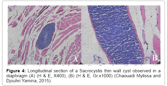
Figure 4: Longitudinal section of a Sacrocystis thin wall cyst observed in a diaphragm (A) (H & E, X400), (B) (H & E, Gr.x1000) (Chaouadi Mylissa and Djouhri Yamina, 2015).
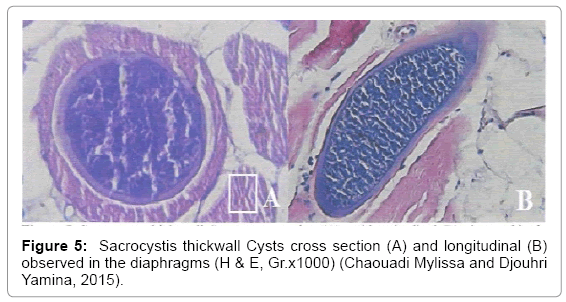
Figure 5: Sacrocystis thickwall Cysts cross section (A) and longitudinal (B) observed in the diaphragms (H & E, Gr.x1000) (Chaouadi Mylissa and Djouhri Yamina, 2015).
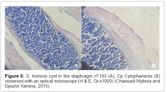
Figure 6: S. hominis cyst in the diaphragm n0 193 (A); Cp Cytophaneres (B) observed with an optical microscope (H & E, Gr.x1000) (Chaouadi Mylissa and Djouhri Yamina, 2015).
Overall prevalence of microscopic Sarcocystis cysts
On the 200 cattle studied, histological analysis revealed the presence of cysts in 160 samples sarcosporidiens corresponding to a prevalence of 80% (Figure 7). Prevalence of sarcosporidiens cysts according to the muscle the Sarcocystis cysts were found in 121 diaphragms on 200, and in 106 esophagi on 150 corresponding to a prevalence of 60.5% and 70.7% respectively (Figure 8). The test “chi-squared” of independence demonstrates that there is a significant difference (p ≥ 0.05) between the presences of cysts in the two muscles. The esophagi were more parasitized by Sarcocystis as diaphragms.
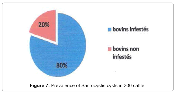
Figure 7: Prevalence of Sacrocystis cysts in 200 cattle.
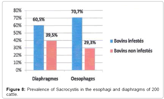
Figure 8: Prevalence of Sacrocystis in the esophagi and diaphragms of 200 cattle.
Prevalence of cysts sarcosporidiens depending on the type of wall
In cattle infected with Sarcocystis cysts, we observed cases of mono infections (cysts thin wall only or wall just thick) as well as instances of double mixed infections or infestations (cysts thin wall and thickwalled cysts simultaneously). Of the 160 infected cattle, 158 (98.8%) were infested with cysts thin wall; while 7 cattle (4.4%) had cysts thick wall (Figure 9). Of the 7 cattle infested with cysts thick wall, only 2 were only infested with cysts thick wall.
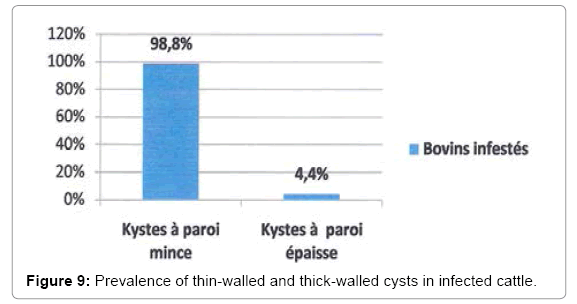
Figure 9: Prevalence of thin-walled and thick-walled cysts in infected cattle.
Prevalence of sarcosporidiens cysts with thin and thick wall in the infested diaphragm and esophagus
Whatever the parasitized muscle, the thin-walled cysts were predominant in the two types of muscles, with a high prevalence; 94.2% and 100% in the diaphragm and into the esophagus, respectively; In contrast, the prevalence of cysts with thick wall was very low 1.7% and 0% in the diaphragm and esophagus respectively. Totally the cysts with thick wall were observed on diaphragm, but no thick wall cysts were observed in the esophagus. The recorded frequency for mixed infestations is very low (4.1%) in the diaphragm and zero (0%) in the esophagus (Figure 10).
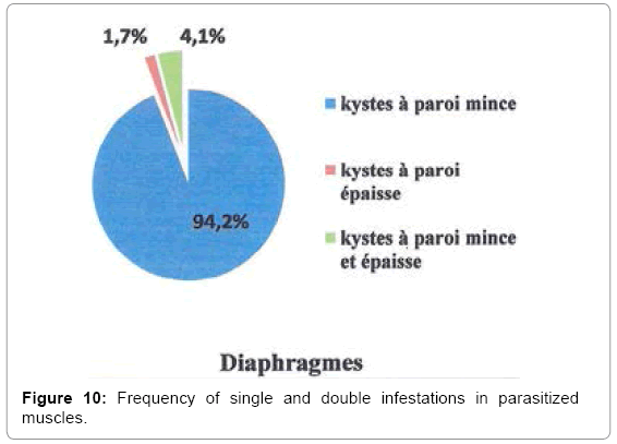
Figure 10: Frequency of single and double infestations in parasitized muscles.
Intensity of parasitism of diaphragms and esophagi by Sarcocystis spp.
A total of 1430 cysts of Sarcocystis spp. was demonstrated in the diaphragms 675 cysts thin walled, 10 cysts thick wall were noted, while in the esophagus, the presence of 745 cysts thin wall were observed and no cyst wall thickness (Figure 11). The majority of infected samples had between 1 and 3 cysts with a thin wall in the diaphragm and esophagus, and fewer had between 4 and 10 cysts with a thin wall (Table 1). Finally, a minority of samples contained more than 10 thin wall cysts. 66 and 68 respectively cysts were found in a diaphragm and esophagus. By cons, cysts with thick wall were located all one diaphragm and the number did not exceed 2 cysts per sample.
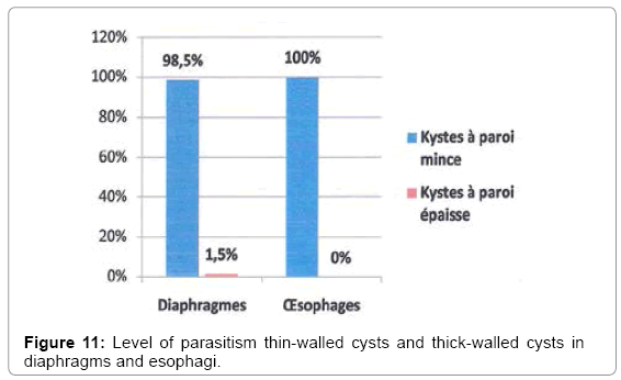
Figure 11: Level of parasitism thin-walled cysts and thick-walled cysts in diaphragms and esophagi.
| Muscles | 1 to 3 cysts | 4 to 10 cysts | More than 10 cysts | |||
| Thin-walled cysts | Thick-walled cysts | Thin-walled cysts | Thick-walled cysts | Thin-walled cysts | Thick-walled cysts | |
| Diaphragms | 78 | 7 | 27 | 0 | 15 | 0 |
| oesophagi | 57 | 0 | 30 | 0 | 19 | 0 |
Table 1: Level of parasitism by thin-walled cysts and thick-walled cysts in diaphragms and esophagi.
Study of risk factors on the prevalence of Sarcocystis cruzi cysts
Prevalence of S. cruzi cysts depending of sex: On the 160 carcass of cattle infested with Sarcocystis, 80 were males and 80 females. The cysts of Sarcocystis cruzi were present in 79 carcass males and in 79 female carcasses. The thin-walled cysts of S. cruzi had the same frequency (98.75%) in both sexes (Figure 12).
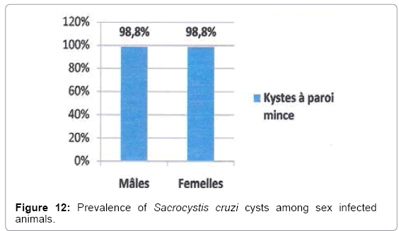
Figure 12: Prevalence of Sacrocystis cruzi cysts among sex infected animals.
Prevalence of sarcosporidiens cysts with thin wall depending to age: In carcass cattle infected with Sarcocystis spp, all cattle aged less than 2 years (47) and 111 on 113 cattle over 2 years showed cysts of Sarcocystis cruzi; or a prevalence of 100% and 98.2% respectively (Figure 13).
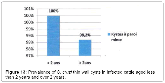
Figure 13: Prevalence of S. cruzi thin wall cysts in infected cattle aged less than 2 years and over 2 years.
Comparison of prevalence obtained by the two methods of diagnosing sarcosporidiosis (enzymatic digestion and histological analysis)
Histological analysis revealed 80% of cattle infected with Sarcocystis spp. While the technique of enzymatic digestion revealed 95% (Table 2). The test “chi-squared” shows that there is a significant difference (p ≥0.05) sensitivity between the enzymatic digestion technique and histologic technique.
| Samples | Enzymatic digestion | Histology | Total |
|---|---|---|---|
| Infested | 190 (95%) | 160 (80%) | 350 |
| No infested | 10 (05%) | 40 (20%) | 50 |
| Total | 200 | 200 | 400 |
Table 2: Comparison of the prevalence of the two methods of Sacrocystis diagnosing (histology and enzymatic digestion) in 400 samples analysed.
Search by macroscopic examination of Sarcocystis
In Algeria, no macroscopic cyst was observed until now in our slaughterhouses whether by veterinary inspectors or during specific studies conducted as part of Magister or graduation memory. Indeed, in 2002 Nedjari et al. had noted an absence of macroscopic cysts in 573 oesophagus of cattle taken in slaughterhouses of Hussein Dey. Harhoura et al. [2] who studied 170 diaphragms and 170 esophagi of cattle in the slaughterhouse of Rouïba also had noted the absence of macroscopic cysts. Dekkiche [7] and Lardjane et al. [8] observed no macroscopic cyst on the 63 and 110 beef carcasses at slaughterhouse of El Harrach, and Ruisseau, respectively. The absence of these cysts was also found in other parts of the country, including Boussebata et al. [9] during the inspection of 103 cattle carcass in slaughterhouses of East regions in Algeria. Our results are similar to those authors. Indeed, of the 200 cattle carcass inspected we have observed no macroscopic cyst. In addition, studies conducted in other countries have achieved the same results, notably in Iran, where no macroscopic cyst was identified in 02 different studies. The first study by Nourollahi Fard et al. [10] on samples of esophagus, hearts, tongues and skeletal muscle taken from 480 cattle to the slaughterhouse of the city of Kerman. The second Nourani [11] done by Hossein et al. on samples of diaphragm and esophagus 100 cattle taken at the slaughterhouse of Isfahan. Similarly, Aldemir and Güçlü [12] observed no macroscopic cyst inspecting samples frogs, esophagus and diaphragm by 100 cattle sampled in a slaughterhouse in the province of Konya in Turkey. Contradictory results were noted by Latif et al. [4]. These authors found in Baghdâd (Iraq), a prevalence of 0.2% of gross cysts, after an examination of esophagus, hearts, diaphragms and skeletal muscle 1080 cattle. Just as Nahed [13] in Egypt had observed a macroscopic cysts in 3% of 61 bovine carcass inspected. The greatest prevalences were recorded in China by Shi and Zhao detected, giant cysts in 64.78% of 159 cattle carcass in a slaughterhouse inspected in Jilin province. Authors conducted experimental infections with giant cysts on défintifs hosts. These experiences have enabled the authors to establish with certainty that the macroscopic cysts were those of S. hirsuta. For example, Böttner et al. [14] have infested experimentally 2 cats, 2 dogs and a human volunteer by macroscopic cysts of about 8 mm long and 1 mm wide, isolated from skeletal muscle of cattle. This was confirmed after demonstrating that the macroscopic cysts that were ingested by cats, dogs and humans were infesting for cats only. The same experiment was performed by the team of Aissi and Col [15] who showed oocysts S. cruzi after infestation of cats, dogs and primates by portions of muscle infested with microscopic cysts. In our present study, the absence of visible cysts is probably related to the absence of the feline species. This hypothesis could be explained by the fact that cattle are rarely in contact with cats in pastures and farms in Algeria. In France, some authors observed significant prevalence of bovine carcasses suffering from eosinophilic myositis [16,17]. Indeed, in 2013, in slaughterhouses countries of the Loire, it was the cause of 347 total entries. It frequently involves cattle healthy and valuable butcher, as the blonde of Aquitaine. In Algeria, the main cattle breeds are the black pie, red pie, which would explain the absence of eosinophilic myositis carcases inspected during our study.
Sarcocystis search by microscopic examination
Sarcocystis overall prevalence by enzymatic digestion technique: According Euzéby [18], Fassi-Fehri et al. [19] and Harhoura et al. [2], the infestation of these muscles by Sarcocystis similarly. Also, we have in our study, mixed the esophagus and diaphragms for the production of enzymatic digestion. Microscopic examination of our samples revealed a prevalence of 95% in the 200 cattle, and only 10 negative cattle (5%). The majority of studies on bovine sarcosporidiosis had related high prevalences. In Iran, Nourollahi Fard et al. [10] found 100% of the muscle samples 480 cattle infected with Sarcocystis (heart, esophagus, tongues and skeletal muscles). In Egypt, Nahed et al. [13] revealed the presence of bradyzoites in 60% of cattle taken from different slaughterhouses in Cairo and Giza. In Algeria, similar results have been reported by Harhoura et al. [2] which reported a prevalence of 100% by examining samples of 170 cattle esophagus and diaphragm. A few years later, Dekkiche [7] found 88.52% of the 61 samples of cattle esophagus and diaphragm positive Sarcocystis. Similarly Lardjane et al. [8] reported a prevalence of 89.9% on 110 cattle samples of esophagus and diaphragm. Similarly for Boussebata et al. [9] found that 93.2% of cattle parasitized by Sarcocytis. These authors have also worked on samples of esophagus and diaphragms. In France, Mary [20], revealed a prevalence of 97% by the enzymatic digestion of muscle samples (esophagus, diaphragms, hearts and skeletal muscle). In our study, the heavy infestation of cattle by Sarcocystis could be attributed to extensive pollution of pastures and farms by Sarcocystis sporocysts excreted in the faeces of definitive hosts. The infestation rate Sarcocystis spp. is remarkably high ubiquitously. First, the dog, the cat and the man could be behind this strong infestation they released in large quantities of cysts in faeces [21]. Second, an intense multiplication of tachyzoites could be the cause. Furthermore, a study in the north of Western Australia by Savini et al. [3] showed that the flocks were less infested by sarcosporidia. These authors found a prevalence of 31.2% of cattle infested by Sarcocystis spp. This low prevalence can be explained by an unfavorable climate for the survival of cysts in fact the northern part of Western Australia is characterized by a very arid climate; the survival of cysts is not favored in this environment [3].
Study of risk factors on the prevalence of Sarcocystis
Age factor: In our study, the prevalence of microscopic cysts of Sarcocystis spp. in cattle was not influenced by age. In Algeria, our results are similar to those of Harhoura et al. [2] Dekkiche [7] Lardjane et al. [8] and Boussebata et al. [9] who did not find any influence of age on the prevalence of Sarcocystis. In Iran, Nourollahi Fard et al. [10]; Najafiyan et al. [22], Fassi-Fehri et al. [19] in Morocco, also noted the absence of the influence of age on the prevalence of Sarcocystis. However, other authors found that there was an influence of age on the prevalence of Sarcocystis. Indeed, in France, The study by Flandrin [23] and Guénégan [24] showed that there is a relationship between the age of cattle and foreclosure rates for sarcosporidiosis. Indeed, all concerned entries of cattle, while no calf has been entered for that reason. Seneviratna et al. [25], Park et al. [26] found the absence of infection in calves aged less than a year, while older cattle were infected. Guénégan [24] after, noted that low risk of being exposed to infection in young cattle can be explained by the fact that they are a majority of their living in closed buildings. So the direct or indirect contact with domestic or wild carnivores are a priori virtually nonexistent. Thus, access to pasture is limited to suckler cows. According Fukuyo et al. [27], this increased parasitism with age may be due to the fact that the animal suffered repeated infestations, which would lead to a gradual accumulation of cysts in muscle with age. However, Savini et al. [3] obtained conflicting results. According to them, acquired immunity with age of the host will reduce the number of cysts over time. Thus, the prevalence of Sarcocystis spp. fell significantly in older.
Sex Factor: In this study, the prevalence of microscopic Sarcocystis cysts in cattle is not affected by gender. The same results were found by Harhoura et al. [2]. Dekkiche [7] who found no influence of sex on the prevalence of Sarcocystis. In Iran, Fard et al. [10], Najafiyan et al. [22] also noted the absence of the sex influence on the prevalence of Sarcocystis. However, from Edwards [17,28], Corner et al. [29] and, it seems that there is a significant difference between the degree of infestation of males and females. Indeed, in their studies were more females than males infested. Edwards [28]; justifies this high rate of infection in females by muscle tropism of Sarcocystis vary by sex. The parasite localizes preferentially at the level of the uterus. This hypothesis is supported in his view by the occurrence of abortions reported in cattle in acute sarcosporidiosis. In France, Bertin [30] found that females were also more infested than males. According to last, this could be of the type of farming (access to pastures, feeding modalities) slightly different for males and females. However, Savini et al. [31] noted a higher prevalence in males than in females.
Search of Sarcocystis cysts by histological study
Overall prevalence of microscopic Sarcocystis cysts: Histological examination of the muscles showed that 80% of the 200 cattle were infested with cysts of Sarcocystis spp. Our results are similar to those obtained by Harhoura et al. [2] who found a prevalence of 90.8% of 120 cattle slaughtered at the abattoir Rouiba and are also consistent with those obtained by Woldemeskel and Gebreab [32], Ethiopia, who noted a prevalence of 82% on samples of diaphragm, masseter, heart and esophagus cattle. Godoy et al., Venezuela, estimated the rate of infestation by Sarcocystis 92.8% of 630 samples of myocardium examined cattle. Mary [20] in France detected the presence of Sarcocystis spp cysts in 89% of studied cattle. Fassi-Fehri et al. [19] in Morocco have detected the presence of Sarcocystis spp cysts in 100% of cattle. Nourani et al. [11] in Iran, 92% of cattle infested with cysts of Sarcocystis spp.
Prevalence of cysts according to the organ: In our study the esophagus was the organ the most frequently infested with cysts of Sarcocystis spp. Monitoring of the diaphragm, with infestation rates of 70.50% and 60.50% respectively. Our results are similar to those found by Aldemir and Güçlü [12] in Turkey which revealed a higher prevalence of microscopic Sarcocystis cysts in the esophagus with 92% of positive cases, followed by the heart and diaphragm with rates infestation of 84% and 63% respectively. Moreover, these authors found that the esophagus also contained the highest number of Sarcocystis cysts. Fassi Fehri and et al. [19] also found that the organ most regularly infested is the esophagus, followed by the diaphragm and masseter. However, our results contradict those obtained by Harhoura et al. [2] who observed a predominance of cysts in the diaphragm rather than in the esophagus with infection levels of 84% and 63.3% respectively. Mary in 2005 has noted a strong infestation in the myocardium followed by the masseter and diaphragm. The esophagus seems to be the organ most infested and according to Fassi-Fehri et al. [19], esophagus probably has the best conditions for the development of the parasite; it is the organ most sensitive, the most faithful and therefore safest for the diagnosis of sarcosporidiosis.
Prevalence of cysts depending on the type of wall: In our study, histological examination showed that 98. 8% of infected cattle were infested with thin-walled cysts (S. cruzi), and only 4. 4% harbored thick-walled cysts (S. hirsuta and/or S. hominis). In Algeria, Nedjari [1] noted the presence of S. cruzi cysts in 60.2% of cattle, while 39.8% had S. hirsuta and / or S. hominis cysts. A few years later, Harhoura et al. [2] reported the presence of S. cruzi cysts in 85.8% of the analyzed cattle, while the thin-walled cysts were present in 25% of cattle. In a study conducted in Turkey, Aldemir and Güçlü [12] noted the presence of S. cruzi in 74% of cattle, the presence of S. hominis and S. hirsuta was reported respectively in 15% and 3% of cattle. In Argentina, Moré et al. [33] observed that 71.5% of cysts were with a thin wall, while 23.1% had a thick wall. According Ruas et al. [34]; the prevalence of S. cruzi is also enhanced by the ability and cyst removal time by dogs. However, some authors noted a predominance of thick-walled cysts of S. hominis. In France, Dimitri [35] found that 88.6% of cattle were infested by cysts with a thick wall of S. hominis 61% had the pecies S. cruzi while 1.6% contained cyst wall thickness S. hirsuta. In Belgium, Vangeel et al. [36] found a high prevalence of S. hominis cysts (97.4%) in samples of raw ground beef. In our study, we identified S. hominis in a single cattle based on morphological observation of cytophanères by optical microscopy. These were visible, they were long, cylindrical (fingerlike), almost perpendicular to the surface of the wall, characteristic of S. hominis (zoonotic species). According Nedjari [1] Woud et al. [37] the distinction between S. hirsuta cysts and S. hominis may be possible using an optical microscope by using the morphological criteria of the wall, as the provision of cytophanères. The presences of S. hominis mean in our study that bovine number 193 was directly exposed to human feces. Thus, the possibility that this animal had grazed grass in areas polluted by the discharge of sewage homes is not to be discarded, especially in rural areas where there is a lack of infrastructure for routing and the treatment of these waters.
Prevalence of cysts sarcosporidiens thin-walled and thick-walled
Prevalence of thin-walled cysts: In our study, the thin-walled cysts S. cruzi were more frequently found in the esophagus and diaphragms with high prevalence (94.2%) and (100%), respectively. Our results are homologous to those obtained by Harhoura et al. [2]. They noted that the thin-walled cysts S. cruzi were largely present at the oesophagus (60.8%) and diaphragms (79.8%). Ruas et al. [34] revealed infestation rate by 100% S. cruzi cysts in the two bodies. However, other studies suggest that S. cruzi cysts preferentially localized in the esophagus; as the study of Moré et al. [33] who noted more frequent infection of the esophagus by S. cruzi cysts compared to the diaphragm, with prevalences of 71% and 28%. Similar results were observed in France, by Flandrin [23] found that a preferential localization of S. cruzi in the esophagus with a prevalence of 57%.
Prevalence of cysts thick-walled: In our study, thick-walled cysts were located only at the diaphragm with a very low prevalence. Our results are homologous to those of Ruas et al. [34]. These authors have shown that S. hirsuta cysts were found only in the diaphragms with low prevalence (1.4%). Similarly Harhoura et al. [2] showed that the cyst wall thickness S. hirsuta and/or S. hominis were present in the diaphragms (23.5%) and in the esophagus with a lower prevalence (7.5%).
Mixed infections: In our study, histological examination revealed the presence of mixed infections in one cattle. We noted the presence of two types of cysts with a 4.1% frequency in diaphragms. Many studies have revealed the presence of double infestations within one pet, in sheep [6]; the Asian buffalo [38] but also cattle. Indeed, Harhoura et al. [2] noted the presence of mixed infections as well as in the esophagus in the diaphragm with respective frequencies of 5% and 19.3%. Mary [20] also found 27% of cattle infested by simultaneously cysts thinwalled and thick-walled and localized much in the myocardium. Cases of double infestations have also been reported in wild animals such as white-tailed deer [39] and the armadillo [40-43]. The presence of mixed infections indicates that cattle may be infected simultaneously by several species of Sarcocystis.
Study of risk factors on the prevalence of species of Sarcocystis spp.
Intensity of parasitism diaphragm and esophagus of infested cattle: We noted the presence of 1430 cysts of Sarcocystis spp. among 160 infected cattle. First, we found a large number of thin-walled cysts in the diaphragms (675 cysts) and esophagus (745 cysts). These results are comparable to those of Harhoura et al. [2] who noted the presence of 619 and 694 thin-walled cysts in esophagus and diaphragm, respectively. Furthermore, we found 10 thick-walled cysts only all located at the diaphragm. Unlike our results, Harhoura et al. [2] have noted 109 and 26 thick-walled cysts in the diaphragms and oesophagi respectively. We found that the majority of the diaphragm and esophagus samples of infected animals showed less than 10 cysts. However a minority was heavily parasitized, between 11 and 68 cysts. Diaphragms and esophagus having a large number of cysts probably increase the risk of infestation of definitive hosts. Indeed, the study of Aissi et al. [15] on experimental infection in dogs, cats and monkeys with bovine mixed meat contaminated with two types of cysts have revealed that a low concentration of cysts in meat was not sufficiently infective for guests final.
Study of risk factors (sex and age) on the prevalence of thinwalled cysts: S. cruzii: Histological examination showed no influence of sex and age factors on the overall prevalence of thin-walled cysts. The study of Harhoura et al. [2] showed no influence of sex factor on the overall prevalence of cysts thin-walled and thick. Furthermore, these authors revealed the existence of the influence of age on the prevalence of the two types of cysts that increase with the age of cattle.
Comparison of prevalence obtained by the two methods of diagnosis
In our study, we compared the sensitivity of the two diagnostic methods used in the sarcosporidiosis studied on 200 cattle. Our results showed a high sensitivity of enzymatic digestion with a detection rate of 92% compared to the histological technique with 80% of positive cases. In fact, several authors have proved the high sensitivity of enzymatic digestion in detecting Sarcocystis. By comparing the sensitivity of the two methods, Vercruysse et al. [44] and Mary [30] had more positive cattle digestion by histology. Harhoura et al. [2] demonstrated that the artificial digestion with pepsin was more sensitive in detecting infestations Sarcocystis that examination of histological sections. These authors found a detection rate of 100% by enzymatic digestion and 90.8% by histology. In our study, the search for Sarcocystis by pepsin digestion method was performed in 20 g of muscle. This has increased our chances of finding the parasite. Whereas with the technical histological detection of cysts was performed in an area of 1 to 2 cm2 tissue only, thereby limiting the search field. According Desportes-Livage and Datry [15], the high sensitivity of the pepsin digestion technique can also be explained by the fact that mature cysts Sarcocystis contain thousands of bradyzo?tes that can be released by digestion cysts. However, negative results we obtained by histological technology would be related to the low number of Sarcocystis cysts in the examined samples. In some studies, Beyazit et al. [9,41] were statistically no significant differences in sensitivity between the enzymatic digestion and the histological method in the detection of species of Sarcocystis. According Gajadhar and Marquardt [42-45] even the enzymatic digestion can have drawbacks that may underestimate the prevalence and intensity of infection. In some cases, the tissues are not fully digested and pests can be trapped and disposed of with the unfiltered material.
In our study we found a high prevalence of Sarcocystis (95%) in cattle carcass at slaughterhouses El Harrach (Algiers) by enzymatic digestion method. Histological examination has allowed us to identify two types of Sarcocystis cysts. Cysts thin wall predominant corresponding to S. cruzi and a cyst wall thickness corresponding to S. hominis (cytophanères observed by light microscopy) and other cysts probably corresponding to S. hirsuta. Our study reveals the important role of the dog in the circulation of Sarcocystis, and the role of man seems minimal in the transmission of the parasite in cattle. This study is a preliminary work will be supplemented by other studies in other Algiers region abattoirs to determine the true prevalence of cattle sarcosporidiosis in Algiers slaughterhouses and the importance of each Sarcocystis species.
We sincerely thank Professor Hadj-AMAR R., Director of the Laboratory of Pathological Anatomy and Cytology Centre University Hospital of Bab El Oued for here help.