Journal of Cell Science & Therapy
Open Access
ISSN: 2157-7013
ISSN: 2157-7013
Commentary - (2018) Volume 9, Issue 3
The mechano-sensitive mouse Piezo1 channel function as key eukaryotic mechano-transducers. However, the structure and mechano-gating mechanism of the mechano-sensitive mouse Piezo1 channel with small molecule agonist remain unknown. New studies have now provided critical insights into the high-resolution cryoelectron microscopy structure and mechano-gating of the mechanosensitive mPiezo1 channel with agonist (Figure 1).
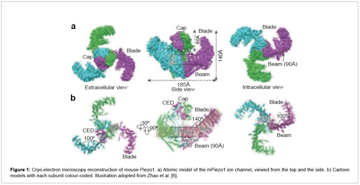
Figure 1: Cryo-electron microscopy reconstruction of mouse Piezo1. a) Atomic model of the mPiezo1 ion channel, viewed from the top and the side. b) Cartoon models with each subunit colour-coded. Illustration adopted from Zhao et al. [6].
Piezo family of genes are mechanically activated Ca2+ permeable non-selective cationic channels that mediate touch perception, proprioception, vascular development and brain development. They have been established as the long-sought-after mechano-sensitive cation channels in mammals [1]. Considering their physiological importance, both loss-of-function and gain-of-function mutations of Piezo family of genes have been associated with disease such as xerocytosis and lymphatic dysplasia [2,3]. Mammalian Piezos are large transmembrane proteins that are composed of about 2547 amino acids with 38 transmembrane-helix topology. Mouse Piezo1 has been previously determined, revealing its three-bladed, propellerlike trimeric architecture comprising a central pore module and three extended peripheral wings [4]. The complex mPiezo1 protein might be divided into the peripheral blades and a central cation selective pore module [5]. These features make structure and function studies of Piezo proteins highly challenging, partly explaining the slow progress in our understanding of the molecular basis of mechano-gating properties of this novel of ion channels. In the latest researches have now made major breakthroughs in demystifying Piezo channels.
In the first latest research, the present study shows that the threebladed, propeller-like cryo-EM structures of mPiezo1 and functionally reveal its mechano-transduction components. The mPiezo1 protein comprises 9 repetitive units of 4 transmembrane helices segments, assembling into the highly curved peripheral blade-like structure, which is critical for mechano-sensing and transduction [6]. The last transmembrane encloses a hydrophobic pore, followed by three intracellular fenestration sites and side portals comprising poreproperty- determining residues. The central region that forms a ~90 Å-long intracellular beam-structure, which bridges the blade to the pore. These results indicate that Piezo1 possesses an unprecedented 38-transmembrane topology and designated mechano-transduction components, enabling a lever-like apparatus for effectively transducing force from the mechano-sensing blade to the ion-conducting pore, enabling long-distance mechano-gating mechanism (Figure 2).
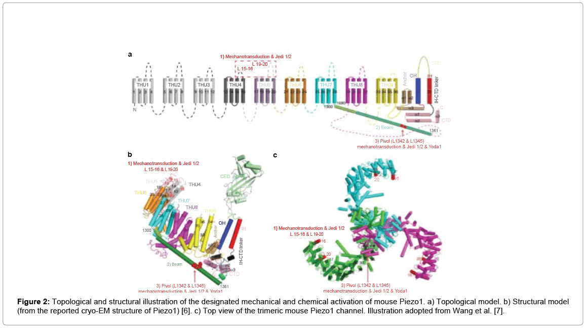
Figure 2: Topological and structural illustration of the designated mechanical and chemical activation of mouse Piezo1. a) Topological model. b) Structural model (from the reported cryo-EM structure of Piezo1) [6]. c) Top view of the trimeric mouse Piezo1 channel. Illustration adopted from Wang et al. [7].
In the second latest research, the present study shows that a novel Piezo1 chemical activators (Jedi) that directly bind to Piezo1 and modulate its mechano-sensitivity and ion selectivity. Jedi act through the N-terminal extracellular loop regions that might reside in the distal blade-structure activates Piezo1 through the extracellular side of the blade instead of the C-terminal ion-conducting pore, suggesting a longrange allosteric gating. Remarkably, Jedi-induced activation of Piezo1 requires the key mechano-transduction components, including the two extracellular loops (L15-16 and L19-20) in the distal transmembrane helical units or the mutating single (L1342/L1345) residues in the proximal end of the beam [7]. The research further demonstrates that Jedi1 and Yoda1 (Yoda1 is a useful pharmacological tool [8]) act through distinct mechanisms to modulate Piezo1. Piezo1 chemical activators (Jedi and Yoda1) appear to activate and modulate Piezo1 via acting on different loci along the blade-beam gating pathway. The extracellular loops (L15-16 and L19-20) mutants completely lose their ability to Jedi1/2 but remained Yoda1-induced a preferentially potentiated their poking currents, but not the stretching currents. In contrast, the L1342/L1345 mutants are insensitive to both Jedi and Yoda1. The data suggest that Jedi1/2 act on the upstream blade, while Yoda1 acts at the downstream beam. These results indicate that Piezo1 utilizes the beam as a lever-like apparatus for effectively transducing force from the mechano-sensing blade to the ion-conducting pore, enabling long-distance mechano-gating.
Piezo proteins serve as a principal type of mechano-transduction channels. The cryo-electron microscopy structure of mouse Piezo1 and functionally shows that it forms a three-bladed, propeller shaped trimeric complex. The research identifies 9 repetitive units consisting of 4 transmembrane helices each, which assemble into a highly curved blade-like structure. The mPiezo1 structure described represents the first high-resolution of this structurally and functionally novel of channel. Toward exactly understanding the structure-function relationship of this sophisticated type of mechano-sensitive channels, future efforts should also be oriented toward obtaining Piezo structures in high-resolution and making depth structure functional characterizations. The present disclosure relates to a bio-medicine, and more particularly to use of regulator used for activating Piezo in preparation of a medicament.