Advanced Techniques in Biology & Medicine
Open Access
ISSN: 2379-1764
ISSN: 2379-1764
Review - (2025)Volume 13, Issue 1
In this article, an electronic converter is introduced, which can be used to connect the damaged part of the spinal cord to the input and output of the converter in order to transmit an electrical signal and revive the damaged part. At first the configuration of spinal cord and then structure of electrical transducer are introduced after that connections between each other is clarified.
There are no reliable estimates of the global prevalence of Spinal Cord Injury (SCI), but the global annual incidence is estimated to be 40-80 per million people. Young people are most at risk, followed by older people with a male to female ratio of at least 2:1 [1]. This condition can have both traumatic and nontraumatic causes. Regardless of the etiology, the injury can cause long-term disabilities, ranging from motor and sensory deficits to autonomic dysfunction and neurohormonal disorders. Autonomic processes play an important role in hormonal regulation and also influence central metabolism [2]. In Furthermore, the effects on the motor system and the resulting reduced opportunities for physical activity influence energy expenditure [3]. Therefore, the effects of disease mechanisms after SCI are likely to cause changes in body composition, potentially affecting body weight [4]. The negative effects of low and high Body Mass Index (BMI) have been well described [5]. It has been suggested that increased BMI/weight may affect mobility and participation in SCI patients. Furthermore, an association between BMI and leisure-time physical activity has been found [6]. In summary, the findings suggest a bidirectional relationship between various measures of nutritional status and activity level. The stages of rehabilitation and return to daily life can influence body composition [7]. Therefore, it may be beneficial to measure changes in body composition throughout the course of rehabilitation. Recent studies have found that BMI remains stable during the first few years after trauma, although sample sizes are lacking to detect patterns related to functional level [8]. However, other studies have found a pattern of initial weight loss followed by either sustained weight loss, weight gain or weight stability, with long-term follow-up after 5 years suggests that increases in BMI occur in all ethnic groups [9]. Additional knowledge about weight changes during and after rehabilitation is needed to prevent negative effects on the health and outcomes of patients with SCI. A global consensus recommends criteria such as BMI, inflammation, weight change and body composition for the diagnosis of malnutrition in clinical practice [10]. A possible association between the pattern of weight change after SCI and the degree of disability has not yet been described [11]. Examination of the relationship between body composition and disability in SCI patients is often based on the American Spinal Injury Association disability scale (ASIA or AIS) and body weight or BMI [12,13]. The AIS scale carefully describes the characteristics of SCI according to the degree and level of disability at the level of physical function and is classified on a 5- point scale [14]. Although the AIS describes important symptoms of injury, it may not be sufficient to characterize the functional capacity to support participation and activity. Spinal Cord Independent Measurement (SCIM) is used to assess daily functioning. Activities and scales are divided into self-care, respiratory and sphincter management and mobility [15]. Using SCIM to investigate patterns of weight change may provide further insight into health-related factors when living with a spinal cord injury. The first objective of this study is to describe the structure of the spinal cord. Next, we will explain the function of the spinal cord from an electrical perspective, showing electrical devices as transducers [16].
Structure of spinal cord
The spinal cord is the most important structure between the body and the brain. The spinal cord extends from the foramen magnum where it is continuous with the medulla to the level of the first or second lumbar vertebrae. It is a vital link between the brain and the body and from the body to the brain. The spinal cord is 40 cm to 50 cm long and 1 cm to 1.5 cm in diameter. Two consecutive rows of nerve roots emerge on each of its sides. These nerve roots join distally to form 31 pairs of spinal nerves. The spinal cord is a cylindrical structure of nervous tissue composed of white and gray matter, is uniformly organized and is divided into four regions: Cervical (C), Thoracic (T), Lumbar (L) and Sacral (S), each of which is comprised of several segments (Figure 1). The spinal nerve contains motor and sensory nerve fibers to and from all parts of the body. Each spinal cord segment innervates a dermatome [17].
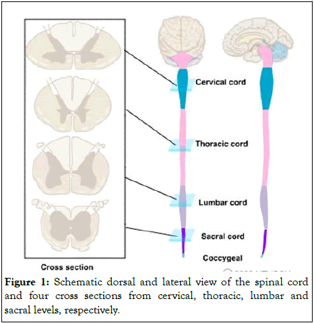
Figure 1: Schematic dorsal and lateral view of the spinal cord and four cross sections from cervical, thoracic, lumbar and sacral levels, respectively.
General features
• Similar cross-sectional structures at all spinal cord levels.
•It carries sensory information (sensations) from the body and some from the head to the Central Nervous System (CNS) via afferent fibers and it performs the initial processing of this information.
•Motor neurons in the ventral horn project their axons into the periphery to innervate skeletal and smooth muscles that mediate voluntary and involuntary reflexes.
•It contains neurons whose descending axons mediate autonomic control for most of the visceral functions.
•It is of great clinical importance because it is a major site of traumatic injury and the locus for many disease processes.
Although the spinal cord constitutes only about 2% of the Central Nervous System (CNS), its functions are vital. Knowledge of spinal cord functional anatomy makes it possible to diagnose the nature and location of cord damage and many cord diseases [18].
Segmental and longitudinal organization
The spinal cord is divided into four different regions: The cervical, thoracic, lumbar and sacral regions. The different cord regions can be visually distinguished from one another. Two enlargements of the spinal cord can be visualized: The cervical enlargement, which extends between C3 to T1; and the lumbar enlargements which extends between L1 to S2. The cord is segmentally organized. There are 31 segments, defined by 31 pairs of nerves exiting the cord. These nerves are divided into 8 cervical, 12 thoracic, 5 lumbar, 5 sacral, and 1 coccygeal nerve (Figure 2). Dorsal and ventral roots enter and leave the vertebral column respectively through intervertebral foramen at the vertebral segments corresponding to the spinal segment [19].
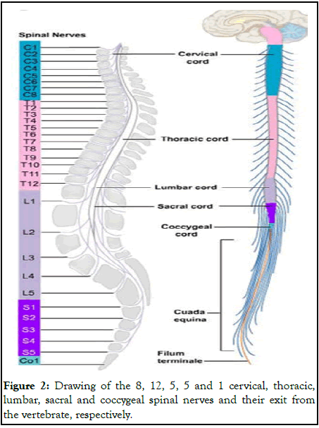
Figure 2: Drawing of the 8, 12, 5, 5 and 1 cervical, thoracic, lumbar, sacral and coccygeal spinal nerves and their exit from the vertebrate, respectively.
The cord is sheathed in the same three meninges as is the brain: The pia, arachnoid and dura. The dura is the tough outer sheath, the arachnoid lies beneath it and the pia closely adheres to the surface of the cord (Figure 3). The spinal cord is attached to the dura by a series of lateral denticulate ligaments emanating from the pial folds [20].
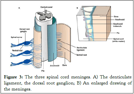
Figure 3: The three spinal cord meninges. A) The denticulate ligament, the dorsal root ganglion; B) An enlarged drawing of the meninges.
During the initial third month of embryonic development, the spinal cord extends the entire length of the vertebral canal and both grow at about the same rate. As development continues, the body and the vertebral column continue to grow at a much greater rate than the spinal cord proper. This results in displacement of the lower parts of the spinal cord with relation to the vertebrae column. The outcome of this uneven growth is that the adult spinal cord extends to the level of the first or second lumbar vertebrae and the nerves grow to exit through the same intervertebral foramina as they did during embryonic development. This growth of the nerve roots occurring within the vertebral canal, results in the lumbar, sacral and coccygeal roots extending to their appropriate vertebral levels. All spinal nerves, except the first, exit below their corresponding vertebrae. In the cervical segments, there are 7 cervical vertebrae and 8 cervical nerves. C1-C7 nerves exit above their vertebrae whereas the C8 nerve exits below the C7 vertebra. It leaves between the C7 vertebra and the first thoracic vertebra. Therefore, each subsequent nerve leaves the cord below the corresponding vertebra. In the thoracic and upper lumbar regions, the difference between the vertebrae and cord level is three segments. Therefore, the root filaments of spinal cord segments have to travel longer distances to reach the corresponding intervertebral foramen from which the spinal nerves emerge. The lumbosacral roots are known as the cauda equina. Each spinal nerve is composed of nerve fibers that are related to the region of the muscles and skin that develops from one body somite (segment). A spinal segment is defined by dorsal roots entering and ventral roots exiting the cord, (i.e., a spinal cord section that gives rise to one spinal nerve is considered as a segment) (Figure 4).
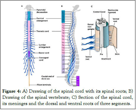
Figure 4: A) Drawing of the spinal cord with its spinal roots; B) Drawing of the spinal vertebrate; C) Section of the spinal cord, its meninges and the dorsal and ventral roots of three segments.
Structure of electrical transducer
Information of signal transmitted from brain via spinal cord is alternative with determined frequency and analogue type, therefore a converter must be designed to first convert analogue signal to digital code (input) and finally decode information to readable information for spinal cord (output). To turn this reasoning into reality we need an ADC in input connected to spinal cord directly in order to alter analogue code to digital code (Figure 5).
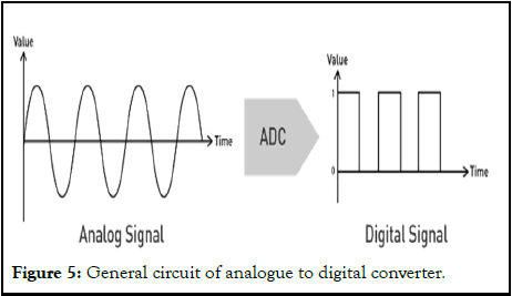
Figure 5: General circuit of analogue to digital converter.
To show more accurate than the configuration of ADC converter, Figure 6 is illustrated.
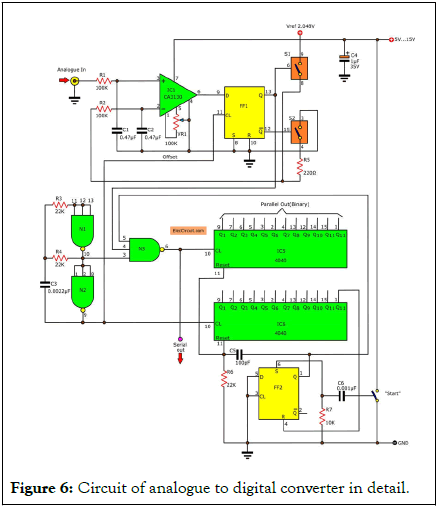
Figure 6: Circuit of analogue to digital converter in detail.
For reminder, to sense the signal of sent from spinal cord to ADC, an electrical sensor in necessity to receive that. After ADC and in the third stage we need a decoder to turn output information from ADC into understood signal as shown in Figure 7.
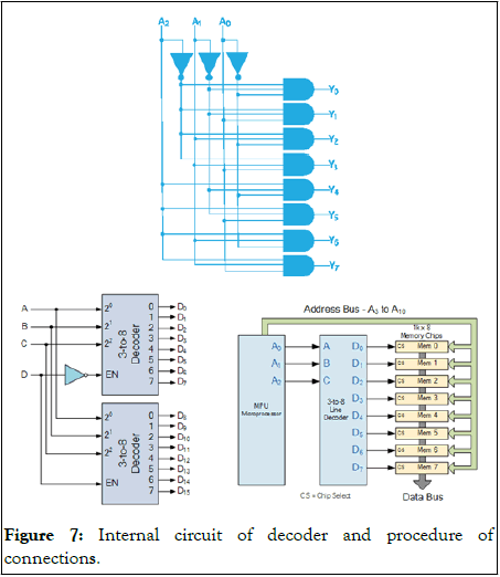
Figure 7: Internal circuit of decoder and procedure of connections.
For summarizing entirely, signals are transmitted from spinal cord to sensor, then information from sensor is transmitted to ADC and then ADC to decoder, eventually signal is transmitted to another point of spinal cord in according to Figure 8.
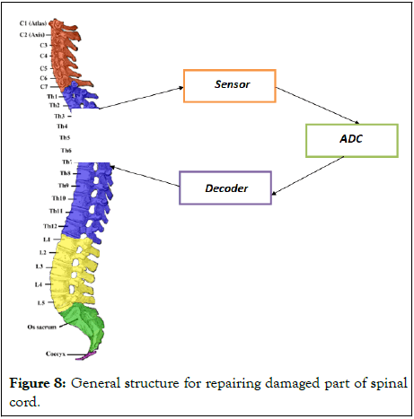
Figure 8: General structure for repairing damaged part of spinal cord.
[Crossref] [Google Scholar] [PubMed]
[Crossref] [Google Scholar] [PubMed]
[Crossref] [Google Scholar] [PubMed]
[Crossref] [Google Scholar] [PubMed]
[Crossref] [Google Scholar] [PubMed]
[Crossref] [Google Scholar] [PubMed]
[Crossref] [Google Scholar] [PubMed]
[Crossref] [Google Scholar] [PubMed]
[Crossref] [Google Scholar] [PubMed]
[Crossref] [Google Scholar] [PubMed]
[Crossref] [Google Scholar] [PubMed]
[Crossref] [Google Scholar] [PubMed]
[Crossref] [Google Scholar] [PubMed]
[Crossref] [Google Scholar] [PubMed]
[Crossref] [Google Scholar] [PubMed]
[Crossref] [Google Scholar] [PubMed]
[Crossref] [Google Scholar] [PubMed]
[Crossref] [Google Scholar] [PubMed]
[Crossref] [Google Scholar] [PubMed]
Citation: Ghanbari Z, Shirzad E (2025) Repairing a Part of the Spinal Cord with Electrical Transducers in Order to Continue Living Normally. Adv Tech Biol Med. 13:457.
Received: 18-Dec-2023, Manuscript No. ATBM-23-28529 ; Editor assigned: 20-Dec-2023, Pre QC No. ATBM-23-28529 (PQ); Reviewed: 03-Jan-2024, QC No. ATBM-23-28529 ; Revised: 07-Mar-2025, Manuscript No. ATBM-23-28529 (R); Published: 14-Mar-2025 , DOI: 10.35248/2379-1764.25.13.457
Copyright: © 2025 Ghanbari Z, et al. This is an open-access article distributed under the terms of the Creative Commons Attribution License, which permits unrestricted use, distribution, and reproduction in any medium, provided the original author and source are credited.