Advanced Techniques in Biology & Medicine
Open Access
ISSN: 2379-1764
ISSN: 2379-1764
Research Article - (2015) Volume 3, Issue 3
Multi drug resistance of pathogens to drugs has become a universal problem. The aim the study was to investigate if plants’ natural products could offer alternative solutions to the problems. Roots of the plant were collected from Keiyo District; air dried in the shade then ground into fine powder and extracted with methanol, analytical grade, which eventually evaporated using a rotor evaporator. Known amount of the resultant material was measured and dissolved using 1 ml of dimethyl sulphoxide and topped with deionized water to form a known concentration. Serial dilutions of the drug were further made. Disc diffusion methods was used in the microbial studies with registered microorganisms at the National Public Health Laboratory, Nairobi. There were indications that the plant’s extracts possessed reasonable antimicrobial activities. The other portion of the extracts was subjected to wet bench studies which revealed the presence of several classes of compounds that are known to have curative properties. It was evident that the local inhabitants are justified to use the plant in the management of various that inflict them and it was recommended that the plants extracts cytotoxicity in higher animals be evaluated so as to justify its continued used in folklore medicine. It was also recommended that the compounds in the plant be further elucidated to assess their potential as sources of new compounds that could be used by the pharmaceutical concerns in new drugs development.
Keywords: Multi-drug resistance, Indigofera homblei, Keiyo, Kenya
Some of the Indigenous people of the North Rift Valley of Kenya, the Keiyo, use plants as traditional medicine. Indigofera homblei, which is indigenous to the area, is used in traditional medicine in that, about100g of freshly harvested roots, boiled in a litre of water, decanted, extracts about 250g drunk once daily till recovery from gonorrhea and stomach ailments. Freshly harvested roots may also be chewed in the same doses for the same conditions. The stem of the plant is also used as toothbrush by the some communities in Kenya [1].
Currently there is emergence of pathogens that are multi drug resistant to newly discovered synthetic antibiotics at the rate that researchers find it difficult coping up [2]. Such scenarios call for conventional approach to their management. It is, however, unfortunate that the antibiotics that are available in market are too expensive such that virtually majority of the common populations in developing can hardly afford them [3]. Testing and validating the safe use of the locally and traditionally available drugs, like the one given herein, could offer solutions to the problems.
The plant, Indigofera homblei Bak.f. and Martin, Fabaceae, (and locally called Kipkoros) is softly woody shrub to 2.5m, a 3 angled or winged stem; leaflets are usually 11-17, on rachis 14cm;receme many flowered, with stiff brown hairs, to 16cm long; sepals densely brownbrown sepals, same long up to 12mm, pods hairless 4-5mm hanging on long stalk, 6-8 seeded, to 45mm long. In Kenya, rare to scattered tree grassland and forest edges. The species is listed as endangered (Agnew, 2013). Indigofera arrecta A. Rich.which is widely distributed in Kenya is used for curative purposes by several the communities in Kenya to manage some microbial conditions and I. spinosa Forsk. is used against sore throat by certain communities in Western Kenya (Kokwaro, 1993).
Plant samples were collected from Keiyo District about 40 Km East of Eldoret town. The stem bark, root bark and leaves were sampled and authenticated by a trained taxonomist then confirmed at the Herbarium of the Kenya National Museums by Mbalu Mbale where voucher specimens were then deposited. The samples were air-dried in the shade and ground into a fine powder using a grinding m ill.
Methanol extraction
The National Cancer Institute (NCI) protocol was followed during extraction of bioactive molecules [4]. The ground material (100 g) were soaked in methanol for 72 h. with occasional shaking, after which the extracts were filtered and concentrated using rotary evaporator at 45°C under reduced pressure and finally dried to a gummy material in a desiccator containing anhydrous calcium chloride. The crude extracts were then used for bioassay. Generally all the co-authors participated in the extraction and microbial studies at the department of Chemistry, Kenyatta University for extraction and characterization, and National Public Health Laboratories where the microbial studies were done.
Phytochemical screening
A standard qualitative procedure was used to detect classes of compounds present in the active extracts as described by Chhabra (1989) and Harbone (1973) [5,6]. The classes of compounds that were screened were alkaloids, flavonoids, Anthocyanins, anthraquinones, Tannins, coumarins, saponins, steroids and reducing compounds. These classes of compounds were chosen for phytochemical screening as they have been associated with antimicrobial activities. Positive reactions were indicated by a (+) mark w ith different concentrations as (+) for low; (++) for moderate and (+++) for high concentrations. All the negative reactions were indicated by a (-) mark.
Bioassay culture strains
Microorganisms used were obtained from Kenya National Public Health Laboratories (KNPHL) and Kenya Medical Research Institute (KEMRI). These included standard and local clinical isolates from patients. Microorganisms that were used included: three Gram positive bacteria, Staphylococcus aureus (ATCC 20591), a reference strain, Enterococcus faecalis (KNPHL 016) and Bacillus subtilis (Type K [11]), both clinical isolates. Three Gram negative bacteria; Pseudomonas aeruginosa(ATCC 27853), a reference strain, Salmonella typhi (Type K[I]) and Klebsiella pneumoniae (KNPHL 002) both clinical isolates were also used. Two yeasts, Candida albicans (ATCC 90028) and Cryptococcus neoformas (ATCC 66031), both reference strains; two fungi, Trichophyton mentagrophyte (KMR 100) and Microsporum gypseum (KMR 101), both clinical isolates were used in studying the antifungal activities of the plant .
The Multiple Drug Resistant (MDR) strains Staphylococcus aureus (KNPHL 003) and Klebsiella pneumoniae (KNPHL 001) both clinical isolate were used in the study The test strains of bacteria were kept refrigerated at 4°C in Muller-Hinton (Merck, Germany) agar slants during the experimental period and were subcultured and incubated for 24h at 37°C then tested biochemically for purity before each use. Candida albicans cultures were maintained on Potato Dextrose Agar (PDA) at 4°C. Yeasts were subjected to two successive transfers in PDA broth and tested for purity and viability using biochemical and morphological characteristics before each use as described by Elgayyar et al., (2000).
Screening for antimicrobial activity
Antimicrobial efficacies were tested using the filter paper disc diffusion method [7]. A solution of each extract was prepared by dissolving 200 mg in 1 ml of methanol and 10 ml of the solution that were dispensed onto 6 mm sterile filter paper discs and dried (2 mg/ disc). The Muller-Hinton and Potato Dextrose Agar (PDA) were used in the culture of bacteria and fungi, respectively. Each media was prepared by weighing the quantities recommended by the manufacturer and dissolving in recommended quantities of distilled water. The media was then sterilized using an autoclave set at 121°C for 15 minutes and poured onto sterile Petri dishes then allowed to cool on a clean bench.
Each plate was inoculated with 0.1 ml of bacterial and yeast cultures directly from the 24h broth culture diluted to match 0.5 and 1.0 McFarland standard, respectively (108 Colony Forming Units (CFU)/ ml) and fungi diluted to match 1.0 McFarland standard (108 spores/ ml). The discs loaded with the extracts were then placed onto the seeded plates. The bacterial and yeast cultures were incubated at 37o C for 24 and 48 h, respectively, while fungi were incubated at 25°C for 5 days. After the incubation period the zones of inhibition were measured and recorded in mm as described by Elgayyar et al., (2000)[7].
Negative control plates had discs with sterile distilled water and methanol. Antimicrobial sensitivity and resistance were confirmed by use of standard discs containing ampicilin (10 μg), chloramphenicol (30 μg), erythromycin (15 μg), gentamycin (10 μg), ciprofloxacin (10 μg), tetracycline (30 μg), amikacin (30 μg) and an additional oxacilin (1 μg) for S. aureus (oxoid, London). The standards for fungi were discs containing fluconazole. MIC Accuracy was checked against standard antibiotics; gentamycin for bacteria and fluconazole for fungi. Sterility of media and growth of the organism was controlled by use of broth only in negative control tubes and broth plus microorganism in question in the positive control tubes. All the controls were subjected to the same conditions as the tests, Minimum Inhibitory Concentrations (MIC) and Minimum Bactericidal/Fungicidal Concentrations (MBC/ MFC) the active extracts from the antimicrobial screening were tested for Minimum Inhibitory Concentrations (MICs) and Minimum Bactericidal/Fungicidal Concentrations (MBsC/MFCs). MIC is the lowest concentration of an antimicrobial that will inhibit the visible growth of a microorganism after overnight incubation. The minimum bactericidal concentration (MBC) is the lowest concentration of an antibacterial agent required to kill a particular bacterium and Standard conditions are not available for evaluating the Minimum Fungicidal Concentrations (MFCs) of antifungal agents but can be defined as the concentration at which there will be no appearance of the Fungus.
The MICs were determined using a two-fold serial dilution method in a peptone water solution for bacteria and PDA broth for yeast and fungi for the active extracts to give a final extract concentration of between 1.95 and 8000 μg/ml. Each tube was then inoculated with 0.1 ml of standardized bacterial suspension (1×10⁸ CFU/ml) and fungal suspension (1×10⁸spores/ml). The cultures were incubated at 37°C for 24h for bacteria, 48h for yeast and at 25°C for 5 days for moulds.
The first tube showing no growth was taken as the MIC. MBC and MFC were determined by sub-culturing 0.1 ml of all the tubes showing no growth on Nutrient Agar (NA) for bacteria and PDA plates for yeast and moulds. After 24 h incubation at 37°C of bacteria, the first p late showing no growth was considered as the MBC, while after 48 h at 37°C and 5 days at 25°C for yeast and fungi, respectively. The first plate showing no growth was taken as the MFC (Michael et al., 2003). The negative controls of the disc diffusion testing was done by use of methanol that showed no inhibition, while positive control was done by use of Standard antibiotic discs (Oxoid).
The extracts showed substantive activities on the test organisms as indicated on the Table 1, Table 2 and Table 3. In plate 2 The control cultures did not show any growth except in the case of multidrug resistant strains (Figure 2).
| Micro-organism | Root | Stem | Leaf | control | ||||||
| Zone | MIC | MBC | Zone | MIC | MBC | Zone | MIC | MBC | ||
| (mm) | µg/ml | µg/ml | (mm) | µg/ml | µg/ml | (mm) | µg/ml | µg/ml | ng | |
| S.aureus(Ref) | 15 | 500 | >8000 | 14 | 1000 | >8000 | 13 | 500 | 8000 | n |
| S.aureus(MDRSA) | 13 | 1000 | >8000 | 11 | 2000 | >8000 | 11 | 1000 | 8000 | ng |
| E. faecalis(L) | 11 | 1000 | 8000 | 10 | 2000 | 8000 | 10 | 2000 | 8000 | ng |
| B.subtilis(L) | 14 | 1000 | 2000 | 14 | 1000 | >8000 | 12 | 1000 | 1000 | ng |
| P. aeruginosa(Std) | 9 | 4000 | >8000 | 6 | nd | nd | 9 | 2000 | 8000 | ng |
| S.typhi(L) | 9 | 4000 | >8000 | 6 | nd | nd | 9 | 4000 | 8000 | ng |
| K.pneumoniae(L) | 9 | 4000 | >8000 | 9 | >8000 | nd | 6 | nd | nd | ng |
| K. pneumoniae (MDRS) |
9 | >8000 | nd | 6 | nd | nd | 6 | nd | nd | ng |
L: Drug-sensitive local clinical strain; MDRS: Multiple-drug resistant local strain; Ref: Reference strain; nd: not done; ng: no growth detected; MDRSA: Multi drug resistant Staphylococcus aureus
Table 1: A ntibacterial activities of I. homblei methanol extracts showed antimicrobial activities on several microorganisms.
| Fungi | Root | Stem | Leaf | control | ||||||
| Zone | MIC | MFC | Zone | MIC | MFC | Zone | MIC | MFC | ng | |
| (mm) | µg/ml | µg/ml | (mm) | µg/ml | µg/ml | (mm) | µg/ml | µg/ml | ng | |
| C.albicans(Ref) | 6 | nd | nd | 6 | nd | nd | 6 | nd | nd | ng |
| C.neoformas(Ref) | 6 | nd | nd | 6 | nd | nd | 6 | nd | nd | ng |
| T.entagrophytes (L) |
13 | 250 | 1000 | 10 | 500 | 4000 | 14 | 250 | 1000 | ng |
| M.gypseum(L) | 17 | 250 | 1000 | 12 | 500 | 2000 | 17 | 125 | 500 | ng |
L: Drug-sensitive local clinical strain; Ref: Reference strain; nd: not done
Table 2: Antifungal activities of methanol ext racts of I. homblei.
| Plant part | ||||||||||
| Leaves | 1 | 2 | 3 | 4 | 5 | 6 | 7 | 8 | 9 | 10 |
| Stem | + | + | - | +++ | - | - | + | - | +++ | + |
| Root | - | ++- | - | ++ | + | - | + | +++ | -++ | + |
1: Flavonoids; 2: Coumarins; 3: Alkaloids; 4: Tannins; 5: Saponins; 6: Anthocyanins; 7: Anthraquinones; 8: Red compounds; 9: Sterols; 10: Polyoses; + : low concentration; ++ : moderate; +++ : high; - : not detected
Table 3: Phytochemical screening results of I. homblei methanol extracts.

Figure 1: A branch of Indigofera homblei (Photo Self, Keiyo Valley 2012).
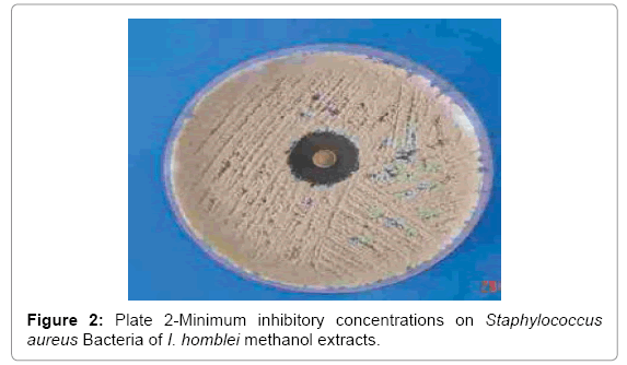
Figure 2: Plate 2-Minimum inhibitory concentrations on Staphylococcus aureus Bacteria of I. homblei methanol extracts.
The methanolic extract of the root showed MICs of 250 μg/ml against both M. gypseum and T. mentagrophytes and the MFCs were 1000 μg/m l against both as presented in Tab le 2. The stem extracts had an MIC of 500 μg/ml against both dermatophyte and MFCs of 2000 μg/ml against M. gypseum and 4000 μg/ml on T. mentagrophytes. The methanolic extract of the leaf exhibited the highest inhibition properties w ith MICs of 125 μg/ml against M. gypseum and 250 μg/m l against T. mentagrophyte The MFCs of 500 and 1000 μg/ml against M. gypseum and T. mentagrophytes were observed.
The phytochemical screening results of the sequential extracts (Table 2) indicated high presence of alkaloids (+++) in both the ethyl acetate and methanol fractions of the root and stem bark extracts of I. homblei.
All parts of I .homblei exhibited presence of flavonoids with stem and leaf having high (+++) concentration while the root had moderate (++) and these results are presented in Table 3. The tannins were moderate (++) and reducing compounds and polyoses high (+++) in all the three parts. The leaf displayed high (+++) amounts of coumarins while stem.
On the basis of the result of the current investigation, it is possible to point out that the broad antibacterial activity of I. homblei methanol extracts towards bacterial strains used may be ascribed apparently to the presence of tannins which were present in all the parts of the plant tested. This is because tannins have been associated with antibacterial activities before (Seigler, 1998). Saponins which were detected in the stem and root have been reported by Seigler (1998; Hassan et al., 2010; Maatalah et al., 2012) as having generalized antibiotic properties and therefore possible to have contributed to the activities [8,9]. The antimicrobial activities of flavonoids, also present in all parts of the plant tested, are well documented (Quiroga et al., 2004; Makkar et al., 2009; João-Henrique et al., 2014) and therefore a possibility of having contributed synergistically or additively to the antibacterial activities exhibited [10,11]. The study showed that extracts of all parts possess antibacterial activity and more importantly, against MDRSA that showed MICs of between 1000 and 2000 μg/m l.
Drug resistance, currently, is a real problem in treatment of infectious diseases, and therefore, there is need to carry out more tests of the extracts of this plant against MDRSA (Arias, and Murray, 2009; Thomas et al., 2012)) [12]. Among the bacteria tested, B. subtilis was the most sensitive bacterium to I. homblei extracts. The M BC for B. subtilis was 1000 and 2000 μg/ml of the methanolic leaf and root extracts, respectively. All the other bacteria had MBC of 8000 μg/ml. The big difference between the MIC and the MBC (most ratios of MBC/MIC > 4) indicates that I. homblei is possibly bacteriostatic. There was no significant difference between the antibacterial activity of the root and stem, root and leaf and stem and leaf (P < 0.05) against Gram-positive bacteria. There was also no significant difference between the root and leaf in their activity against Gram-negative bacteria (p < 0.05). In general there was no significant difference in the antibacterial activity of all the plant parts against Gram-positive and Gram-negative bacteria (P < 0.05).
The antifungal activity of I. homblei was high as indicated by the MICs of 250 and 500 μg/ml against the tested fungi. These MICs are high when compared with the MICs of the Std antifungal drugs but considering that these are crude extracts, if the compounds responsible for the activity are identified and isolated probably the activities might be increased. The activity of I. homblei methanol extract against filamentous fungi could be ascribed to the presence of flavonoids in all parts of the plant. Flavonoids have been associated with antifungal activity before (Quiroga et al., 2004; Urmila et al., 2011) [10,13]. The current upsurge of fungal infections due to HIV/AIDS scourge, there is need to focus on plants that have activity against these microorganisms so as to supplement the conventional drugs currently in use. This is with a view to contributing towards the possible discovery of new biologically active compounds with decreased or no toxicity and side effects present in the current antifungal drugs. There were no significant differences between the activity of the root and stem (p < 0.05), root and leaf (p < 0.05) and stem and leaf (p > 0.0 the average zone of inhibition was calculated for the 3 replicates. A clearing zone of 9 mm for Gram-positive and gram-negative bacteria and 10 mm for fungi or greater was used as the criterion for designating significant antibacterial and antifungal activity (Faizi et al., 2003) [14].The in vitro MIC results were classified as per Pessini et al. (2003)[15]. The extracts that displayed MIC lower than 100 μg/ml, the antimicrobial activity was considered very high; from 100-500 μg/m l, high, 500-1000 μg/m l, moderate; 1000-4000 μg/ml, low and anything above this, the extracts were considered inactive for both bacteria and fungi [16-20].
Nine out of twelve traditional medicinal plants were found to have antimicrobial activity. There are antimicrobial agents in plants. Medicinal plants I. homblei has activity against MDRSA. that is within the ranges of conventional antibiotic and this justifies the fact that the plant contain classes of metabolites like alkaloids, saponins, flavonoids and tannins that possess wide range of antimicrobial activities ranging from antibacterial to antifungal. The plant has a wide range of antibacterial and antifungal activities that cause medical conditions in human and therefore justify their usage as medicine by the local population.
Studies should be extended to other related species that showed antimicrobial activity. Conservation efforts should be put in place and directed at plants that scarce I .homblei which are also most sought after and are facing an imminent decimation. Sustainable harvesting of medicinal plants that have showed activity should be encouraged through environmental edu cation. There is need for the in vivo studies on this plant to determine if the active constituents will display the same activity in vivo. Testing for toxicity, side effects, serum-attainable levels, pharmacokinetic properties and diffusion in different body sites are necessary initially in small Laboratory animals. The determination of adequate dosage for proper administration is necessary. It is essential to check for the presence of immune boosting or antiviral properties of the plants, now that showed it reasonable antimicrobial activity. Kenya should put structures in place for sustainable marketing of plants that possess antimicrobial activities. The results obtained from the plant studied should be considered for further studies aimed at isolating and identifying the single active principle(s) that could be used for the basis of drug discovery and development. There should also be a toxicity evaluation to justify its continued use as traditional medicine. Evaluation of possible synergism or additive antimicrobial activity among these extracts should be explored. The fact that there was strong activity against MDRSA , the extracts, should make this plant a candidate for further studies aimed at getting effective, safe and cheap drugs against multi drug resistant pathogens. There should be a collaborative work among scientists and after all the evaluations finds it safe, the species could be commercially produced.