Journal of Ergonomics
Open Access
ISSN: 2165-7556
ISSN: 2165-7556
Research Article - (2016) Volume 6, Issue 2
The efficiency priority working environments without considering the physical property of workers make the workload of workers increased and make the efficiency of workers decreased unintentionally under an aging society with a declining birth-rate. Therefore, it is necessary for plant managers to design the safe and efficient working environment considering the physical characteristic. This study suggested the method to simulate various motion patterns in the lifting operation with the parameters obtained from the actual lifting operations. This study also evaluated the muscle force of each muscle during simulated motions using the musculo-skeletal model considering the role of antagonistic muscles and biarticular muscle. Therefore, this study evaluated the quality of various lifting operation patterns from the muscle force point of view. As a result, this study succeeded to simulate various motions considering the role of antagonistic muscles and biarticular muscle related to the physical characteristic. The results of this study will make it possible to allocate workers optimally and result in a productivity improvement of industry.
Keywords: Movement simulation; Antagonistic muscles; Biarticular muscles; Lifting operation; Musculo-skeletal model
The efficiency priority working environments without considering the physical property of workers makes the workload of workers increased and makes the efficiency of workers decreased unintentionally under an aging society with a declining birth-rate. Therefore, it is necessary for plant managers to design the safe and efficient working environment considering the physical characteristic. Previously, computer human models that duplicate the properties and the functions of human have been developed. These existing computer human models are used for analysis of motions or movement simulation. Unfortunately, they are not enough to realize the muscle function considering human properties [1] although some existing computer human model can evaluate muscle activities [2]. Crowninshield et al. [2] outlined an optimization method to estimate muscle forces. However, the optimization method unfortunately usually does not consider the functions of antagonistic muscles and biarticular muscles. Therefore, the some estimated muscle forces are zero by optimized although these muscle forces are not actually zero. Human has the unique coordinate system of muscles consisted of antagonistic muscles and biarticular muscles [3-6]. Antagonistic muscles are the muscles that act in opposition to the prime movers or restriction of a rotational motion about joint. Biarticular muscles are the muscles that work simultaneously on two joints. Using the computer human models considering these human properties is effective for product design, movement simulation, rehabilitation and sports to evaluate muscle forces.
Iwata et al. [7] analysed the lifting operation from the aspect of dynamics including motion pattern, trajectories of center of gravity and angle variations of joints. They also estimate net forces and net moments during lifting operation. However, the method to optimize motion patterns considering the role of muscles has not been suggested. This study suggests the method to simulate various motion patterns in lifting operation with the parameters obtained from the actual lifting operations. This study also evaluates the muscle force of each muscle during simulated motions using the musculo-skeletal model considering the role of antagonistic muscles and biarticular muscle. This study evaluates the simulated various motion patterns in lifting operation by estimating the muscle force of each muscle of upper limb and lower limb.
The purpose of this study is to propose the method to evaluate the quality of various lifting operation patterns simulated with movement simulation based on the muscle force of each muscle. Then this study makes it possible to propose the motion patterns or the working conditions suitable for the physical properties of workers. Therefore, this study will make it possible to allocate workers optimally and result in a productivity improvement of industry.
Human segment model
This study assumed lifting operations to be 2-dimensional motion. This study used a human rigid segment model that consists of 8 rigid body segments (1-Foot, 2-Leg, 3-Thigh, 4-Torso, 5-Upper arm, 6- Forearm, 7-Hand, 8-Head) as shown in Figure 1, where the link length of segment i is represented by li , the contact point between segment i and i -1 is represented by Oi and the angle between segment i and horizontal direction is represented by θi . This study did not split the torso in the upper trunk and the lower trunk because this study evaluates the muscles on the upper limb and the lower limb.

Figure 1: 2-dimensional human segment model.
Movement simulation
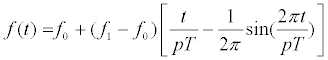
The variations of the angles of the joints during the lifting operations were suggested to be approximated by the function described as follow [8].
 (1)
(1)
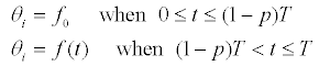
Then, T is the duration of the angle variation, f0 is the angle at t = 0 [s] (initial posture) and f1 is the angle at t = T [s] (final posture). p is the variable that changes the duration of the angle variation. The variable p equals 1 in the standard lifting operation. For example, when the value of the variable p is changed to 1/2, the duration of the angle variation is changed to 1/2 and the velocity of the angle variation is changed to be doubled. When the value of the variable p of all joints is changed to 1/2 in the lifting operation, the lifting operations is conducted at twice the speed. Furthermore, when the value of the variable p of the angle related to lower body,θ 1, θ 2, θ 3 is changed to 1/2 and that of upper body, θ 4, θ 5 θ 6 θ 7θ 8 is still 1, the motion pattern that human gets off his hip before lifting up the object can be simulated. Thus, the function of the angle variation can change the relationship among the joints and simulate various motion patterns of lifting operations.
This study simulated various motion patterns of the lifting operations with the motion of the human body separated to the upper and lower bodies.
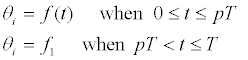
The angles of the upper body are related toθ 4, θ 5 θ 6 θ 7θ 8 . The angles of the lower body are related to θ 1, θ 2, θ 3 The motion pattern that human gets off his hip (moves lower body) before lifting up the object (moving upper body) is assumed to be pattern A and the motion pattern that human gets off his hip (moves lower body) after lifting up the object (moving upper body) is assumed to be pattern B. Then, the pattern A can be simulated by changing the variable of each p joint, which is assumed to be  , with the function (1) and determining the angle variation of each joint described as follows.
On the angles of the upper body, θ 4, θ 5 θ 6 θ 7θ 8 :
 (2)
(2)
On the angles of the lower body, θ 1, θ 2, θ 3 :
 (3)
(3)
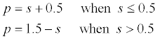
On the other hand, the pattern B can be simulated by determining the angle variation on the upper body,θ 4, θ 5 θ 6 θ 7θ 8 with the formula (3)and that on the lower body, θ 1, θ 2, θ 3 with the formula (2). As the value of the variable is changed to approaching 1/2, the motion pattern is approaching to the pattern A or the pattern B. As the value of the variable is p changed to approaching 1, the motion pattern is approaching to the standard motion pattern. Then, the control variable is defined to be ( ).
).
The variable p is defined as follows to simulate the pattern A as the control variable s is changed to approaching 0 and the pattern B as the control variable s is changed to approaching 1.
 (4)
(4)
When the control variable s is 0.5, the standard motion pattern is simulated. This study regards the movement simulation method as effective because the various motion patterns of the lifting operations can be simply simulated with one control variable s .
Estimate muscle forces with musculo-skeletal model
This study evaluates various lifting operation patterns simulated by the method described in section “Movement Stimulation” with the muscle forces in upper limb and lower limb. As there are many muscles in the upper and lower limbs, it is difficult to model all the contributory muscles. Therefore, this study used a musculo-skeletal model that includes 6 representative muscles of the upper limb and 6 representative muscles of the lower limb in sagittal plane as shown in Figure 2. The following muscles are included: 1-deltoid anterior (Da), 2-deltoid posterior (Dp), 3-brachialis (Br), 4-lateral head of triceps brachii (Tla), 5-long head of biceps (Blo) and 6-long head of triceps brachii (Tlo) at the upper limb and i-gluteus maximus (GMAX), ii-iliopsoas (IL), iii-biceps femoris and short head (BFSH), iv-vastus lateralis (VAS), v-semimembranosus (SM), vi-rectus femoris (RF) at the lower limb.

Figure 2: Muscle arrangements at the human upper and lower extremities (Upper limb: 1-deltoid anterior (Da), 2-deltoid posterior (Dp), 3-brachialis (Br), 4-lateral head of triceps brachii (Tla), 5-long head of biceps (Blo), 6-long head of triceps brachii (Tlo) , Lower limb: i-gluteus maximus (GMAX), ii-iliopsoas (IL), iii-biceps femoris and short head (BFSH), iv-vastus lateralis (VAS), vsemimembranosus (SM), vi-rectus femoris (RF).
This study estimates muscle forces during lifting operation with the musculo-skeletal model considering the roles of antagonistic muscles and biarticular muscles suggested by Oshima et al. [9]. The model suggested by Oshima et al. [9] including three pairs of the antagonistic muscles in the upper limb and lower limb is shown in Figure 3A and Figure 3B. Oshima et al. [9,10] also defined that these muscles act on the distal extremity and the maximum force of each muscle acts on the distal extremity as F’mf1, F ’me1,F ’mf2, F ’me2, F ’ mf3, F’me3 as shown in Figure 3A and Figure 3B. Then, the maximum output force distribution on the distal extremity is geometrically a hexagon from maximum force of each muscle. They also verified the maximum output force distribution on the distal extremity is a hexagon in the experiment. They also investigated the vector of the output force on the distal extremity iss related to the muscles activation pattern as shown in Figure 3C [9,10]. For example, when the vector of the output force is direction a as maximum in Figure 3A, the muscle forces are defined as 100% of maximum muscle force for muscles f1, e2 and e3 and 0% of maximum muscle force for muscles e1, f2 and f3. Therefore, the distribution of each muscle force is determined by the vector of the output force on the distal extremity and the muscle activation pattern as shown in Figure 3C.

Figure 3: Musculoskeletal model considering the role of antagonistic muscles and biarticular muscles [9] (A = Upper limb: f1-deltoid anterior (Da), e1-deltoid posterior (Dp), f2-brachialis (Br), e2-lateral head of triceps brachii (Tla), f3-long head of biceps (Blo), e3-long head of triceps brachii (Tlo) , B = Lower limb: lf1- gluteus maximus (GMAX), le1-iliopsoas (IL), lf2-biceps femoris and short head (BFSH), le2-vastus lateralis (VAS), lf3- semimembranosus (SM), le3-rectus femoris (RF), C = Muscle activation level related to the output).
The vector of the output force on the distal extremity is necessary to be calculated to estimate the distribution of each muscle using the musculo-skeletal model described as above. The vector of output force on the distal extremity can be calculated from the net moments at joints. Figure 4A shows the relationship between the output force on the distal extremity and the net moments of the shoulder joint and the elbow joint. The output force of x axis is represented by f x, that of y axis is represented by f y, the angle of the shoulder and the elbow joints are represented by θ shouler and θ elbow, the link length of the upper arm and the forearm are represented by l upper and l fore and the net moments of the shoulder and elbow joints are represented by M shoulder and M elbow. The relationship between the output force and the net moments is represented as follows. The vector of the output force can be calculated by solving the simultaneous formulas.

Figure 4: Relationship between output force on the distal extremity and net moment (A = Upper limb, B = Lower limb).
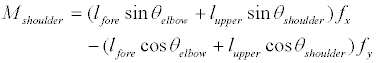 (5)
(5)
 (6)
(6)
The distribution of each muscle force can be estimated with the obtained output force on the distal extremity (f x, f y) applied to the model in Figure 3. Then the muscle forces can be estimated with the scale of the vector of the output force on the distal extremity.
Figure 4B similarity shows the relationship between the output force on the distal extremity and the net moments of the hip joint and the knee joint. The output force of x axis is represented by f x, that of y axis is represented by f y, the angle of the ankle and the knee joints are represented byθ ankle andθ knee, the link length of the thigh and the leg are represented by l thigh and l leg and the net moments of the hip and elbow joints are represented by M hip and M knee. The relationship between the output force and the net moments is represented as follows. The muscle forces can be estimated from the output force.
 (7)
(7)
 (8)
(8)
Identification of parameter for simulation of lifting operation
It is necessary for simulation of lifting operation described in section “Movement Stimulation” to identify the parameters, T (duration of the angle variation), f0(angle at t = 0 [s] (initial posture)) and f1 (angle at t = T [s] (final posture)). This study determined these parameters from the following experiment.
One participant (height: 172.0 cm, mass: 66.0 kg), after informed consent, participated in this study. Participant conducted lifting operation recorded at 125 frames per second using the CCD camera (SONY Corp. XC-009). The conditions of the lifting operations were that the height of lifting was 1.0 [m] and the mass of the object was 10 [kg] (The shape of the object was cuboid with handgrips.). The positional data of the joints obtained from the captured images were smoothed using a Butterworth filter (cut-off frequency 6 Hz) [11,12]. Table 1 shows the parameters T , f 0 and f1 determined from the recorded motion.
| Segment | T (sec) | f0 (degree) | f1 (degree) |
| ①Foot (θ1) | 1.93 | 180 | 180 |
| ②Leg (θ2) | 53.5 | 82.9 | |
| ③Thigh (θ3) | 175 | 103.4 | |
| ④Body (θ4) | 31.5 | 77.1 | |
| ⑤Upper arm (θ5) | -103 | -62.8 | |
| ⑥Forearm (θ6) | -36 | 18.1 | |
| ⑦Hand (θ7) | -39.4 | -7.7 | |
| ⑧Head (θ8) | 56.8 | 81.2 |
Table 1: Parameters for movement simulation determined from experiment (T: Duration of the angle variation, : f0angle of initial posture, f1: angle of final posture).
Identification of parameter for estimation of muscle forces
It is necessary for estimation of muscle forces described in section Estimate muscle forces with musculo-skeletal model to identify the maximum muscle force of each muscle. The maximum muscle force of each muscle can be obtained from the maximum output force distribution on the distal extremity. The participant needs to output all directions with the maximum forces to measure the maximum output force distribution. However, the results of these trials are susceptible to error due to the muscle fatigue. Then, this study used the method of measuring the maximum output force distribution suggested by Oshima et al. [13]. The method can describe the maximum output force distribution in the shape of hexagon geometrically with the measured forces of only four directions. The aluminium frame with a handgrip on a three component dynamometer (KYOWA Corp. LSMB- SAI) shown in Figure 5A was used to measure the maximum output forces of the upper limb. The aluminium frame with the jig fixing a foot on a three component dynamometer (KISTLER Corp. 9257B) shown in Figure 5B was used to measure the maximum output forces of the lower limb. The posture of the upper and lower limbs of participant can be adjusted with the three component dynamometers and seat changed. Tables 2A and 2B shows the maximum muscle force of each muscle determined from the experiment.

Figure 5: Measuring equipment of the output force distribution on the distal extremity (A = upper limb, B = lower limb).
Simulation of lifting operation and evaluation of muscle forces
Various motion patterns in lifting operation were simulated with the method described in section “Movement simulation” and the parameters determined in section “Identification of parameter for simulation of lifting operation”. The evaluation of various motion patterns was conducted. This study simulated 5 motion patterns with the control variable s , which determines the motion pattern, changed to 0, 0.25, 0.5, 0.75 and 1. The motion pictures of each simulated motion are shown in Figure 6. This study assumed that the object always worked on the vertical direction because the acceleration of the horizontal direction of the object was small enough to be ignored. Furthermore, this study assumed all weight of the object was applied just after lifting up although the object still attached on the ground. Therefore, the differences between the estimated muscle forces and the actual ones were occurred at that moment. However, these differences are considered small enough to be ignored to evaluate the lifting operation. The muscle force of each muscle was estimated on each simulated motion with the method described in section “Estimate muscle forces with musculo-skeletal model”.

Figure 6: Motion patterns of lifting operation simulated with the control variable (s = 0, 0.25, 0.5, 0.75, 1).
The net moment of each joint were calculated on each simulated motion with the human rigid segment model and Newton’s equation. Then, the weight of each segment is calculated from the mass of the participant with the distribution measured by Ae et al. [14]. The condition of the lifting operations was that the mass of the object was 10 kg during lifting operation. The results of the net moments of each simulated motion are shown in Figure 7. The net moments of the knee joint (flexion as a positive direction), the hip joint (extension as a positive direction), the shoulder joint (flexion as a positive direction) and the elbow joint (flexion as a positive direction) that have a great effect on the lifting operations are shown in Figure 7. The results of the normalized muscle forces of each simulated motion of the upper and lower limbs estimated by the method described in section “Estimate muscle forces with musculo-skeletal model” are shown in Figure 8.

Figure 7: Net moments of knee, hip, shoulder and elbow joints.


Figure 8: Estimated %MVC of each muscle (A = Upper limb, B = Lower limb).
The results of the muscle forces of the upper limb showed that muscles f1 (Da), f2 (Br) and f3 (Blo) were activated greatly. The results of the muscle forces of the lower limb showed that muscles lf1 (GMAX), lf2 (BFSH) and lf3 (SM) were activated greatly.
The muscle forces of muscle f2 (Br), which works for the flexion of the elbow joint, were not greatly different among the simulated motions. The muscle forces of muscle f1 (Da), which works for the flexion of the shoulder joint, and muscle f3 (Blo), which works for the flexion of the shoulder and elbow joints simultaneously, were larger as the control variable s was larger, where the motion pattern was changed to the motion that human lifts up the object forward. The reason of this is considered that the horizontal length between the shoulder joint and the object was longer as human lifts the object forward. The reason is also considered that the external forces on the distal extremity derived from the object were larger with the acceleration of the object larger as human gets off his hip faster with the control variable s larger such as 0.75 and 1.
The muscle forces of muscle le2 (VAS), which works for the extension of the knee joint, were small on each simulated motion. The results that muscle le3 (RF), which is biarticular muscle and works for the extension of the knee joint, was not activated showed that only muscle le2 (VAS) were used for the extension of the knee joint on each simulated motion. Although muscle le2 (VAS) was not activated greatly, muscle le2 (VAS) is considered to contribute to the extension of the knee joint mainly because the maximum muscle force of muscle le2 (VAS) was large (939.2 [N] from experiment in Table 2). However muscle le2 (VAS) is considered not to have a great effect on the evaluation of various lifting operations because the muscle forces of muscle le2 (VAS) were not greatly different among the simulated motions.
| A.) Muscle | Maximum Forces [N] |
|---|---|
| f1 (deltoid anterior (Da)) | 154.8 |
| e1 (deltoid posterior (Dp)) | 118.8 |
| f2 (brachialis (Br)) | 290.8 |
| e2 (lateral head of triceps brachii (Tla)) | 169.4 |
| f3 (long head of biceps (Blo)) | 94.7 |
| e3 (long head of triceps brachii (Tlo)) | 94.7 |
| B.) Muscle | Maximum Forces [N] |
| lf1 (gluteus maximus (GMAX)) | 185.4 |
| le1 (iliopsoas (IL)) | 255.1 |
| lf2 (biceps femoris and short head (BFSH)) | 171.8 |
| le2 (vastus lateralis (VAS)) | 939.2 |
| lf3 (semimembranosus (SM)) | 65.1 |
| le3 (rectus femoris (RF)) | 144.6 |
Table 2: Maximum force of each muscle (A = upper limb, B = lower limb).
The muscle forces of muscle lf1 (GMAX), which works for the extension of the hip, and muscle lf3 (SM), which is biarticular muscle and works for the extension of the hip, were larger as the control variable s was smaller, where the motion patterns were changed to the motion that human gets off his hip forward. Especially, muscles lf1 (GMAX) and lf3 (SM) were activated greatly on the motions with the control variable s = 0 and 0.25. The results of this showed that the motion that human gets off his hip forward was a back-straining work. The back-straining has been clarified to cause disorders on back such as a hernia of intervertebral disk and a spondylolysis in the lifting operations. Thus, muscles lf1 (GMAX) and lf3 (SM) are very important to evaluate various lifting operations.
The averages of normalized muscle force of muscles f1 (Da) and f3 (Blo) on the upper limb and muscles lf1 (GMAX) and lf3 (SM) on the lower limb, which have great effects during the lifting operations, are shown in Figure 9 in order to discuss the relationship among the muscles on various simulated lifting operations. The results showed that the muscles on the upper limb were more activated, but the muscles on the lower limb were less activated as the motion patterns were changed to the motion that human lifts up the object forward (s = 1) from the motion that human gets off his hip forward (s = 0). Thus, the strain of the upper limb was more, but that of the lower limb was less on the motion that human lifts up the object forward. However, the results, that muscles lf1 (GMAX) and lf3 (SM) were activated over 80% of maximum muscle force during motion that human gets off his hip forward (s = 0), are considered that the motion that human lifts up the object forward (s = 1) is better to reduce the possibility of working injuries during the lifting operations.

Figure 9: Average of %MVC of each muscle on the various lifting operations.
Furthermore, the proposed method can figure out if the participant can operate under the given conditions beforehand because the proposed method can estimate the muscle force of each muscle in the operation. For example, when the maximum muscle force of each muscle is temporarily decreased to 60% of the default maximum muscle force, the estimated muscle force at the peak of muscle f1 (Da) is over 100% of maximum muscle force under the same condition (s = 1) as shown in Figure 10. Therefore, it can be figured out that the participant cannot conduct the operation under the same condition. Then, it can be proposed that the weight of the object is reduced or other participant whose maximum muscle force is larger is switched to conduct the operation under the same condition.

Figure 10: Estimated %MVC of muscle f1 (Da) compared between the default maximum muscle force and the maximum muscle force decreased to 60% of the default in the lifting operation of s = 1 (In the maximum muscle force decreased to 60% of the default, estimated muscle force at the peak is over 100% of maximum muscle force).
Thus, the proposed method to simulate various motions in computer simulation and evaluate the motions considering the role of muscles will make it possible to allocate workers optimally and result in a productivity improvement of industry.
This study suggested a way to simulate various motion patterns in lifting operation with the parameters obtained from the actual lifting operation. This study also evaluated the muscle force of each muscle during simulated motions. The results of this study succeeded the creation of various motions considering the role of antagonistic muscles and biarticular muscle that is the physical characteristic.
The novelty of this study is to propose the method to evaluate the quality of various lifting operation patterns simulated with movement simulation based on the muscle force of each muscle. The results of this study will make it possible to propose the motion pattern or the working condition suitable for the physical properties of workers. Therefore, the proposed method can figure out if workers can operate under the given conditions beforehand because the proposed method can estimate the muscle force of each muscle in the operation. This study will continue to evaluate not only the lifting operation but also other motions. Furthermore, this study will continue to estimate not only muscle forces but also muscle fatigue. Then this study will make it possible to allocate workers optimally and result in a productivity improvement of industry.