Journal of Medical Diagnostic Methods
Open Access
ISSN: 2168-9784
ISSN: 2168-9784
Research Article - (2015) Volume 4, Issue 4
Background: Matrix-assisted laser desorption ionization-time-of-flight mass spectrometry (MALDI-TOF) provides fast and accurate identification of microorganisms. Several reports have speculated about its use in epidemiological typing. However, there is no systematic comparison with standard typing methods, such as pulsed-field gel electrophoresis (PFGE). This study evaluates the potential use of MALDI-TOF for the analysis of suspected outbreaks of vancomycin-resistant enterococci (VRE), compared to PFGE.
Methods: During a suspected outbreak of VRE, including five patients, on an intensive care unit, all isolates were analyzed by PFGE as well as by MALDI-TOF. A series of 24 spectra for each extracted isolate were created. Obtained spectra were smoothed, baseline corrected and calibrated. Subsequently, main spectra (MSP) were created using a Microflex mass spectrometer (Bruker-Daltonik) and the MALDI Biotyper 3.0. Mass peaks were compared manually; a dendrogram was automatically generated. All isolates were tested in triplicate to assess measurement fluctuations.
Results: Analysis of the MSP mass peaks and the dendrogram could clearly identify three different strains which were in agreement with the PFGE. Two clusters, reflecting four isolates and two occurred transmissions, were clearly identified. Measurement fluctuations did not affect the strain typing.
Conclusions: MALDI-TOF provided results concordant to PFGE and that in a fraction of time and costs. However, Bruker's algorithm for automated dendrogram creation is partly consisting of identification log scores making its resolution too low for exact typing analysis. Hence, fully automated analysis is not possible. However, the obtained results show some potential for the use of MALDI-TOF as an epidemiological tool for future outbreak management.
Keywords: MALDI-TOF MS; Strain typing; Outbreak analysis; VRE; Epidemiology
Numerous studies have shown that matrix-assisted laser desorption ionization time-of-flight mass spectrometry (MALDI-TOF MS) provides fast and accurate identification of microorganisms [1-3]. It has been shown to identify a broad spectrum of organisms, including aerobic Gram-positive [4-7] and Gram-negative bacteria [8-11], mycobacteria [12], anaerobic bacteria [7], and yeasts [13]. Compared to conventional biochemical methods, this technique is rapid and reproducible with minimal consumable costs [14,15]. Supposedly, because MALDI-TOF MS detects a broad spectrum of proteins, the discrimination between closely related species and the classification of organisms at the subspecies level appears to be feasible [16]. Recently published studies provided promising results, supporting the thesis that MALDI-TOF MS is an important technology for the subtyping of clinical isolates [17-20]. Though those works have speculated about its usefulness in taxonomy and epidemiological typing, no comparison with genomic typing methods such as SmaI macrorestriction analysis in pulsed-field gel electrophoresis (PFGE) have been performed regularly. Indeed, hierarchical typing of highly recombinogenic species like enterococci can be difficult [21]. Thus, similar typing methods have different discriminatory power in different species and new methods such as MALDI-TOF need to be evaluated per species.
This study evaluates the usefulness of MALDI-TOF as a novel tool for the epidemiological analysis of suspected outbreaks of vancomycinresistant enterococci (VRE) and compares it to PFGE.
VRE are prevalent worldwide and resistance rates are increasing [22,23]. In central Europe, especially in south-west of Germany, increasing rates of VRE have been previously found [22-25]. This study was conducted at the Heidelberg University Hospital, which is a one of Europe’s largest hospitals with 2,200 beds, ~93,000 in-patients and ~220,000 out-patients per year.
A recent clustering of VRE (Enterococcus faecium , Van A type) on an intensive care was studied in detail; all strains were used for MALDI-TOF MS and PFGE analysis. The aim of this study was to assess the usefulness and accuracy of MALDI-TOF MS for epidemiological typing and real-time management of suspected VRE outbreaks in the clinical setting.
Sample acquisition
Clinical isolates were recovered from rectal or wound swabs and in one case from a urine sample. Swabs (Eurotubo®, Amies Viscosa, Deltalab) were inoculated on Columbia 5% sheep blood (COS) agar plates (BD, USA) as a growth control and spiked into a Vancomycin (4 mg/l) containing broth culture (Enterococcsel™ broth, BD, USA); then incubated for 48 h under aerobic conditions at 36°C. When VRE broth culture turned black due to indicator change (esculin cleavage), quantitative real-time polymerase chain reaction (qRT-PCR) for detection of Vancomycin-resistance genes vanA and vanB was performed, as described elsewhere [26]. When qRT-PCR was positive for vanA or vanB, subsequent culture affirmation with MALDI-TOF identification, and susceptibility testing with VITEK 2 (BioMérieux) were performed. All tests needed to provide concordant results to confirm VRE positive status. In all cases, no divergent results were obtained.
The urine sample was inoculated automatically on MacConkey, COS and CPS-3 plates (BioMérieux), using the PREVI® isola streaker (BioMérieux). Enterococci were identified by typical morphology on the chromogenic CPS-3 plates, MALDI-TOF identification and subsequently susceptibility testing by VITEK 2 (BioMérieux) was performed. In case of detected vancomycin-resistance, qRT-PCR was performed to confirm van gene presence.
Sample preparation for MALDI-TOF MS, spectrum generation and analysis
Four to five VRE colonies of a fresh overnight culture were used for sample preparation. The VRE isolates were suspended in 300 μl double-distilled water, and subsequently 900 μl ethanol were added. Samples were then thoroughly mixed, centrifuged at 13,000xg for 2 min, supernatants were removed, and the pellets were dried. VRE isolates underwent an ethanol/formic acid extraction procedure, as described elsewhere [3]. After centrifugation at 13,000xg for 2 min, 1 μl of the supernatant containing the bacterial extract was transferred on a MALDI target plate and allowed to dry at room temperature. Subsequently, the sample was overlaid with 1 μl of MALDI matrix (saturated solution of α-cyano-4-hydroxy-cinnamic acid in 50% acetonitrile-2.5% tri-fluoro-acetic acid) and dried again.
Measurements were performed using a Microflex mass spectrometer (Bruker Daltonik, Bremen, Germany) and the MALDI Biotyper 3.0 software. Spectra were recorded in the positive linear mode (laser frequency 20 Hz; ion source 1, voltage at 20 kV, ion source 2, voltage at 18.5 kV; lens voltage 8.5 kV; mass range 2000-20137 Da). A series of 24 spectra for each isolate were created. The Escherichia coli strain DH5α was used as an external control. Obtained spectra were smoothed, baseline corrected and calibrated at the peak of the 50S ribosomal protein L36 of E. faecium at 4427 kDa (UniProt Database). Subsequently main spectra (MSP) were created, containing the average peak mass, average peak intensity, and frequency information. Automated dendrogram creation was based on cross-wise MSP matching, using standard default settings with a critical distance level of 300. Each MSP is compared to all MSPs, at which high matching score values are attributed to similar MSPs. Matching scores are used to calculate normalized distance values; species with distance levels under 500 have been described as reliably classified [27]. Additionally, mass peaks were compared manually using the MALDI Biotyper and Flex Analysis 3.0 software.
All isolates were tested in triplicate from fresh overnight cultures following the above described protocol to analyze the influence of measurement fluctuations on mass peaks creation for the assessment of the methods robustness in practice.
Pulsed-field gel electrophoresis
PFGE was performed on all patients and their VRE positive contacts. Contacts were defined as in-patients who shared a room for more than 2 hours with the index patients. Chromosomal DNA was digested with SmaI (Fermentas) and analysed by PFGE in a CHEF-DR II apparatus (BioRad Laboratories). The preparation of genomic DNA of the isolates was performed as previously described [26]. Restriction fragments were separated on 1% Agarose gels. Gels were run at 6 V/cm in 0.5x Tris-Borate-EDTA buffer at 14°C. Optimal separation was achieved by using a 5-35s pulse-time linear gradient for 22 h [28]. Results were interpreted following the Tenover criteria [29].
The VRE strains were isolated from three rectal swabs, one urine sample and one wound and rectal swab. All were Vancomycin-resistant Enterococcus faecium , carrying the vanA gene. Comparison of the mass peaks of the created MSPs (Figure 1) and the created dendrogram (Figure 2) could clearly identify three different strains which were in agreement with that inferred from the PFGE (Figure 3) analysis. C1 was the index case. Two transmissions occurred, according to the Tenover criteria; the first from patient C1 to C2 and the second from patient A1 to A2. Strain B was not related to the other strains. In the dendrogram, strain B is separated from the others but closer related to strain C than A (Figure 2). The PFGE confirms that strain B is not clonally related to A or B. The results were in agreement with the bed occupancy history; patient A1 shared a room with patient A2 and C1 with C2, respectively. The analysis of triplicate measurements of the same strains showed minor variations in the obtained mass spectra (Figure 4). However, significant differences in mass peaks for epidemiological typing were still detectable for all strains and allowed reliable differentiation of the outbreak strains in all three measurements (Figure 5).
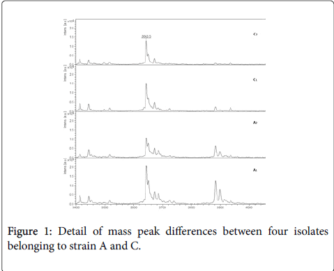
Figure 1: Detail of mass peak differences between four isolates belonging to strain A and C.
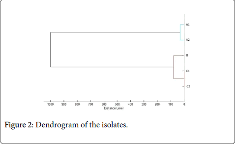
Figure 2: Dendrogram of the isolates.
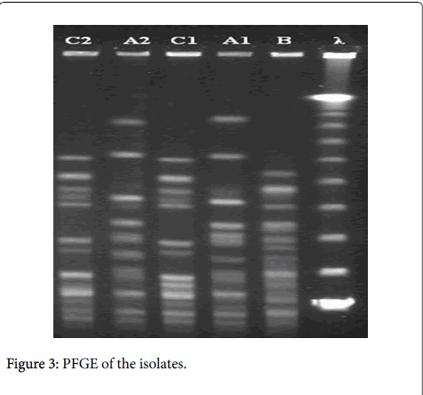
Figure 3: PFGE of the isolates.
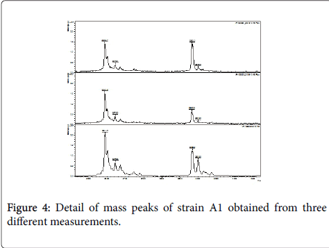
Figure 4: Detail of mass peaks of strain A1 obtained from three different measurements.
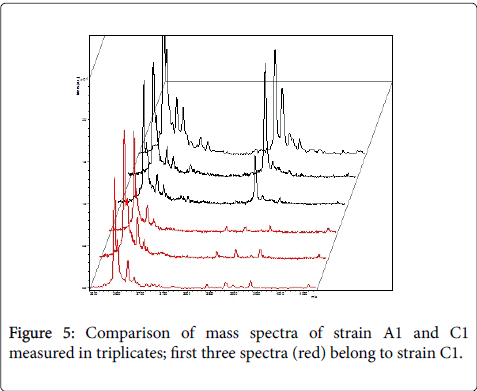
Figure 5: Comparison of mass spectra of strain A1 and C1 measured in triplicates; first three spectra (red) belong to strain C1.
MALDI-TOF MS provided results concordant to PFGE and that in a fraction of time and costs. Repeated measurements did not interfere with the typing of the strains, suggesting the methods robustness. The manual comparison of the mass spectra provided reliable results for the typing of strains, however, is time consuming and prone to human errors when sample size increases. Additionally, fully automated analysis is not possible yet. The dendrogram creation alone, without manual comparison of obtained mass spectra, is prone to mistakes. The algorithm for automated dendrogram creation is based on cross-wise MSP matching, partly including the comparison of identification logscores. Given that the identification log-scores are based on cut-off values (scores ≥1.7 are considered as reliable identification of genus and scores ≥2.0 as reliable identification of species), a difference of 0.1 is hardly relevant between scores of 1.7 to 1.9, but becomes crucial between 1.9 and 2.0. Furthermore, MSP peaks are analyzed on m/z position only and no measure of intensity is taken into account; albeit that intensity measures could negatively affect the robustness of measurement due to wide variations in activity and manner of bacterial metabolism. Though, in the investigated outbreak in this study, the dendrogram was concordant to PFGE, we would not recommend it to be used solely. Lasch and colleagues showed that MALDI TOF can differentiate hospital-associated E. faecium clones (CC17) from other E. faecium strains with 87% accuracy [30]. Thus, typing appears to be possible but discriminatory power is yet insufficient for routine use. The development of an automated analysis program based on a modified and improved biomathematical algorithm to compare mass spectra is needed to warrant a use in routine epidemiological typing. The further improvement of the MALDI-TOF MS technology and the usage of TOF/TOF systems could further improve its resolution and discriminatory power in epidemiological typing. Based on the obtained results of this study, MALDI-TOF MS cannot be neglected as a method for epidemiological typing of VRE strains and usage in real-time management of suspected outbreaks, however, further research is needed to prove its robustness in daily practice.
We thank Konrad Bode for his support during the initial study planning. Partial results were presented at the 3rd German Conference on Infection Control and Microbiology in Essen.