Biochemistry & Pharmacology: Open Access
Open Access
ISSN: 2167-0501
ISSN: 2167-0501
Research Article - (2015) Volume 4, Issue 4
Glutinone (1), coixol (2), friedelin (3), glutinol (4), and betulinic acid (5) were isolated from the plant Scoparia dulcis. Their structures were identified using mass and 1D- and 2D- NMR techniques. All the compounds were tested for their immunomodulatory potential in oxidative burst assay. Compound 1 showed a significant inhibitory effect on the release of reactive oxygen species (ROS) from zymosan activated cells from whole blood (IC50 = 4.3 ± 0.6 μg/mL) as well as from isolated PMNs (IC50 = 5.0 ± 0.3 μg/mL) as compared to standard drug ibuprofen in whole blood (IC50 = 11.2± 1.9 μg/ mL) and in isolated PMNs (IC50 = 2.5± 0.6 μg/mL) shows that it is more active in whole blood as compared with isolated PMNs. Compound 1 when further tested for its effect on pro-inflammatory cytokines TNF-α, IL-1β and on nitric oxide (NO), was found to moderately inhibiting the production of TNF-α (19%) at a concentration 25 μg/mL. On the other hand a weak inhibitory effect of this compound was also observed on the production of IL-1β and NO production, whereas, compounds 2-5 showed no effect (IC50 = >100) on the release of ROS from zymosan activated cells.
<Keywords: Scoparia dulcis; Glutinone; Immunomodulatory; Reactive oxygen species (ROS); Pro-inflammatory cytokines
The genus Scoparia belongs to the family Scrophulariaceae. The genus comprises 32 species of flowering plants, native to Central Asia, and also found in India, Nepal, Pakistan, Taiwan, Brazil, Nigeria, and Nicaragua [1]. S. dulcis has been used in indigenous system of medicine since hundreds of years [2]. Local and tribal people in Nepal, India, and Pakistan have been using S. dulcis in the management of diabetes [3]. Significant anti-hyperglycemic activity of aqueous extract of S. dulcis has been reported earlier [3-5]. The plant has also been used for treatment of stomach problem, hypertension, inflammation, bronchitis, hemorrhoids, hepatosis, and as analgesic and antipyretic. Many phytochemicals have reported from various parts of S. dulcis contributing to the medicinal value of the plant [6]. Anti-inflammatory activity of the 70 % ethanolic extract of S. dulcis on Carrageenan induced paw edema in mice have been reported [7]. The antiinflammatory activity of water and ethanolic extracts of S. dulcis was also reported in in vivo [8].
Immunomodulation with natural and synthetic agents for the treatment of various chronic conditions, such as cancers, atherosclerosis, rheumatoid arthritis, and diabetes and transplantation rejection have the focus of major attention [9-12]. The cells of the immune system communicate in a complex fashion. During an innate immune response first line of defense is mediated by professional phagocytes, including polymorphonuclear neutrophils (PMNs), monocytes, and macrophages. Upon activation, phagocytes migrate to the site of inflammation, and produce various mediators, such as chemokines, cytokines, and ROS to eradicate the infectious agents. Phagocytosis is the elimination of pathogens through a process called oxidative burst. Although normally it has a protective role, the over expression of inflammatory mediators, such as ROS, NO and proinflammatory cytokines due to an in appropriate immune response can cause damage to body tissues, and leads to various chronic illnesses. Inhibitors of ROS, TNF-α, IL-1β, etc. are being used for the treatment of various clinical conditions and compounds having capability to modulate immune response and target the mediators of inflammation with less toxicity have great therapeutic value [11,13].
In the present study, the immunomodulating potential of five compounds, isolated from S. dulcis was investigated. During this study, all isolated compounds were screened for the inhibition of oxidative burst using luminol-induced chemiluminescence techniques. The compound found to have promising effect in this assay, was further studied for their effect on other parameters, including effect on proinflammatory cytokines, and nitric oxide.
General experimental
JASCO DIP-360 digital polarimeter was used for the measurement of optical rotations. The infrared (IR) spectra were recorded on JASCO A-302 infrared spectrophotometer. 1D- and 2D -NMR spectra were recorded on a 500 MHz on Bruker Avance-500 nuclear magnetic resonance spectrometer. The low-resolution EI was performed on Finnigan MAT 311 mass spectrometer. The HREI-MS was recorded on the Finnigan MAT 95 XP mass spectrometer. The column chromatography was performed on silica gel (230-400 mesh, Merck), while TLC was carried out on pre-coated preparative silica gel plates (GF-254, 20 × 20 cm, 0.5 mm thick, Merck).
Plant material
Whole plant material of Scoparia dulcis Linn. was collected from Chitwan district of Nepal in July 2013. The plant was identified by Dr. Rita Chhetry, Research Officer, National Herbarium and Plant Resources, Ministry of Forests and Soil Conservation, Godawari, Nepal. A voucher specimen SD 2812 was deposited to the same department.
Extraction and isolation of compounds
The shade dried whole plant of Scoparia dulcis (1.7 kg) was extracted with 80% methanol water (4.0 L) for three times. The concentrated methanolic extract (72.0 g) after evaporation of solvent was then dissolved in distilled water (1.5 L). The aqueous layer was then subjected to solvent-solvent extraction. First the aqueous layer was extracted with n-hexanes (each 1.5 L volume of aqueous layer three times with 1.5 L of n-hexanes). After evaporation of n-hexanes, 15.0 g of the crude hexane fraction was obtained. The aqueous layer was then extracted with CH2Cl2 and 2.0 g of crude CH2Cl2 fraction was obtained. The aqueous layer was then extracted with ethyl acetate and 10.0 g of crude ethyl acetate fraction was obtained. The dichloromethane (DCM) fraction (2.0 g) was subjected to column chromatography in order of increasing polarity of ethyl acetate in hexanes, which yielded many sub-fractions. Out of them, sub-fraction B (800 mg) obtained by 10% EtOAc/hexanes was further subjected to silica gel column chromatography using 10% EtOAc/hexanes, as an eluting agent which yielded compound 2 (45.0 mg). The hexanes fraction (15.0 g) was eluted in column chromatography using silica gel with n- hexanes and ethyl acetate. In order of increasing polarity of ethyl acetate in n-hexane yielded five sub-fractions. The sub-fraction A (800 mg), obtained by pure hexanes, was further subjected to column chromatography using pure hexanes as eluting agent to obtain the pure compound 1 (40.0 mg). The sub-fraction B (750.0 mg), obtained by 5% EtOAc/hexanes, was again subjected for column chromatography on silica gel using 5% EtOAc/hexanes as eluting agent to obtain compounds 3 (35.0 mg), and 4 (50.0 mg). The sub-fraction E (900.0 mg), obtained from 30% EtOAc/ hexanes, was further subjected to column chromatography using 20% EtOAc/hexanes to obtain compound 5 (50.0 mg).
Determination of ROS by chemiluminescence assay
Oxidative burst studies using chemiluminescence technique was performed as described by [14]. The assay was performed on whole blood from healthy human volunteers and on isolated neutrophils using luminol as a probe, and zymosan as an activator. Briefly 25 μL of whole blood (1:20 dilution in HBSS++) [Sigma, St. Louis, USA] or isolated neutrophils (1x10⁶ cells/mL) were incubated with 25 μL of different concentrations of compounds (1, 10 adnd 100 μg/mL) each in triplicate in white half area 96 well plates [Costar, NY, USA]. The plate was incubated at 37 ºC for 15 min in the thermostat chamber of luminometer [Labsystems, Helsinki, Finland]. 25 μL of (7×10⁻⁵ M) luminol [Research Organics, Cleveland, OH, USA], and 25 μL serum opsonized zymosan (SOZ) (2 mg/mL) [Fluka, Buchs, Switzerland] was then added into the wells Table 1. The plate was then read in luminometer for 50 min, and results were recorded as total integral readings as relative light units (RLU).
| Compound | NO % inhibition | TNF-α % inhibition | IL-1β % inhibition | Oxidative Burst (IC50 µg/mL) | |
|---|---|---|---|---|---|
| Whole blood | PMNs | ||||
| 1 | 13 ± 0.7 | 19 ± 1.0 | 3.3 ± 3.5 | 4.3 ± 0.6 | 5.0 ± 0.3 |
| L-NMMA | 65.5 ± 1.1 | - | - | - | - |
| Ibuprofen | - | - | - | 11.2 ± 1.9 | 2.5 ± 0.6 |
Table 1: Effect of glutinone (1) on nitric oxide (NO), pro-inflammatory cytokines TNF-α and IL-1β, and on luminol enhanced myeloperoxidase dependent oxidative burst,using zymosan activated PMNs and mice peritoneal macrophages.
Nitric oxide (NO) assay
The assay was performed as previously described by [15]. Briefly (1x10⁶ cells/mL) macrophages from J774.2 cell line (European Collection of Cell Cultures, UK) were plated in 24-well tissue culture plates. Cells were activated by adding 30 μg/mL of E. coli lipopolysaccharide (LPS) (DIFCO Laboratories Michigan, USA) and treated with test compound at a concentration of 25μg/mL. The supernatant was collected after 48 hours for analysis. Nitrites (a stable product of nitric oxide) concentration in supernatant was measured using the Griess reagent.
Cytokine assay
Effect of compound on the release of pro-inflammatory cytokines TNF-α and IL-1β was performed by method previously described by [16]. Briefly THP-1 cells (European Collection of Cell Cultures, UK) were differentiated by adding 20 ng/mL of phorbol myristate acetate (PMA), (SERVA, Heidelberg, Germany) for 24 hours at 37ºC in 5% CO2 and then stimulated with 50 ng/mL E. coli Lipopolysacchride B (DIFCO Laboratories, Michigan, USA). 25 μg/mL of test compound was then added for four hours, and plate was incubated at 37ºC in 5% CO2. Cytokines quantification in supernatants was performed using the human TNF-α and IL- 1β Duo Set (R and D Systems, Minneapolis, USA), according to manufacturer’s instructions.
Toxicity assay
Cytotoxicity activity of compound 1 was evaluated on 3T3 cell line by using the standard MTT (3-[4, 5-dimethylthiazole-2-yl]-2, 5-diphenyl-tetrazolium bromide) colorimetric assay according to Mosmann T [17]. In this assay compound 1 showed no toxic effect (IC50 = 200 μM).
Compound 1 was isolated from hexane fraction of methanloic extract of S. dulcis Linn. The EI-MS showed a [M+.] at m/z 424 and a base peak at m/z 274. The molecular formula C30H40O, was deduced from the HREI-MS which showed a [M+.] at m/z 424.3721 (calcd for C30H40O = 424.3705) and the 13C-NMR spectra (BB and DEPT). The IR spectrum indicated the presence of carbonyl (1706 cm-1), and olefinic groups (1631 cm-1).
The 1H-NMR spectrum of 1 displayed singlets for protons of eight methyl groups at δ 0.79 (H-25), 0.93 (H-29), 0.96 (H-30), 1.01 (H-26), 1.07 (H-27), 1.14 (H-28), 1.20 (H-23), and 1.22 (H-24). A downfield signal at δ 5.6 br m was assigned to olefinic H-6. Position of double bond between C-5/C-6 was inferred from the base peak at m/z 274 (EIMS) due to the retro Diels -Alder cleavage of the B ring, which was further supported by the HMBC spectrum, in which H-23 and H-24 showed HMBC correlation with carbonyl C-3 (215.6) and olefinic C-5 (142.4). Structure of the compound was further confirmed from 2D-NMR spectra (COSY, HSQC, HMBC, and NOESY). All the spectral data of the compound 1 was unambiguously matched with reported data in literature [18]. Structure of compounds, coixol (2), friedelin (3), glutinol (4), and betulinic acid (5) were identified by comparing their spectral data with reported data in literature [19-22].
Among all tested compounds, compound 1 (glutinone) (IC50 = 4.30 μg ⁄ mL) exerted potent inhibition of oxidative burst from whole blood cells, whereas, remaining compounds showed no effect (IC50 > 100 μg ⁄ mL) (Figure 1). Compound 1 was also found to be a potent inhibitor (IC50 = 5.0 μg/mL) of intracellular reactive oxygen species (ROS), when tested on zymosan activated isolated human PMNs using luminol as probe. Cytotoxicity activity of compound 1 was evaluated on 3T3 cell line by using the standard MTT assay was performed as described in materials and methods. In this assay compound 1 showed no toxic effect (IC50 = 200 μM) (data not shown).
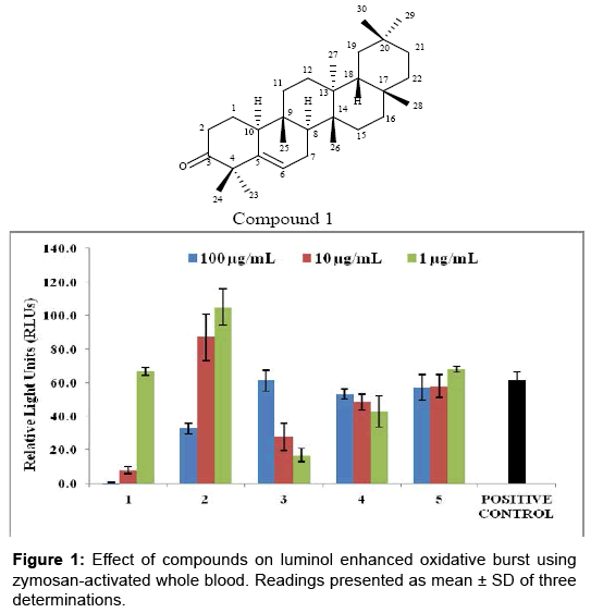
Figure 1: Effect of compounds on luminol enhanced oxidative burst using zymosan-activated whole blood. Readings presented as mean ± SD of three determinations.
Glutinone (1) was further studied for its effect on the production of pro-inflammatory cytokines TNF-α and IL-1β. In case of TNF-α, moderate level of inhibition was observed (19%) at a concentration of 25 μg/mL compared to positive control. 1-100 μg/mL concentration was used out of which, 25 μg/mL was seen effective for partial inhibition TNF-α and IL-1β after a dose dependent treatment of compound. Very weak inhibition was observed when it was tested for IL-1β (3.3%). The standard drug used TNF-α inhibition is pentoxifillin (IC50 = 98.4 μg/mL). Glutinone (1) was also evaluated for its effect on nitric oxide production in cellular assay. Low level of inhibition was observed (13%), when compared to standard L-NMMA (65.5%) at concentration of 25 μg/mL.
Among all tested compounds, 1 was found to be the potent inhibitor of ROS production. It also moderately inhibited TNF-α and showed weak inhibition on IL-1β and NO. The anti-inflammatory effect of compound 1 was reported earlier. Previously it was reported to reduce inflammation in carrageenan induced rat paw oedema [23] and found to inhibit protaglandlin E2 (PGE2) in mouse peritoneal cells and thromboxane B2 (TXB2) in human platelets [24]. Present study describes the potent suppressive effect of this compound on myeloperoxidase dependent intracellular ROS production in human PMNs. Luminol is used as a probe in this assay, as having a low molecular weight it can cross the cell membrane, and hence can detect both intra and extra cellular ROS, produced by the cells [25]. Previously reported anti-inflammatory activities of 1 are in agreement with in current results as a reciprocal relationship between COX and ROS. The mediators derived from ROS and prostanoids from COX, such as PGE2, are well known in promoting inflammation and enhancing pathogenesis of various inflammatory diseases including cardiovascular disorders and hypertension [26]. The In vivo reduction in inflammation in rat paw oedema by this compound [27] might be due to its strong suppressive activity on derivatives of COX [24] and in this study on ROS. Suppression of other inflammatory markers, including pro-inflammatory cytokines TNF-α, IL-1β and on NO also accounts for reduction in inflammation. Furthermore, this compound found to be non-toxic when tested on mouse 3T3 fibroblast cells.
Most inflammatory conditions are associated with oxidative stress and hyper activation of COX mediators. Current studies showed a strong relationship between mediators of both pathways [26]. The compound like glutinone (1) which have ability to block the mediators of both pathways might be of therapeutic value against inflammatory diseases. However, further studies to unrevealed exact underlying molecular mechanism, as well as detailed in vivo studies and clinical trials are needed to evaluate the effects of this compound in reducing inflammation.
Among all five tested natural compounds isolated from S.dulcis, compound 1 (glutinone) exerted potent inhibition of oxidative burst from whole blood cells whereas, remaining compounds showed no effect. Compound 1 showed potent inhibition of intracellular reactive oxygen species (ROS), when tested on zymosan activated isolated human PMNS using luminol as probe. Compound 1 showed moderate level of inhibition on the production of proinflammatory cytokines TNF-α and IL-1β and weak inhibition was observed when it was tested for IL-1β. Glutinone showed low level of inhibition for its effect on nitric oxide production. Current study demonstrated the antiinflammatory potential of gultinone and it may be the lead compound for further drug discovery process.
Mr. Khaga Raj Sharma is thankful to the Nepal Academy of Science and Technology (NAST), Nepal, for providing Ph. D. fellowship and Financial support for the publication by South Asian University.
Note: We would like to add the acknowledgement of South Asian University for the assistance of publication fees