Advanced Techniques in Biology & Medicine
Open Access
ISSN: 2379-1764
ISSN: 2379-1764
Mini Review - (2016) Volume 4, Issue 1
3-mercaptopyruvate sulfurtransferase (MST) transfers sulfur from 3-mercaptopyruvate (3MP) or thiosulfate to a sulfur acceptor. We developed MST-knockout (MST-KO) mice to clarify the physiological role of MST. They showed increased anxiety-like behaviors. In the prefrontal cortex and hippocampus, levels of serotonin (5-hydroxytryptamin, 5-HT) and/or serotonin metabolite, 5-hydroxyindoleacetic acid were higher in MST-KO mice than in wild-type mice and further 5-HT2A receptor mRNA was increased. Experiments using MST-KO mouse revealed that MST produces hydrogen polysulfides from 3MP and sodium sulfide and hydrogen trisulfide was also enzymatically produced.
Keywords: 3-Mercaptopyruvate sulfurtransferase, Knockout mice, Mercaptolactate-cysteine disulfiduria, Serotonin, 5-Hydroxyindoleacetic acid, Serotonin receptor, Anxiety-like behavior
3-mercaptopyruvate sulfurtransferase (MST) is a universal enzyme in prokaryotes and eukaryotes. In 1998, we reported the distribution of MST in rat [1]. MST was expressed in numerous somatic cells and was found to be present in both cytosol and mitochondria [1] (Figure 1a). In the kidney, MST levels were higher in the cortex and proximal convoluted tubules as compared to that in the medulla and distal convoluted tubules, respectively [1] (Figure 1b). In the liver, MST levels were higher in hepatocytes localized around central veins as compared to that in the portal areas [1] (Figure 1c). In the heart muscle, MSTs were found in the myocardial cells around the perinuclear area [1] (Figure 1d). In the lung, MST levels were higher in the bronchiolar epithelial cells, including ciliated and non-ciliated cells, as compared to that in the alveolar cells [1] (Figure 1e). In the brain, MSTs were present in the stellate-shaped glial cells but not in the neural cells [1] (Figure 1f). In the testis, MSTs were detected in the leydig cells [1].
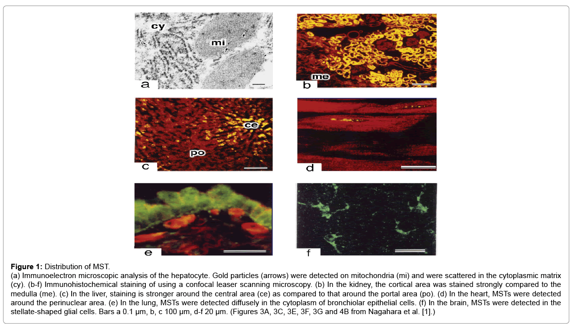
Figure 1: Distribution of MST.
(cy). (b-f) Immunohistochemical staining of using a confocal leaser scanning microscopy. (b) In the kidney, the cortical area was stained strongly compared to the medulla (me). (c) In the liver, staining is stronger around the central area (ce) as compared to that around the portal area (po). (d) In the heart, MSTs were detected around the perinuclear area. (e) In the lung, MSTs were detected diffusely in the cytoplasm of bronchiolar epithelial cells. (f) In the brain, MSTs were detected in the stellate-shaped glial cells. Bars a 0.1 μm, b, c 100 μm, d-f 20 μm. (Figures 3A, 3C, 3E, 3F, 3G and 4B from Nagahara et al. [1].)
MST catalyzes the enzymatic transfer of sulfur atoms from either 3-mercaptopyruvate (3MP) or thiosulfate to a sulfur acceptor. Alternatively, MST transfers sulfur to cyanide or to thiols to produce thiocyanate and hydrogen sulfide (H2S), respectively [2,3]. Various physiological roles of H2S have been reported, including its ability to act as a neurotransmitter. In the hippocampus, H2S increases the activity of N-methyl D-aspartate receptors resulting in long-term potentiation, which suggests that H2S is an endogenous neuromodulator in the brain [4]. In the vascular system, H2S activates potassium channels in the smooth muscles resulting in their relaxation and hyperpolarization of their membrane potential in thoracic aorta, portal vein, and ileum, particularly in association with nitric oxide [5,6]. In astrocytes, H2S activates transient receptor potential (TRP) channels, resulting in calcium influx [7].
A defect of MST causes mercaptolactate-cysteine disulfiduria (MCDU) [8], which in causes mental retardation in severe MCDU patients [9,10]. However, the pathogenesis of MCDU is not known. To investigate the mechanisms underlying this defect, we recently developed MST-knockout (MST-KO) mice [11]. We report the recent study that compare MST-KO mice with wild-type mice and the study that was carried out in wild-type mouse cells and MST-KO mouse cells.
The characteristics of MST-KO mice
MST-KO mice demonstrated increased anxiety-like traits in various behavioral tests [11]. In the open field test, MST-KO mice moved less during the first 9 min as compared to wild-type mice [11] (Figure 2a). In the light/dark transition test, as compared with wild-type mice, MST-KO mice spent longer time in the dark chamber as opposed to the light chamber and demonstrated a longer latency of first transition from the dark chamber to the light chamber [11] (Figures 2b and 2c). In the elevated plus-maze test, MST-KO mice tended to spend less time in the open arms and to move shorter distances than wild-type mice [11] (Figure 2d).
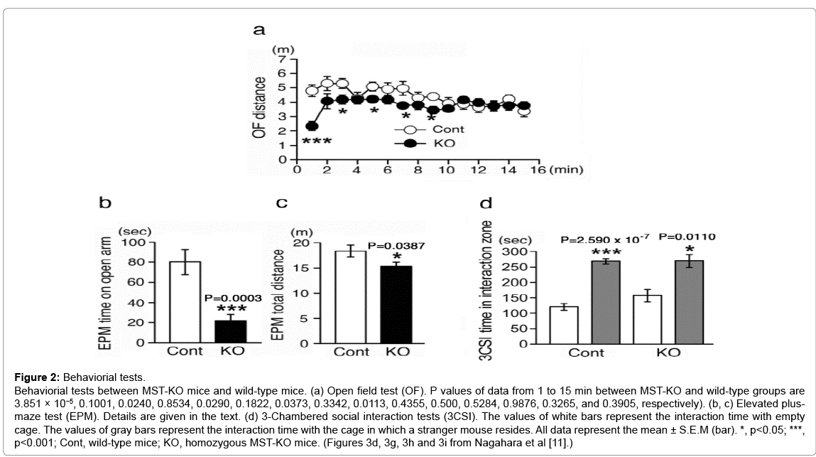
Figure 2: Behaviorial tests.
Behaviorial tests between MST-KO mice and wild-type mice. (a) Open field test (OF). P values of data from 1 to 15 min between MST-KO and wild-type groups are 3.851 × 10⁻⁵, 0.1001, 0.0240, 0.8534, 0.0290, 0.1822, 0.0373, 0.3342, 0.0113, 0.4355, 0.500, 0.5284, 0.9876, 0.3265, and 0.3905, respectively). (b, c) Elevated plusmaze test (EPM). Details are given in the text. (d) 3-Chambered social interaction tests (3CSI). The values of white bars represent the interaction time with empty cage. The values of gray bars represent the interaction time with the cage in which a stranger mouse resides. All data represent the mean ± S.E.M (bar). *, p<0.05; ***, p<0.001; Cont, wild-type mice; KO, homozygous MST-KO mice. (Figures 3d, 3g, 3h and 3i from Nagahara et al [11].)
In the prefrontal cortex, levels of serotonin (5-hydroxytryptamin, 5-HT) and 5-hydroxyindoleacetic acid (5-HIAA, a metabolite of 5-HT) in MST-KO mice were higher than those in wild-type mice [11] (Figure 3a). In the hippocampus, 5-HIAA levels were also increased in MST-KO mice as compared to wild-type mice but the 5-HT levels did not differ [11] (Figure 3b). It is reasonable that expression of 5-HT2A receptor mRNA was increased in MST-KO mice as compared to wild-type mice [12] (Figures 3c and 3d). Because MCDU has been reported to be associated with mental retardation in humans [9,10], MST defects may have a connection with depression, although this has not been proven.
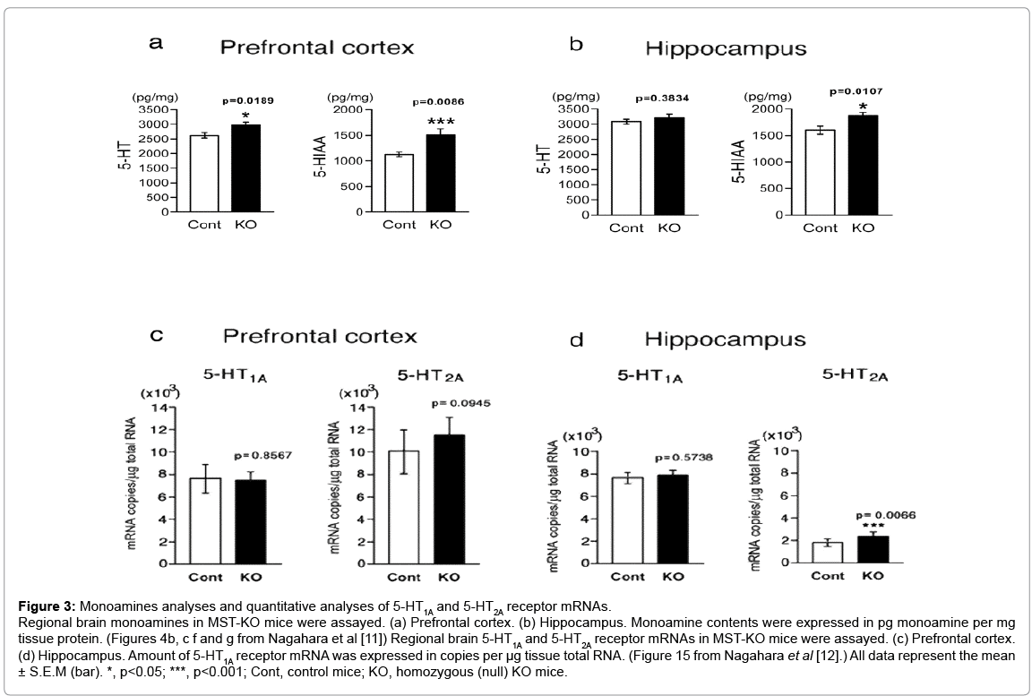
Figure 3: Monoamines analyses and quantitative analyses of 5-HT1A and 5-HT2A receptor mRNAs.
Regional brain monoamines in MST-KO mice were assayed. (a) Prefrontal cortex. (b) Hippocampus. Monoamine contents were expressed in pg monoamine per mg tissue protein. (Figures 4b, c f and g from Nagahara et al [11]) Regional brain 5-HT1A and 5-HT2A receptor mRNAs in MST-KO mice were assayed. (c) Prefrontal cortex. (d) Hippocampus. Amount of 5-HT1A receptor mRNA was expressed in copies per μg tissue total RNA. (Figure 15 from Nagahara et al [12].) All data represent the mean ± S.E.M (bar). *, p<0.05; ***, p<0.001; Cont, control mice; KO, homozygous (null) KO mice.
No morphological or histological differences were observed between MST-KO and wild- type mice [11]. Hematoxylin and eosin and Nissl staining of the cerebrum and cerebellum did not reveal any histological differences between MST-KO and wild-type mice (Figure 4). Similarly, hematoxylin and eosin staining of liver and kidney did not reveal any difference between MST-KO and wild-type mice [11]. Although MST-KO mice developed normally and did not appear to have anomalies [11], further investigation is necessary.
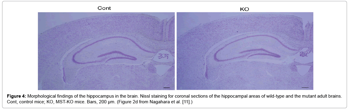
Figure 4: Morphological findings of the hippocampus in the brain. Nissl staining for coronal sections of the hippocampal areas of wild-type and the mutant adult brains. Cont, control mice; KO, MST-KO mice. Bars, 200 μm. (Figure 2d from Nagahara et al. [11].)
Determinations of polysulfide-producing enzymes
Experiments using MST-KO and wild-type mouse brain cells demonstrated that MST produces hydrogen polysulfide (H2Sn) [13]. H2Sn has a greater ability to activate TRP channels in astrocytes than H2S [14] and has been reported to activate TRP ankyrin 1 channels in sensory neurons [14,15], leading to acute pain [15]. In addition, H2Sn induces kelch-like ECH-associated protein 1 to form oxidized dimers and nuclear factor-like 2 to translocate to the nucleus [16]. In Neuro2A cells, H2Sn increases Akt phosphorylation and may protect cells from oxidative damage [16] while also functioning to inhibit cell growth and the promotion of neurite outgrowth [17]. In addition, H2Sn oxidatively modifies phosphatase and tensin homolog in intact cells [18]. Moreover, it has been reported that H2Sn promotes protein kinase G Iα to form the active homodimer and induces vasodilation [19].
Hylin and Wood suggested that enzymes from rat liver produced polysulfides from 3MP in 1959 [20]. In 2015, Kimura et al. confirmed that MST produced hydrogen trisulfide (H2S3) and H2S from 3MP in brain cells [13]. Wild-type mouse brain cells produced greater H2S3 and H2S from 3MP than MST-KO mice [13]. They also reported that MST produced hydrogen disulfide (H2S2), as opposed to hydrogen pentasulfide (H2S5), from 3MP in brain cells [13]. When exposed to 3MP, levels of H2S2 in wild-type mouse brain cells increased but those of H2S5 did not, whereas the exposure of 3MP did not affect levels of either H2S2 or H2S5 in MST-KO mouse brain cells [13]. In addition, Kimura et al. found that MST and its homolog rhodanese produced H2S3 from H2S in brain cells [13]. Both wild-type and MST-KO mouse cells produced H2S3 by sodium sulfide exposure with MST-KO mouse cells producing approximately two-thirds of that produced by wild-type mouse cells, suggesting that other enzymes may produce H2S3 [13]. This was subsequently confirmed by rhodanese mutant experiments [13].
In conclusion, comprehensive and systematic research using MSTKO mice is ongoing with the hope of better understanding the multiple roles of MST in the near future.