Journal of Geology & Geophysics
Open Access
ISSN: 2381-8719
ISSN: 2381-8719
Review Article - (2016) Volume 5, Issue 6
The article represents an overview and generalization of the microprobe and thermomagnetic data on the distribution, content and composition of metallic iron in Pleistocene lacustrine sediments of the Darhad Basin, northern Mongolia, in the Upper Miocene sediments of Lake Baikal, epicontinental sediments of Miocene-Late Jurassic, and Early Cambrian ages in different regions of North Eurasia and in Miocene-Late Jurassic oceanic sediments of North- Western Atlantic. Iron particles percentage varies from ~10-5 to 0.05%, their distribution is bimodal with the distinct zero mode, and the accumulation of iron particles is inversely linked with the rate of sedimentation. Pyrite and pyrrhotite are widespread in the studied sediments, and the distribution of metallic iron does not depend on the presence of pyrite (i.e., on redox conditions). The variations in the Curie temperature of iron from 700°C up to 780°C. The nickel admixture in native iron forms three groups, 1) pure iron (main part of particles), 2) iron with mode 5-6% Ni (kamacite), and 3) Fe-Ni alloy with mode 50% Ni (rare), were found in all studied sections. The global pattern of the distribution and composition of the iron particles clearly indicates that their origin is associated with interplanetary cosmic dust. At the same time, the particles of Fe-Ni alloy are very rare, and their concentration does not correlate with the content of other iron particles. Most likely, the particles of Fe-Ni alloy are mainly due to impact events. Correlation between the concentrations of iron particles and terrestrial minerals (magnetite) is absent as a rule. Such correlation (r=0.3-0.7) testifies the redeposition of iron particles.
Keywords: Pleistocene, Magnetization, Sedimentation, Lithology
Particles of metallic iron are often found in sedimentary rocks, but concentration is normally very low. Metallic iron in deep-water ocean sediments is usually considered to be of extraterrestrial origin [1-3]. However, many sediments contain iron particles related to volcanic activity, bacterial activity, and metamorphism eTc. Therefore, it is important to find signs of metallic iron of extraterrestrial origin.
In recent years, we have studied the spreading and composition of metallic iron particles in the Pleistocene lacustrine sediments of the Darhad Basin, northern Mongolia, in the Upper Miocene sediments of Lake Baikal, in epicontinental sediments of Miocene, Oligocene, Eocene, Cretaceous, Late Jurassic, and Early Cambrian age in different regions of North Eurasia and in ocean sediments of the North- Western Atlantic [4-10]. This article represents a result of a review and generalization of thermomagnetic and microprobe analyzes data for mentioned above articles and regions.
Thermomagnetic analysis (TMA) was carried out in the Paleomagnetic Laboratory of the Geological Department of Kazan State University, using Curie express balance (D.M.Kuzina is measurement executor). It included measurement of the specific magnetization of samples in the magnetic field of 200-500 mT at room temperature (M20) and its temperature dependence M(T) up to 800°C. The heating rate was 100°C/min. The obtained thermomagnetic curves were used to determine the Curie temperatures (Tc) of magnetic minerals in the samples. The accuracy of determination of Curie temperature is ~5- 10°C. To evaluate the content of magnetic mineral in the sample, an M(T) curve was extrapolated from each Curie temperature to room temperature to determine the specific saturation magnetization of mineral with a given Curie temperature. The ratio of the obtained magnetization to the known saturation magnetization of the mineral is the content of this mineral in the sample [11]. The accuracy of this estimation of the mineral content is rather low, but this is not significant on the background of fluctuations in the contents of metallic iron within several orders of magnitude.
Microprobe analysis (MPA) was carried out in the Borok geophysical observatory, Institute of Physics of the Earth RAS (V.A.Tsel’movich is measurement executor), using a Tescan Vega II microprobe with an energy dispersive spectrometer. Operation conditions: accelerating voltage 20 kV, current 0.2 nA, beam diameter ~0.2 μm, and analyzed zone length 1-2 μm. The MPA measurements were made in polished sections, and magnetic fractions. The content of the metallic iron are very low in the sediments, so the magnetic fraction extracted and measured. The samples were ground and sonically dispersed, and their magnetic fraction was separated with a permanent magnet. The fraction was applied over a carbon double-sided adhesive tape and rolled with a glass rod to make the particle surface oriented parallel to the table surface.
TMA and MPA complement each other. In many cases, the magnetic minerals detected by TMA are not identified by MPA and vice versa. Since the TMA was conducted on a small piece of the rock while the MPA handled the magnetic fraction from another piece of the same sample.
Results of TMA and MPA
According to TMA and MPA data in all studied samples there are unevenly distributed throughout sediments: 1) magnetite and cationdeficient magnetite (Tc=575-650°C) is ubiquitous magnetic mineral, and main part of sample magnetization (M20) (correlation coefficient is 0.94); 2) iron hydroxides, such as goethite (Tc=80-150°C, which disappears on the second heating; its portion in M20 is <5%); 3) most of the studied samples show magnetization growth at >500°C, expressed as a “pyrite” peak caused by the oxidation of Fe-sulfides (pyrite, pyrrhotite and hydrotrolite) to magnetite [12,13]. Relatively low correlation between the contents of magnetite and pyrite (its peak height) suggests the independent formation of these minerals. Magnetite is mainly a terrigenous mineral, which got into the sediments independently of their redox conditions, and pyrite is mainly an authigenic mineral formed in the sediments under reducing conditions. 4) Hematite (Tc~675°C) is present in many samples but in a very low contents. 5) Metallic iron particles present everywhere in all deposits, but in very low contents. Further article analyzes the behavior of metallic iron in the sediments of different regions [6].
Metallic iron particles are extremely scarce in the studied sediments and are chaotically distributed (Fig. 1). Iron was reliably determined only in 26 samples. The content of iron is usually lower than 10-3%. Relatively high iron contents are observed in the depth ranges 64.73-68.73 and 83.20-87.07 m (Figure 1), the presence of iron and its content correlate neither with the height of the pyrite peak (correlation coefficient is 0.08) nor with the content of magnetite (correlation coefficient is -0.11, Figure 2), i.e., do not depend on the redox conditions in the sediments. A group of samples lacking iron particles (the so-called “zero” group) include 79% of the studied samples, if all unreliable data on the presence of native iron are used for the estimation, and 90%, if only the reliable data are taken into account (Figure 3a). And zero group absent for terrestrial magnetite (Figure 3b). For detailed MPA investigation of metallic iron particles and other minerals, we chose nine samples.
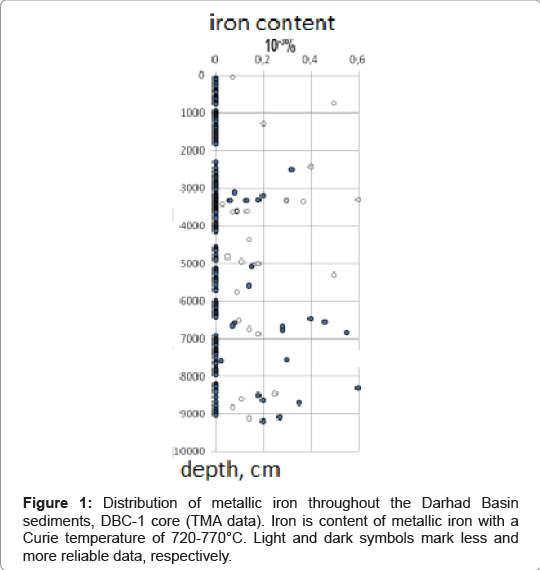
Figure 1: Distribution of metallic iron throughout the Darhad Basin sediments, DBC-1 core (TMA data). Iron is content of metallic iron with a Curie temperature of 720-770°C. Light and dark symbols mark less and more reliable data, respectively.
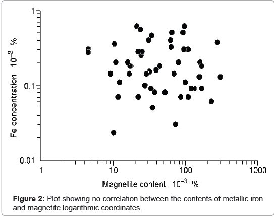
Figure 2: Plot showing no correlation between the contents of metallic iron and magnetite logarithmic coordinates.
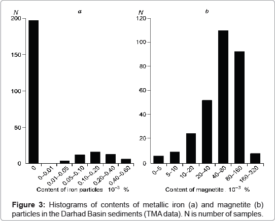
Figure 3: Histograms of contents of metallic iron (a) and magnetite (b) particles in the Darhad Basin sediments (TMA data). N is number of samples.
The MPA data show the presence of metallic iron particles, mostly pure (Figures 4 and 5). Only a minor part of samples contains metallic iron with Ni, Cr and Si impurities, particles of FeCr alloy containing 10- 15% Cr and particles of more complex composition, for example FeCrNi alloy containing Cr, 15-17%; Ni, 10-12%. There are also magnetite balls, apparently, of extraterrestrial origin (Figure 5) [7].
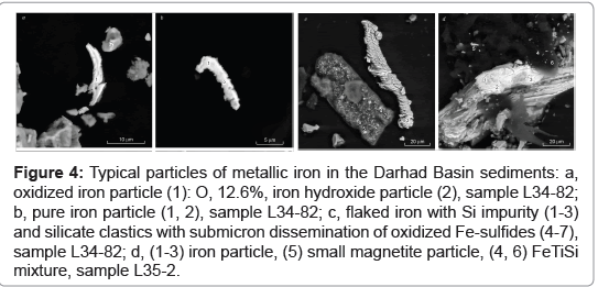
Figure 4: Typical particles of metallic iron in the Darhad Basin sediments: a, oxidized iron particle (1): O, 12.6%, iron hydroxide particle (2), sample L34-82; b, pure iron particle (1, 2), sample L34-82; c, flaked iron with Si impurity (1-3) and silicate clastics with submicron dissemination of oxidized Fe-sulfides (4-7), sample L34-82; d, (1-3) iron particle, (5) small magnetite particle, (4, 6) FeTiSi mixture, sample L35-2.
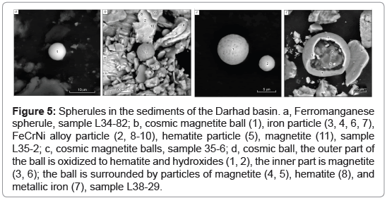
Figure 5: Spherules in the sediments of the Darhad basin. a, Ferromanganese spherule, sample L34-82; b, cosmic magnetite ball (1), iron particle (3, 4, 6, 7), FeCrNi alloy particle (2, 8-10), hematite particle (5), magnetite (11), sample L35-2; c, cosmic magnetite balls, sample 35-6; d, cosmic ball, the outer part of the ball is oxidized to hematite and hydroxides (1, 2), the inner part is magnetite (3, 6); the ball is surrounded by particles of magnetite (4, 5), hematite (8), and metallic iron (7), sample L38-29.
For study, the sediments were sampled from the lower 500- 600 m of the borehole BDP-98. They formed in Miocen age at the paleodelta of the Barguzin River and were characterized by a high rate of accumulation 7-13 cm/ kyr. The studied section is rather uniform by all magnetic parameters. Particles of metallic iron are identified by Tc=710-770°C; they are extremely rare in the studied sediments and are chaotically distributed (Figure 6). Such particles were reliably detected in only five samples. The content of iron is lower than 10-3% (Figure 6).
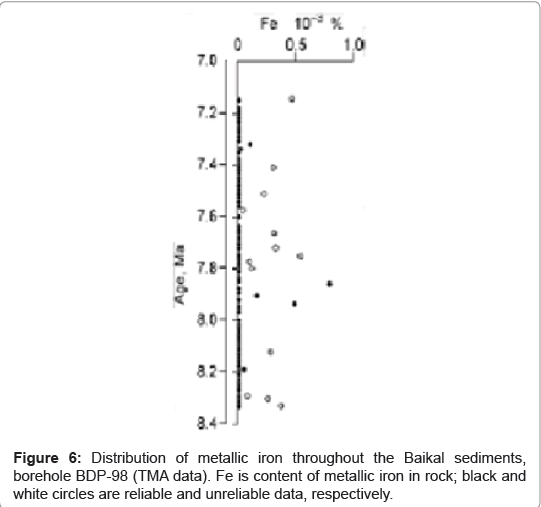
Figure 6: Distribution of metallic iron throughout the Baikal sediments, borehole BDP-98 (TMA data). Fe is content of metallic iron in rock; black and white circles are reliable and unreliable data, respectively.
The distribution of iron content is bimodal, with a distinct “zero” mode (Figure 7). This regularity does not depend on the lithology of sediments and the redox conditions of their accumulation (e.g., the presence or absence of pyrite). Zero groups are absent and distribution is unimodal for terrestrial magnetite from the same rocks (Figure 7). Scarcity of metallic iron in the Baikal sediments distinguishes them from continental (Eurasia) and oceanic (Atlantic) sediments of different ages. It is due to the high rate of sedimentation in the studied interval of BDP-98.
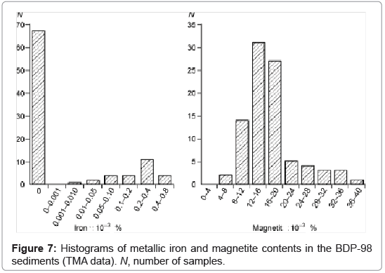
Figure 7: Histograms of metallic iron and magnetite contents in the BDP-98 sediments (TMA data). N, number of samples.
According to the MPA data, all five studied samples contain particles of pure iron (Figures 8a-8e); seldom, Si impurity (Figure 8b) and noticeable Cr impurity (Figures 8f-8k) occur. The iron is oxidized. We think that iron oxidation takes place when extraterrestrial particles pass through the Earth’s atmosphere.
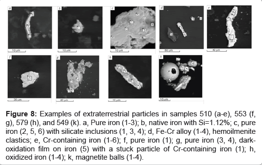
Figure 8: Examples of extraterrestrial particles in samples 510 (a-e), 553 (f, g), 579 (h), and 549 (k). a, Pure iron (1-3); b, native iron with Si=1.12%; c, pure iron (2, 5, 6) with silicate inclusions (1, 3, 4); d, Fe-Cr alloy (1-4), hemoilmenite clastics; e, Cr-containing iron (1-6); f, pure iron (1); g, pure iron (3, 4), darkoxidation film on iron (5) with a stuck particle of Cr-containing iron (1); h, oxidized iron (1-4); k, magnetite balls (1-4).
For study, the Pleistocene-upper Kimmeridge sediments were sampled from the DSDP boreholes 386, 387, 391A and 391C. Deposition was predominantly continuous; the rate of sedimentation varied from 0.2 to 4.7 cm/kyr. Particles of metallic iron are ubiquitously present in the studied sediments. Their concentrations are nonuniformly distributed in the different holes (Figure 9). However, in all cases, there is always a zero group of the samples, in which iron particles are absent (Figure 10a), but zero group is absent and distribution is unimodal for terrestrial magnetite from the same rocks (Figure 10b). The second (nonzero) group represents the main background of the nonuniform distribution of the particles of metallic iron. “Unzero” group is characterized by lognormal distribution. The level of concentrations somewhat varies depending on the sedimentation conditions in the different time intervals (Figure 9). For example, the lowest concentrations (<10-5%) are observed in the Miocene carbonate deposits of gravitational flows (Hole 391A), and the highest are revealed in the lower Berriasian–lower Valanginian sediments (Hole 391C). Against a common background, locally increased concentrations of iron (up to 10-2%) are observed. The shape of the histogram with a clearly pronounced zero group is a specific feature reflecting extraterrestrial nature of the accumulation of iron particles. It is important to note that this specificity doesn’t depend on either the lithological features of the sediments or the redox conditions of their accumulation (for example, it is insensitive to the presence or absence of pyrite).
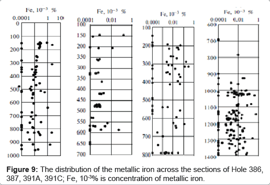
Figure 9: The distribution of the metallic iron across the sections of Hole 386, 387, 391A, 391C; Fe, 10-3% is concentration of metallic iron.
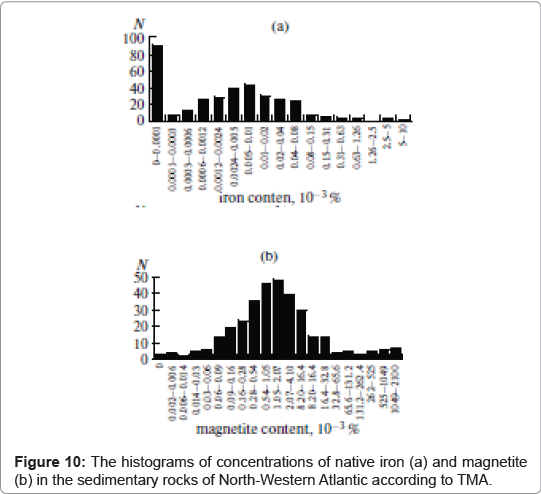
Figure 10: The histograms of concentrations of native iron (a) and magnetite (b) in the sedimentary rocks of North-Western Atlantic according to TMA.
According to the MPA data (Figures 11a-11g), all samples contain particles of pure iron; seldom, Si, S impurity and noticeable Ni, Cr impurity occur. The iron is not infrequently oxidized. The average content of the Ni admixture is almost identical in all the studied sections: 6% (Hole 386), 5.8% (Hole 387), 4.7% (Hole 391A), and 6.4% (Hole 391C). These values are close to the average and modal Ni content in the particles of metallic iron from the epicontinental sediments of Eurasia (see section below). The form of iron particles is typical for all sediments (Figure 11).
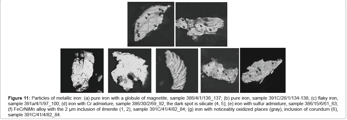
Figure 11: Particles of metallic iron: (a) pure iron with a globule of magnetite, sample 386/4/1/136_137; (b) pure iron, sample 391?/26/1/134-138; (c) flaky iron, sample 391a/4/1/97_100; (d) iron with Cr admixture, sample 386/30/2/69_92, the dark spot is silicate (4, 5); (e) iron with sulfur admixture, sample 386/15/6/61_63; (f) FeCrNiMn alloy with the 2 μm inclusion of ilmenite (1, 2), sample 391?/41/4/82_84; (g) iron with noticeably oxidized places (gray), inclusion of corundum (6), sample 391?/41/4/82_84.
The role of redeposition of iron particles is assessed from the correlation between the concentrations of metallic iron and terrestrial magnetite+titanomagnetite in the sediments. It is found that the effect of redeposition is not observed only in the sediments from Hole 386 (r=-0.105). In the other holes, the coefficient of correlation is 0.28 (387), 0.439 (391A), and 0.307 (391C), which indicates that redeposition of this particles is real. The effect of redeposition is best pronounced in the Miocene deposits of gravitational flows in Hole 391A (where r=0.439), which are marked by the lowest concentrations of iron particles (Figure 9). Obviously, together with the other magnetic minerals, rare particles of iron (both of terrestrial and extraterrestrial origin) were occasionally carried to these carbonate oozes, which were redeposited from the Blake Plateau by geologically instantaneous gravitational flows. In contrast, the most intense jumps in concentration are revealed in the intervals where the correlation between iron particles and other magnetic minerals is absent (Hole 386).
The composition and distribution of particles of native iron in eight sections of the Cretaceous–Danian sediments in the Caucasus, Crimea and Kopet Dagh were studied using TMA. The studied sediments are abundant with particles of metallic iron. The latter are identified in 330 of 571 studied samples in concentrations ranging from ~10- 5% to 0.05%, and their distribution is bimodal (Figure 12). “Unzero” group is characterized by lognormal distribution with the mode in the interval 0.04-0.15 × 10⁻³%. Intervals enriched with the iron particles were revealed in sediments of Seravalian (12-13 Ma), Santonian (84- 86 Ma), and Cenomanian (94-96) age and at the Maastrichtian/Danian boundary (64-66 Ma) (Figure 13) [9].
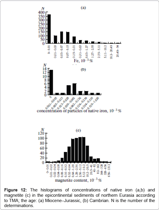
Figure 12: The histograms of concentrations of native iron (a,b) and magnetite (c) in the epicontinental sediments of northern Eurasia according to TMA; the age: (a) Miocene–Jurassic, (b) Cambrian. N is the number of the determinations.
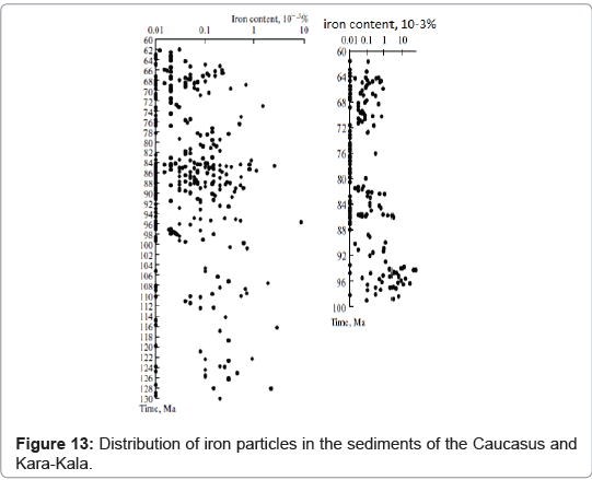
Figure 13: Distribution of iron particles in the sediments of the Caucasus and Kara-Kala.
The iron content increases from west to east. The Cretaceous deposits of the Crimea are very poor in iron; the most is ≤ 10-4%. The Upper Cretaceous carbonate deposits of the Caucasus contain more iron: not infrequently attains 0.5-10 × 10-3% (Figures 13 and 14). The highest iron contents are observed in the East, in the Kara-Kala section, where it attains 0.05% (Figure 13). The appearance and accumulation of iron does not depend on the redox conditions in sediments, which is shown by the lack of correlation of iron with the presence of cationdeficient magnetite (the high-oxidizing conditions), on the one hand, and “pyrite” (the reducing conditions), on the other hand, in the sediments. Tc=680-780°C. A peak of the elevated iron content with nearly constant nickel of 5% was found in all studied sections, i.e., this is a global effect. There is no correlation between the concentration of iron particles and the nickel content in them (r=-0.024).The global pattern of the distribution and composition of the iron particles clearly indicates that their origin is associated with interplanetary dust. At the same time, the particles of Fe-Ni alloy and pure nickel are very rare, and their concentration does not correlate with the content of iron particles. Most likely, the particles of Fe-Ni alloy are mainly due to impact events.
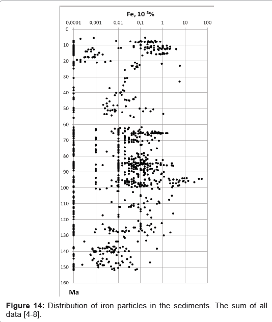
Figure 14: Distribution of iron particles in the sediments. The sum of all data [4-8].
Discussion on extraterrestrial nature of metallic iron particles in terrestrial sediments
Our studies revealed a number of quite simple statistical indications pointing to the extraterrestrial origin of metallic iron in the sediments: 1) Widespread (global) distribution of metallic iron particles in sediments in different regions and ages, from the Quaternary to Cambrian sediments in lakes, seas and oceans, different lithology and redox conditions. The concentration of particles varies widely-from none to 0.05%, mostly between 10-5 and 10-3% (Figures 1, 6, 9 and 13 and the sum of all data on Figure 14 [4-8]). It is logical to associate with interplanetary dust.
2) The bimodal distribution of iron particle concentrations with a necessarily present and clearly expressed “zero” group of the samples, in which iron particles are absent (not detected by TMA). This feature stems from the limited amount of cosmic dust deposited on the surface of the Earth. It is a known fact that 1 m3 of interplanetary space contains about 100000 particles of interplanetary cosmic dust. Therefore, upon deposition of cosmic dust onto the Earth’s surface, only one of ten cm3 contains such a particle, while iron particles are much rarer. The zero group is related to the parts of the sediments that do not contain any iron particles of cosmic origin. This trend is identified in all the studied sedimentary sequences (Figures 3, 7, 10 and 12) and, thus, it is global. This bimodal distribution of particles absent for terrestrial minerals, for example, magnetite (Figures 3, 7, 10 and 12). 3) The absence of a correlation between the concentrations of native iron particles and magnetic minerals (e.g., magnetite) of terrestrial origin. The linear correlation coefficient of less than 0.2 in the case of 12 sections of the 21 studied sections. The presence of such correlation says nothing about the nature of particles, but it is evidenced on existence common conditions of their accumulation in the sediment. And vice versa - the lack of correlation suggests different conditions accumulation of native iron and terrestrial magnetite. The absence of such correlation testifies to the predominantly extraterrestrial origin of the particles of native iron, while their ubiquitous occurrence suggests their relationship to interplanetary cosmic dust. There are examples where r>0.2. And, moreover, there are two examples of when r ≥ 0.5: Selbuhry deposits, Crimea r=0.74 and Cenomanian section of the Kara-Kala, r=0.5, which show a significant contribution of native iron of terrestrial origin, or a significant role of redeposition of native iron. It is the same in terms of the appearance of correlation. 4) Division metallic iron particles composition into three distinct groups. Particles of iron in their content of nickel impurities form three groups (Figure 15): 1) Pure nickel-free iron, such particles form a separate group, 2) A group of Ni-containing iron with a mode 4-6% Ni, these particles are the most common metal in the meteorites and interplanetary cosmic dust. 3) Rare particles Fe- Ni alloy with a nickel content of >20% and mode of 50%.
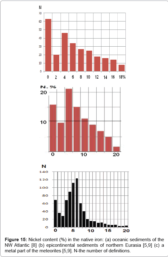
Figure 15: Nickel content (%) in the native iron: (a) oceanic sediments of the NW Atlantic [8] (b) epicontinental sediments of northern Eurasia [5,9] (c) a metal part of the meteorites [5,9]. N-the number of definitions.
The average content of the Ni admixture is almost identical in all the studied sections of sediments from the oceanic sediments of the NW Atlantic (Figure 15a), epicontinental sediments of Eurasia (Figure 15b). The global character of Ni content distribution in metallic iron is also highlighted by the similar pattern of histograms for sediments (Figures 15a and 15b) and Ni content in the metallic part of the meteorites (Figure 15c). Hence, we may conclude that the particles of metallic pure and nickelous iron in the meteorites have a common origin: both are products of the disintegration of planetary material. 5) The predominant particle size less than 100 microns (Figure 16) corresponds to extraterrestrial particles that are preserved passing through the Earth’s atmosphere, whereas for the earth particles there is no upper size limit [14,15]. 6) A negative correlation between the concentrations of iron particles and the rate of sedimentation. Against the scatter of the data, it is clearly seen that the zero group tends to increase with an increase in the sedimentation rate (Figure 17). In particular, the scatter of the data is controlled by the degree of redeposition of iron particles and their probable terrestrial origin, i.e., the situations when these particles participate in the accumulation of the sediments just as the terrestrial particles. In our case, this is reflected by the coefficients of the linear correlation between the concentrations of iron particles and magnetite of 0.3-0.6 [9]. In contrast to the general trend, at the points that are related to the redeposition (the open circles in Figure 17), the dependence of the size of the zero group and the rate of sedimentation vanishes. In the case of terrestrial native iron, we should expect the distribution to follow the Poisson zero-mode distribution, which is supported by the single-mode distribution of the magnetite content having knowingly terrestrial origin in the same sediments [9,10]. The absence of a correlation between the concentrations of terrestrial magnetic minerals (e.g., magnetite) and the rate of sedimentation is clearly seen in Figure 18. The coefficient of linear correlation is 0.032.
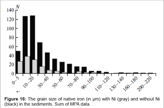
Figure 16: The grain size of native iron (in μm) with Ni (gray) and without Ni (black) in the sediments. Sum of MPA data.
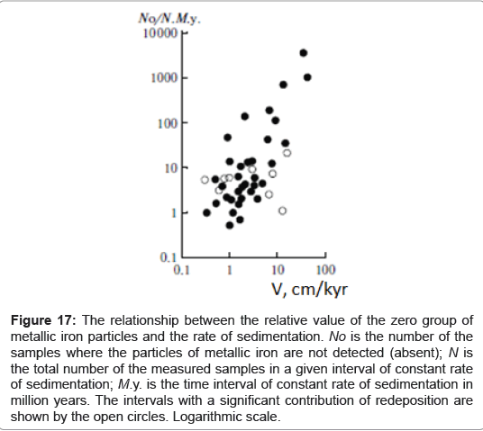
Figure 17: The relationship between the relative value of the zero group of metallic iron particles and the rate of sedimentation. No is the number of the samples where the particles of metallic iron are not detected (absent); N is the total number of the measured samples in a given interval of constant rate of sedimentation; M.y. is the time interval of constant rate of sedimentation in million years. The intervals with a significant contribution of redeposition are shown by the open circles. Logarithmic scale.
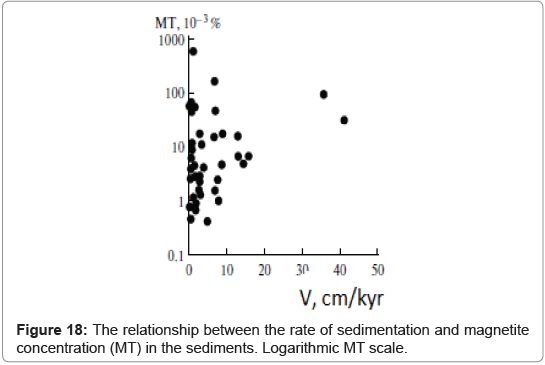
Figure 18: The relationship between the rate of sedimentation and magnetite concentration (MT) in the sediments. Logarithmic MT scale.
Firstly, the main methodical result noted: the use of TMA to 800°C in combination with MPA allow for faster and easily obtain a quantitative evaluation of the content and composition of metallic iron particles in the sediments. Today thermomagnetic analysis is the only simple method to quantify the presence and distribution in sediments the magnetic particles including iron of extraterrestrial origin with high sensitivity, without destruction, crushing the sample and extract from it the magnetic fraction.
Secondly, we note the main result-the justification of extraterrestrial origin metallic iron particles in terrestrial sediments. It is the system of evidences: 1) Widespread (global) distribution of particles, 2) Bimodal particle content distribution with the obligatory distinct zero mode; 3) Lack of correlation between particles concentrations of iron and earth minerals as magnetite; 4) Three independent groups of metallic iron are identified by the content of nickel impurities: pure iron, iron with an admixture of nickel (kamacite), and Fe-Ni alloy; 5) Iron particle size is limited to 100 μm; 6) An inverse relationship between the concentration of iron particles and sedimentation rate. The totality of these factors clearly indicates the extraterrestrial nature of metallic iron particles.