Anatomy & Physiology: Current Research
Open Access
ISSN: 2161-0940
ISSN: 2161-0940
Research Article - (2016) Volume 6, Issue 4
This project investigated the oxidative stress in the blood of pubertal rabbit fed diet containing 2% organic turmeric at post recovery from acute ultraviolet radiation, with the aid of three oxidative stress markers: Malondialdehyde (MDA), Catalase (CAT) and Superoxide dismutase (SOD). Forty unsexed rabbits with age ranging between 4-6 weeks were weight balanced and randomly divided into eight treatment groups: AM: Male rabbits fed with formulated organic feed without turmeric and without exposure to ultraviolet radiation. AF: Female rabbits fed with formulated organic feed without turmeric and without exposure to ultraviolet radiation. E: Female rabbits fed with commercial feed without turmeric and without exposure to ultraviolet radiation. I: Female rabbits fed with organic feed with turmeric before and after radiation C: Male rabbits fed with organic feed with the turmeric and without exposure to ultraviolet radiation. F: Male rabbits fed formulated organic feed without turmeric before radiation and after radiation turmeric was given. G: Male rabbits exposed to radiation without the inclusion of turmeric in their feed throughout. H: Male rabbits fed formulated organic feed with turmeric before radiation and without turmeric after radiation. MDA mean value obtained in the control treatment for male was not significantly (p>0.05) but only numerically higher than that of the treatment G. MDA mean value obtained in the control treatment for female was only numerically higher than that of treatment I. CAT and SOD mean values obtained in treatment G were also only numerically higher than the control treatment in male rabbits. CAT and SOD mean values obtained in treatment I were only numerically higher than the control treatment in female rabbits. The study concluded that the exposure to ultraviolet radiation at the intensity and period in this study did not result in oxidative stress in the blood of pubertal rabbits.
Keywords: Turmeric; Ultraviolet radiation; Malondialdehyde; Superoxide dismutase; Catalase; Rabbit
Natural plant products have been used throughout human history for various purposes. Tens of thousands of these products are produced as secondary metabolites by higher plants as a natural defense against disease and infection. Many of these natural products have pharmacological or biological usefulness that can be of use in pharmaceutical drug discovery and drug design. Medicines derived from plants have played a pivotal role in the health care of many cultures, both ancient and modern [1].
Ultraviolet radiation is an electromagnetic radiation with a wavelength shorter than that of visible light but longer than x-rays, that is in the range between 400 nm and 10 nm corresponding to photon emerges from 3 ev to 124 ev. It can cause chemical reactions and causes many substances to glow or fluoresce. Ultraviolet (UV) radiation is a crucial factor in the etiology and development of a number of body diseases. It is not only skin which is directly exposed to solar light that is affected by UV radiation, through low molecular weight mediators, generated upon irradiation, "non-skin" tissues can also be affected [2]. In a study on oxidative effects of ultraviolet A (UVA) light (320-400 nm) and the antioxidant effects of quercetin, quercetin significantly increased antioxidant enzymes diminished by UVA irradiation. Exposure of rats to UVA light leads to oxidative stress reflected by increased MDA and reduced antioxidant enzyme levels. The administration of quercetin appears to be a useful approach to reduce the damage produced by UVA radiation [3].
Oxidative stress reflects an imbalance between the systemic manifestation of reactive oxide species and a biological system's ability to readily detoxify the reactive intermediates or to repair the resulting damage. Oxidative stress can cause disruptions in normal mechanisms of cellular signaling. Oxidative stress plays critical roles in the pathogenesis of various diseases [4]. In the diabetic condition, oxidative stress impairs glucose uptake in muscle and fat and decreases insulin secretion from pancreatic β cells [5]. Increased oxidative stress also underlies the pathophysiology of hypertension and atherosclerosis by directly affecting vascular wall cells.
Turmeric is a rhizomatous herbaceous perennial plant. It is a native to tropical south Asia and needs temperature between 20ºC and 30ºC (680f and 860) and a considerable amount of annual rainfall to thrive [6]. Its active ingredient is curcumin. Turmeric powder has long been used for medicinal purposes in Asia to treat gastrointestinal upsets, arthritis pain and low energy [6]. Curcuma can significantly inhibit the generation of reactive oxide species (ROS) like superoxide anions. Its derivatives, demethoxy curcumin and bis-demethoxycurcumin also have anti-oxidant effect. Curcumin reduces oxidized proteins in amyloidal pathology in Alzheimer transgenic mice. It also decreases lipid peroxides in rat liver microsomes, erythrocyte membrane and brain homogenates. This brought about by maintaining the activities of anti-oxidant enzymes like superoxide dismutase, catalase and glutathione peroxides. Since reactive oxide species have been implicated in the development of various pathological conditions, curcuma has the potential to control these diseases through its potent antioxidant activity [7].
Hence, this study seeks to determine the effect of Organic turmeric Supplemented-Diet in rabbits acutely exposed to Ultraviolet Radiation: Oxidative Stress in the Blood.
Experimental site
The experiment was carried out at the Rabbit production and Research unit of Teaching and research farm, Ladoke Akintola University of Technology, Ogbomoso, located in the derived savannah zone of Nigeria.
The rabbits were housed in wooden cages reinforced with “1/2” wire-netting above expanded square shaped metal gauze with each hutch measuring 48*30*48 cm under ambient temperature and natural light.
Experimental animals
Forty-five (45) weaned rabbits aged 4 – 6 weeks were purchased from reputable rabbit farm (Onile Ola Farms), Osogbo, Osun State and fed for 4 weeks prior to the commencement of experiment for acclimatization. The rabbits were weight balanced into eight treatment groups, which were randomly allocated to the treatments AM: Five rabbits (males) fed with formulated organic feed without inclusion of turmeric throughout the experimental period and without exposure to ultraviolet radiation (ttrt). AF: Five rabbits (females) fed with formulated organic feed without inclusion of turmeric throughout the experimental period and without exposure to ultraviolet radiation (ttrt). C: Five rabbits (males) fed with formulated organic feed with the inclusion of turmeric throughout the experimental period without exposure to ultraviolet radiation (TTrT). E: Five rabbits (female) fed with ready-made commercial feed (Top feed growers mash) without inclusion of turmeric and without exposure to ultraviolet radiation. F: Five rabbits (male) fed formulated organic feed without turmeric before radiation, after radiation turmeric was given (ttRT). G: Five rabbits (male) exposed to radiation without the inclusion of turmeric in their feed throughout (ttRt). H: Five rabbits (male) fed formulated organic feed withturmeric before radiation and without turmeric after radiation (TTRt). and I: Five rabbits (female) fed formulated organic feed with turmeric before and after rradiation (TTRT).
Experimental diets
Two experimental diets one with and the other without 20% organic turmeric inclusion. The organic turmeric served as the test ingredient for the study. Commercial feed (growers mash-Top feed) was also used without inclusion of organic turmeric.
Sample collection and preparation
Needle and syringe was used to collect 5 ml of blood from the auricular marginal vein of the ear into an ethylenediametetra-acetic acid (EDTA) bottle. Before collection, the restrained rabbit’s ear was cleaned with methylated spirit and massaged for dilation of the vein to occur.
Determination of lipid peroxidation (MDA)
Assessment of lipid peroxidation: This was carried out based on the principle of varshney and kale.
Principles: Estimation of lipid peroxidation is based on the reaction of malondialdehyde (MDA) with thiobarbituric acid (TBA) forming a MDA - TEAR adduct that absorb light strongly at 532 mm.
Procedure: 0.4 ml of reaction mixture i.e. sample already quenched with 0.5 ml of 30%. TCA was added to 1.6 ml of Trihydroxymethyl methylamine potassium chloride at PH of 7.4. 0.5 ml of 8% TBA was added and incubated for 45 minutes at 30ºC to produce a pink colored reaction mixture which was centrifuge at 1400 rpm for 15 minutes. The absorb area of the clear supernatant was then read at 532 nm.
Determination of superoxide dismutase (SOD)
SOD activity was determined by using the method of misra and fridorich.
Principle: The ability of superoxide dismutase to inhibit the auto oxidation of epinephrine at PH 10.2 to adrenochrome makes this reaction a basis for a simple assay of dismutase.
Epinephrine [0]111adrenochrome, 02- (SOD)
Reagents: 0.05 M carbonate buffer (PH 10.2), 0.3 MM Adrenaline
Procedure: 1 ml of sample was diluted in 9 ml of distilled water to make a1 in 10 dilutions. An aliquot of the diluted sample was added to 25 ml of 0.05 M carbonate buffer PH 10.2 to equilibrate in the spectrophotometer and the reaction was initiated by adding 0.3 ml of adrenaline.
The change in absorbance was monitored at 430 nm for 5 minutes.
Determination of catalase (CAT)
Catalase activity was determined according to the method of Aebi.
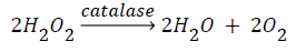
Principle: Catalase catalyzes the decomposition of hydrogen peroxide (H2O2) to water and oxygen. Hydrogen peroxide is formed in the eukaryotic cells as a byproduct of various oxidase and superoxide reactions. Hydrogen peroxide is highly deleterious to the cells and its accumulation causes oxidation of cellular targets such as DNA, protein and lipid leading to mutagenesis and cell death. Removal of the H2O2 from the cell by catalase provides protection against oxidative damage to the cell.
Procedure: 0.1 ml of the sample was pipetted into curette containing 1.9 ml of 50 MM phosphate buffer PH 7.0. The reaction was initiated by addition of 0.1 ml of freshly prepared 30% (w/v) hydrogen peroxide. The rate of decomposition of hydrogen peroxide was measured spectrophotometrically by monitoring the change in absorbance in thermogenesis 10s UV-US spectrophotometer at 240 nm.
Female rabbits
Malondialdehyde (MDA): MDA activity values obtained showed no significant difference (p>0.05) in the oxidative stress caused by the exposure to UV irradiation between the three treatment groups in female rabbits. In MDA, treatment E has the highest mean activity value (6.96) followed by treatment AF and I with lowest mean activity value (6.82).
Catalase (CAT): Treatment E and Treatment AF was significantly lower than treatment I. Treatment I has the highest mean activity value (34.58) while treatment AF has the lowest mean activity value (9.22).
Superoxide dismutase (SOD): SOD values obtained showed no significant difference in the oxidative stress caused by the exposure to UV irradiation between the three treatment groups in female rabbits. Treatment I has the highest mean activity value (14.43), while Treatment AF has the lowest mean activity value (4.98).
Male rabbits
Malondialdehyde (MDA): There was no significant difference between Treatment AM and Treatment H. Treatment AM and Treatment H were significantly higher than other treatments. Treatment F and treatment G showed no significant different but both were significantly higher than treatment C. Treatment H has the highest mean activity value (8.51) while treatment C has the lowest mean activity value (4.71).
Catalase (CAT): There was no significant difference between treatment C and treatment G. Treatment C: and treatment G are significantly higher than treatment AM, F and H. Treatment AM is significantly higher than treatment H. Treatment H is significantly higher than treatment F. Treatment C has the highest mean value activity (60.44) while treatment F has the lowest mean value activity (20.38).
Superoxide dismutase (SOD): Treatment AM, treatment C, treatment G and treatment H showed no significant difference and significantly higher than treatment F. Treatment C has the highest mean value activity (25.74), while treatment F has the lowest mean value activity (5.44). Figures 1 and 2 illustrates graph representation using SOD, CAT and MDA as biomarkers.

Figure 1: A graph showing oxidative stress markers activities in female rabbit.

Figure 2: A graph showing oxidative stress markers activities in male rabbit.
Treatment C has the highest mean value activity (25.74), while treatment F has the lowest mean value activity (5.44). Figures 1 and 2 illustrate graph representation using SOD, CAT and MDA as biomarkers.
Lipid peroxidation (MDA)
In female rabbits, lipid peroxidation (MDA) mean value showed that treatment AF has a higher mean value (9.05) compared to treatment I (6.82). In male rabbits, treatment G (6.05) showed a decrease in mean value compare to treatment AM (7.76), which is contrary to the result obtained by Isabelle et al. [8] which indicated that for oxidative stress to occur the mean value of MDA in patient should be higher than control. Therefore, MDA as an oxidative stress marker indicated no oxidative stress in blood of male and female pubertal rabbits (Table 1).
| Male | Female | ||||||
|---|---|---|---|---|---|---|---|
| Treatment | MDA | CAT | SOD | Treatment | MDA | CAT | SOD |
| A | 7.76ab ± 3.31 | 46.48b ± 3.77 | 18.05a ± 4.39 | A | 9.05 ± 7.71 | 9.22b ± 1.28 | 4.98 ± 3.33 |
| C | 4.71c ± 5.04 | 60.44a ± 3.66 | 25.74a ± 2.42 | E | 6.96 ± 1.25 | 25.88a ± 2.76 | 11.81 ± 2.27 |
| F | 6.36bc ± 6.57 | 20.38d ± 5.25 | 5.44b ± 1.46 | I | 6.36bc ± 6.57 | 20.38d ± 5.25 | 5.44b ± 1.46 |
| G | 6.05bc ± 3.30 | 55.70ab ±2.92 | 24.54a ± 3.08 | ||||
| H | 8.51a ± 8.27 | 34.82c±0.69 | 17.96a ±1.42 | ||||
abc means on the same column with different superscripts differ significantlyMDA: Malondialdehyde, SOD: Superoxide Dismutase, CAT: Catalase
Table 1: Oxidative stress marker level of MDA, CAT and SOD in control and experimental group of male and female rabbit
Catalase (CAT)
In female rabbits, CAT mean value showed that treatment I (34.58) is higher than treatment AF (9.22). In male rabbits, treatment G (55.70) is higher than treatment AM (46.48) which is contrary to the result obtained by Abhinau et al. [9] which indicated that for oxidative stress to occur the mean value of catalase in control should be higher than the patient. Therefore, catalase as an oxidative stress marker indicated no oxidative stress in blood of male and female pubertal rabbits.
Superoxide dismutase (SOD)
In female rabbits, SOD mean value showed that treatment I (14.43) is higher than treatment AF (4.98). In male rabbits, treatment G (24.54) is higher than AM (18.05) which is contrary to the result obtained by Abhinau et al. [9] which stated that the level of mean value of SOD in control experiment should be higher than the patient. Therefore, SOD as an oxidative stress marker indicated no oxidative stress in blood of male and female pubertal rabbits [10].
Oxidative stress did not occur in the blood of male and female pubertal rabbits exposed to acute ultraviolet radiation of 10 minutes for 10 days.
Ethical approval was obtained from the university management and the local government ethics committee under the local government authority of Ogbomosho North local government, Oyo State Nigeria.