Advanced Techniques in Biology & Medicine
Open Access
ISSN: 2379-1764
ISSN: 2379-1764
Review Article - (2016) Volume 4, Issue 2
Recently, Immunohistochemistry (IHC) using specific antibodies has been exploited to detect the localization of stem cells and cancer stem cells (CSCs). Typically, there is a very small population of stem cells in normal and cancerous tissues, making it difficult to investigate their cell type and localization in human specimens. Hence, for pathological detection of human specimens, IHC has certain advantages over immunofluorescence staining. In the present study, we established a method for double-staining cancer stem cell markers and other putative markers associated with human liver cancer. We have summarized and reviewed the methodology for IHC with doublestaining in human liver cancer tissues.
Keywords: Immunohistochemistry, Cancer, Stem cell
Immunohistochemistry (IHC) is a useful technique for detection of proteins in cells and is applicable for diagnosis and research in both human cancer and normal tissues. IHC techniques detect antigens in tissue sections by making use of immunological and chemical reactions [1].
Double-staining experiments aim to determine co-localization, as this method facilitates simultaneous visualization of two markers in one cell or tissue constituent. There are two main methods used for double staining, namely, IHC and immunofluorescence (IF). IF is used to determine the co-localization of target molecules, whereas IHC is preferred when target molecules are not co-localized. IHC provides superior morphological information based on signal intensities compared to IF.
Typically, there is a very small population of stem cells and cancer stem cells (CSCs) in normal and cancerous tissues, respectively [2]. Hence, IHC has several advantages over IF technique including: 1) capability of being repeatedly used in order to search for few positive cells, 2) no reduction in signal intensity, 3) provides superior morphological information, and 4) could be utilized to distinguish between normal and cancerous tissue. Therefore, in cancerous tissues, double-staining IHC is practically suitable for observing stem cell and CSC markers [3,4].
CSCs are a rare subpopulation of tumor cells thought to be responsible for cancer initiation, maintenance, growth, metastasis, recurrence, and resistance to treatments [5]. Cancer tissues are hierarchically organized as they are composed of heterogeneous cell populations including a very small fraction of CSCs. Limited methods are available to purify CSCs owing to tumor cell heterogeneity and lack of specific stem cell markers.
In hepatocellular carcinoma (HCC), putative markers used for the isolation of liver CSCs include epithelial cell adhesion molecule (EpCAM), Thy-1 (CD90), and BMI1. Additionally, a nonimmunological approach based on aldehyde dehydrogenase (ALDH) activity is also used for the isolation of CSCs [6]. EpCAM (CD326), a carcinoma-associated antigen, is a single-pass transmembrane glycoprotein encoded by the tumor-associated calcium signal transducer 1 gene [7]. CD90, a glycosylphosphatidylinositol-anchored glycoprotein, is expressed on many cell types including T cells, thymocytes, neurons, endothelial cells, and fibroblasts [8]. BMI1, a part of the polycomb group genes, is a transcriptional repressor implicated in regulating self-renewal and proliferation of liver stem or progenitor cells [9]. ALDH is a ubiquitously expressed intracellular enzyme that catalyzes the irreversible oxidation of a variety of cellular aldehydes. ALDH activity has been used to isolate liver stem cells [3]. Recently, ALDH1A1, a major isoform of ALDH1, has been reported as a differentiation marker for the detection of HCC. However, it could only detect, well, moderately, or poorly differentiated HCCs, rather than the CSC fraction by IHC. Thus, there are many marker candidates to detect HCCs, but no specific marker exists to detect CSCs. Hence, methods to specifically detect a very small population of CSCs by using multiple markers could be promising for the detection and cure of various types of cancers.
We herein reviewed and summarized a unique, modified doublestaining protocol used in our laboratory for years to detect stem cell markers in human formalin-fixed and paraffin-embedded liver cancer tissue specimens.
Formalin-fixed and paraffin-embedded tissues were used in the present study to carry out double-IHC staining. It is very convenient using archived paraffin-embedded tissue to perform IHC. The most widely used fixative for routine histology is 10% neutral buffered formalin as it can effectively prevent autolysis, preserve tissue and cellular morphology, and helps in the storage of samples for many years. Further, in recent years, the use of antigen retrieval methods has greatly increased the use of formalin-fixed tissue in IHC.
Procedure
The cellular markers used in the present study includes ALDH1A1 (a differentiation marker of liver cancer), BMI1 and CD90 (both putative CSC markers of liver cancer), and Ki-67 (a proliferation marker) as shown in Table 1.
1st staining: 1. Dewax slides with xylene for 5 min each, 5 times.
2. Hydrate samples using an alcohol gradient as follows:
• 4 times in 100% ethanol for 2 min each
• 90% ethanol for 2 min
• 80% ethanol for 2 min
• 70% ethanol for 2 min
3. Soak samples in dH2O for 2 min.
4. Place slides in a Pascal pressure cooker (DAKO, Glostrup, Denmark) in 10 mM citrate buffer (pH 6.0) and heat at 121°C for 1 min.
5. Allow samples to cool at room temperature (RT).
6. Block endogenous peroxidase activity with 10% hydrogen peroxide (H2O2) containing methanol in phosphate-buffered saline (PBS) for 10 min.
7. Wash with PBS for 5 min each, 3 times.
8. Block non-specific sites with 2% bovine serum albumin (BSA) for 40 min at RT.
9. Incubate samples with primary antibody for the optimized time and dilution? at 4°C.
?The optimization time and dilution depend on many factors, such as the type of tissue, primary antibodies, secondary antibodies, and the detection system.
10. Wash with PBS for 5 min each, 3 times.
11. Add secondary antibody conjugated to biotin for 30 min at RT.
12. Wash with PBS for 5 min each, 3 times.
13. Add ABC reagent for 30-45 min at RT (VECTASTAIN ABC Kit, #PK-4000, Vector Labs, Inc., Burlingame, CA, USA).
14. Wash in DPBS for 5 min each, 3 times.
15. Prepare DAB peroxidase substrate in 5 mL dH2O in a glass vial immediately before use.
16. Drop the DAB substrate onto the top of the slides and watch for brown staining.
17. Rinse under cold tap water for 5 min.
2nd staining: 1. Pre-warm 10 mM sodium citrate (pH 6.0) and heat the slides in it at 90-99°C for 10 min to retrieve antigens and quench the primary layer.
2. Rinse slides under cold tap water for 2 min and soak in 50 mM Tris-HCl buffer (pH 8.2) for 5 min at RT.
3. Incubate the sections with Avidin D solution for 15 min.
4. Rinse the sections with the 50 mM Tris-HCl buffer (pH 8.2), and incubate in biotin solution (Avidin/Biotin blocking kit, #SP-2001, Vector Labs, Inc.) for 15 min.
5. Block non-specific sites with 2% BSA for 40 min at RT.
6. Add primary antibody (different from the primary antibody used in 1st staining) diluted with the 50 mM Tris-HCl buffer (pH 8.2) and incubate overnight at 4°C.
7. Wash with 50 mM Tris-HCl buffer (pH 8.2) for 5 min each, 3 times.
8. Add secondary antibody conjugated to biotin and incubate for 30 min at RT.
9. Wash with 50 mM Tris-HCl buffer (pH 8.2) for 5 min each, 3 times.
10. Incubate the slides for 30 min with VECTASTAIN ABC-AP Reagent (AK-5000). (VECTASTAIN ABC-AP Reagent, #AK-5000, Vector Labs, Inc.).
11. Wash with 50 mM Tris-HCl buffer (pH 8.2) for 5 min each, 3 times.
12. Incubate the sections in alkaline phosphatase (AP) substrate solution (SK-5300) (Vector Blue Alkaline Phosphatase Substrate, #SK-5300, Vector Labs, Inc.) for 20-30 min in dark. 13. Wash with 50 mM Tris-HCl buffer (pH 8.2) for 5 min each, 3 times.
14. Rinse the slides under cold tap water for 5 min.
Do not counterstain the slides; if required, a very gentle hematoxylin hue in the nuclei is acceptable.
15. Mount the coverslip using non-xylene-based mounting media.
Double-staining IHC of stem cell markers in human liver cancer tissues was performed based on the above method and under the experimental conditions as shown in Table 1. For double-staining, ALDH1A1 and CD90, ALDH1A1 and BMI1, ALDH1A1 and EpCAM, and ALDH1A1 and Ki67 were used as pairs (Figure 1). In all these pairs, ALDH1A1 was first stained brown by DAB, while CD90, EpCAM, BMI1, and Ki67 were next stained blue by AP. The color reaction of DAB is more sensitive and clearer than that of AP. We selected ALDH1A1 to be stained brown by DAB to distinguish the different kinds of cell types, as ALDH1A1 is expressed by normal hepatocytes and by a large population of HCCs. All double-staining in this experiment was performed in conformity to the above method.
| Antigen | Host | Working dilution | Antigen retrieval | Source |
|---|---|---|---|---|
| ALDH1A1 | Rabbit polyclonal | 1:500 | 10mM sodium citrate buffer (pH 6.0) | Abcam |
| BMI1 | Rabbit monoclonal | 1:200 | 10mM sodium citrate buffer (pH 6.0) | Dako |
| Ki-67 | Rabbit monoclonal | 1:100 | 10mM sodium citrate buffer (pH 6.0) | Abcam |
| CD90 | Rabbit polyclonal | 1:150 | 10mM sodium citrate buffer (pH 6.0) | Epitomics |
| EpCAM | Rabbit polyclonal | 1:150 | 10mM sodium citrate buffer (pH 6.0) | Abcam |
Table 1: Primary antibodies used in this study.
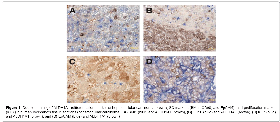
Figure 1: Double-staining of ALDH1A1 (differentiation marker of hepatocellular carcinoma, brown), SC markers (BMI1, CD90, and EpCAM), and proliferation marker (Ki67) in human liver cancer tissue sections (hepatocellular carcinoma). (A) BMI1 (blue) and ALDH1A1 (brown), (B) CD90 (blue) and ALDH1A1 (brown), (C) Ki67 (blue) and ALDH1A1 (brown), and (D) EpCAM (blue) and ALDH1A1 (brown).
ALDH1A1 was not co-expressed with CD90, EpCAM, and BMI1 CSC markers in well to moderately differentiated HCCs. Further, ALDH1A1 was not co-expressed with Ki67, a proliferation marker. These data indicate that ALDH1A1 might be a differentiated cell marker in HCC and the cells expressing this marker are quiescent. Consequently, the double-staining IHC technique is thought to be useful for detecting CSC markers in HCC.
When single staining is conducted on serial sections, ambiguous lesions may disappear, particularly while examining small foci, leaving positive cancer stem cell markers undetected. Therefore, the doublestaining IHC technique is useful for investigating the characteristics of each cancer cell in human cancer tissues.
We would like to thank Masayoshi Shimizu and Reiko Kitazumi for their valuable technical assistance. We thank Editage (www.editage.jp) for their support with the English language editing service. This study was supported by a grant from the Ministry of Education, Culture, Sports, Science and Technology of Japan (#15K11289 and # 26430111).