Journal of Medical Diagnostic Methods
Open Access
ISSN: 2168-9784
ISSN: 2168-9784
Research Article - (2016) Volume 5, Issue 2
Background: The evaluation method for the diagnosis of tinea unguium is mainly microscopy. New antidermatophyte monoclonal antibody have been developed that recognizes the cell wall polysaccharide antigen of filamentous fungi, including dermatophytes, and applied it to immunochromatography. Some scientists focused on this technology, improved it, and determined that this test method provided useful information for a diagnosis of tinea unguium by analyzing large numbers of clinical specimens. However, these studies were carried out by a tentative method.
Objective: Establishment of the usage of this test strip for clinical use and the certification of the stability during long-term storage.
Methods: Various fungi and bacteria were cultured and extracted by extraction buffer to evaluate the reactivity and measurement range of dermatophytes. Trichophyton rubrum was used as a quality control antigen, and detection limits were set at 0.5 μg/ml as weak positive and set at a 100-fold concentration (50 μg/ml) as strong positive. Nail samples, which had already been identified as positive or negative using the test strips, were randomly selected, cut into fine pieces and mixed. Both positive and negative standard nail samples were prepared.
Results: Positive or negative results were obtained from test lines for 5 to 60 minutes at 1 to 30°C. The detection limits of dried 7 samples of dermatophytes were 0.3 to 3 μg/ml. The results of reactivity showed that 8 of dermatophytes were positive. On the other hand, the test strip did not react to Malassezia or Candida species and bacterial strains. Some of Aspergillus, Penicillium, and Fusarium, which are usually not resident microbiota that inhabit nails in healthy persons, showed a positive reaction. The antifungal agents, terbinafine, griseofulvin, and itraconazole did not affect the results. All 3 lots of the test strips met the standards set by the method of quality control after they were kept in storage at 30°C for up to 22 months.
Conclusion: A newly developed dermatophyte-detection device was easy to use, gave rapid results and high reproducibility, and was stable for 22 months at 30°C.
Keywords: Dermatophyte; Monoclonal antibody; Device; Immunochromatography; Tinea unguium
Tinea unguium is a nail disease caused by dermatophytes [1,2]. The evaluation method for a diagnosis is mainly KOH direct microscopy and fungal culture, which comprise the gold standard. It takes 2 to 3 weeks by fungal culture until the results are obtained, and the success rate of the culture is approximately 20 to 70%, which is considered to be relatively low [3]. Therefore, microscopy is usually used to diagnose tinea unguium in clinical practice. However, it requires some skill and experience to correctly diagnose the disease by microscopy, and there is also the problem that it takes a long time to dissolve the nail [4,5].
Noriki et al. have developed an anti-dermatophyte monoclonal antibody that recognizes the cell wall polysaccharide antigen of filamentous fungi, including dermatophyte, and applied it to immunochromatography. They then subjected 37 nail specimens to the test and successfully detected dermatophyte antigen [6,7]. Tsunemi et al. focused on this technology and conducted the test with large numbers of clinical specimens, and then determined that the test method provided useful information for a diagnosis of tinea unguium [4].
However, these studies were carried out by a tentative procedure of use, and to put this test strip into clinical practice widely as an in vitro diagnostics, it was necessary to determine a practical procedure of use along with the basis for it, and clarify simultaneous reproducibility of the results, and long-term storage stability of the test strip.
In this report, we established the usage of this test strip for practical application, and investigated the simultaneous reproducibility and long-term storage stability.
Procedure and principles for measurement with the test strip
Configuration of the test strip:
Test strips used in the current study had a similar structure as the prototype developed by Noriki et al. [6,7] with some modifications, and the structure is shown in Figure 1.
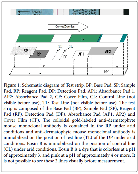
Figure 1: Schematic diagram of Test strip. BP: Base Pad, SP: Sample Pad, RP: Reagent Pad, DP: Detection Pad, AP1: Absorbance Pad 1, AP2: Absorbance Pad 2, CF: Cover Film, CL: Control Line (not visible before use), TL: Test Line (not visible before use). The test strip is composed of the Base Pad (BP), Sample Pad (SP), Reagent Pad (RP), Detection Pad (DP), Absorbance Pad (AP1, AP2) and Cover Film (CF). The colloidal gold-labeled anti-dermatophyte mouse monoclonal antibody is contained in the RP under arid conditions and anti-dermatophyte mouse monoclonal antibody is immobilized on the position of test line (TL) of the DP under arid conditions. Eosin B is immobilized on the position of control line (CL) under arid conditions. Eosin B is a dye that is colorless at a pH of approximately 3, and pink at a pH of approximately 4 or more. It is not possible to see these 2 lines visually before measurement.
The test strip used in this study is composed of the base pad, the sample pad, the reagent pad, the detection pad, the absorbance pad, and the cover film.
Colloidal gold -labeled anti-dermatophyte mouse monoclonal antibody is contained in the reagent pad under arid conditions, and anti-dermatophyte mouse monoclonal antibody is immobilized on the position of test line (TL) and eosin B (Wako Pure Chemical Industories, Ltd.) is immobilized on the position of control line (CL) on the detection pad. Eosin B is a dye that is colorless at a pH of approximately 3, and pink at a pH of approximately 4 or higher.
The production of Colloidal gold-labeled anti-dermatophyte mouse monoclonal antibody and assembly of the test strip was done by Nippon Gene Co. Ltd.
Principle of the measurement:
The sample pad portion is immersed in the sample solution in the test tube, and is allowed to stand. The sample solution is soaked to the test strip and dermatophyte-derived antigen in the sample solution binds to the colloidal gold-labeled anti-dermatophyte antibody to form an immune complex. This immune complex then moves along the detection pad through capillary action, is captured by the immobilized anti-dermatophyte mouse monoclonal antibody on the TL, and a purple test line derived from colloidal gold particles appears (Figure 2, in case of a positive reaction). If there is no dermatophyte-derived antigen in the sample, no immune complex is formed, and the test line does not appear, because the colloidal gold-labeled anti-dermatophyte mouse monoclonal antibody dissolved in the sample solution passes over the TL (Figure 2, in case of negative reaction). Regardless of whether the reaction is positive or negative, a sample solution containing colloidal gold-labeled anti-dermatophyte mouse monoclonal antibody that does not form an immune complex passes over the TL, and a control line appears as the color of the immobilized eosin B on the CL turns pink because the pH comes near neutral. The antigen concentration in the sample and the color density of the test line on the test strip is proportional.
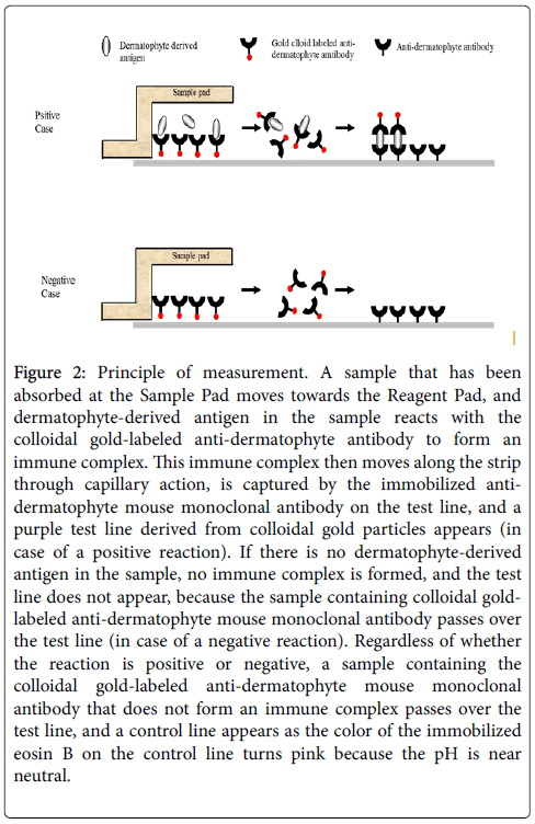
Figure 2: Principle of measurement. A sample that has been absorbed at the Sample Pad moves towards the Reagent Pad, and dermatophyte-derived antigen in the sample reacts with the colloidal gold-labeled anti-dermatophyte antibody to form an immune complex. This immune complex then moves along the strip through capillary action, is captured by the immobilized antidermatophyte mouse monoclonal antibody on the test line, and a purple test line derived from colloidal gold particles appears (in case of a positive reaction). If there is no dermatophyte-derived antigen in the sample, no immune complex is formed, and the test line does not appear, because the sample containing colloidal goldlabeled anti-dermatophyte mouse monoclonal antibody passes over the test line (in case of a negative reaction). Regardless of whether the reaction is positive or negative, a sample containing the colloidal gold-labeled anti-dermatophyte mouse monoclonal antibody that does not form an immune complex passes over the test line, and a control line appears as the color of the immobilized eosin B on the control line turns pink because the pH is near neutral.
The concentration at which the test line cannot be visually confirmed is the detection limit.
Investigation of reactivity and detection range
Table 1 shows the 7 species of dermatophytes used in the study. All of the dermatophytes were cultured for 2 weeks at 28°C on a Sabouraud’s glucose agar (NIHON PHERMACEUTICAL Co. Ltd.) plate. After harvesting the cultured dermatophytes, they were sterilized by an autoclave (for 15 minutes at 121°C). After vacuum drying for 18 hours at room temperature, they were milled in a mortar and fungus powder was prepared. The fungus powder was accurately weighed and suspended in extraction buffer (0.05M phosphate buffer containing detergent and proclin 950Sigma-Aldrich Co. LCC.) as a preservative at a concentration of 0.1 v/v%. The suspension was centrifuged for 5 minutes at 10,000 × g, and the supernatant was obtained. This supernatant was used as the antigen stock solution (30 mg/ml). The antigen stock solution was diluted with the extraction buffer, and various concentrations of the antigen solutions ranging from 0.03 μg/ml to 300 μg/ml were prepared. Two hundred-fifty μl of the antigen solution at each concentration was added to the test tube, and the test strips were placed in each test tube, and after these were allowed to stand for 15 minutes, appearance of the test line was visually examined. The test was repeated 4 times.
| Concentration (μg/ml) | Trichophytons | ||||||
|---|---|---|---|---|---|---|---|
| T.m | T.t | T.v | T.r | M.c | M.g | E.f | |
| 300 | 4/4 | 4/4 | 4/4 | 4/4 | 4/4 | 4/4 | 4/4 |
| 30 | 4/4 | 4/4 | 4/4 | 4/4 | 4/4 | 4/4 | 4/4 |
| 3 | 4/4 | 4/4 | 4/4 | 4/4 | 4/4 | 4/4 | 4/4 |
| 0.5 | 4/4 | 4/4 | 0/4 | 4/4 | 4/4 | 4/4 | 4/4 |
| 0.3 | 4/4 | 4/4 | 0/4 | 0/4 | 0/4 | 4/4 | 4/4 |
| 0.03 | 0/4 | 0/4 | 0/4 | 0/4 | 0/4 | 0/4 | 0/4 |
Table 1: Detection limit of each Trichophyton. T.m: Trichophyton mentagrophytes (Biotechnology Resource Center/National Institute of Technology and Evaluation (NBRC) 32410), T.r: Trichophyton rubrum (NBRC 9185), T.v.: Trichophyton violaceum (NBRC 31064), T.t: Trichophyton tonsurance (American Type Culture Collection (ATCC) 32504), M.c: Microsporum canis (NBRC 32463), M.g: Microsporum gypseum (NBRC 8231), E.f: Epidermophyton floccosum (NBRC 32461).
Unless otherwise noted hereafter, all of the applied sample volume in the test tubes was 250 μl and the tests were repeated and measured 4 times, and results were measured after the test strip had been rested for 15 minutes.
The results were considered as follows. If a line in the test line position was not visible, it was negative, but if a line was visible regardless of the density, it was positive. If the control line did not appear, the test was invalid.
The results were indicated as number of positive measurements/ total number of measurement times. For example, in case of 4 measurements in total, if the results are 3 times positive and once negative, it is expressed as 3/4, and if all results are negative, it is expressed as 0/4. Unless otherwise specified, the subsequent test results are in accordance with this notation.
Preparation of control antigen
Trichophyton rubrum was used as a control antigen because this is the most frequently observed pathogen of tinea unguium, and a 100- fold concentration (50 μg/ml) of the detection limit, 0.5 μg/ml of dried fungi determined in the “Investigation of reactivity and detection range? was prepared and used as a strong positive control (SP). SP was then diluted 100-fold with the extraction buffer at the time of use and used as a weak positive control (WP). The extraction buffer alone was used as a negative control (NC), and used in the subsequent tests.
Simultaneous reproducibility, sensitivity and accuracy studies
Using 3 different lots of the test strips, they were repeatedly tested 4 times with WP, SP, and NC by changing the measurement dates. Each of WP, SP, or NC was added to the test tubes, and the test and control lines were visually examined.
Long-term storage study
The test strips were kept tightly closed with a plate-like desiccant (Ryoko Chemical Co. Ltd.) in an aluminized film bag. Temperature during storage was set at 2 to 30°C. To confirm the storage conditions and the validity period under these conditions, the stability at 30°C was investigated. Three lots of test strips in the aluminized film bag and 3 lots of extraction buffer in 5 ml polyethylene eye drop containers were stored for a certain period of time in a constant temperature incubator. The quality of the test strips was evaluated by the same method used for quality control of the test strip. The test was considered successful when a control line appeared, and the quality of the test strips was regarded as acceptable when 1) all 4 measurements of NC were negative, and 2) all 4 measurements of WP and SP were positive.
Each of WP, SP or NC was added to each test tube and the test and control lines were visually examined. These series of tests were repeated twice on the same day.
Studies of reaction time and reaction temperature
The effect of reaction time and reaction temperature after placing the test strips in the test tubes was examined.
Two hundred-fifty μl each of WP, SP, or NC was added to each test tube, and the temperature was equilibrated by allowing these tubes to stand for 30 minutes in an incubator at each of the set temperatures (0, 15, 25, 37, and 50°C). At the same time, unopened test strips to be used were also allowed to stand for 30 minutes in an incubator at each temperature. The test strips were allowed to stand in each test tube, and the test and control lines were examined at 3, 5, 10, 15, 30, and 60 minutes.
Resting time and resting temperature after removal of the test strip from the test tube
After the end of the test, the effects of resting time and resting temperature were examined after removal of the test strip from the test tube.
Each of WP, SP, or NC was added to each test tube, and the temperature was equilibrated by allowing these test tubes to stand for 30 minutes in the incubator at 25°C. At the same time, unopened test strips to be used were also allowed to stand for 30 minutes in an incubator at 25°C. After the a test strips were allowed to stand in each test tube for 15 minutes, the strip was transferred to a forced circulation-type incubator, where the temperature was set at 0°C, 15°C, 25°C, 37°C, or 50°C, and this timing was considered as a resting time of 0 minute. The test and control lines were examined at 15, 30, and 60 minutes after the resting time of 0 minute.
Effects of temperature and humidity after opening the test strips until use
We investigated whether the performance of the test strip changed after the packaging had been opened and allowed to stand for a certain period of time under various conditions of temperature and humidity.
The test strips were removed from the bag, and then allowed to stand for 20 minutes at 15°C to 50°C in a relative humidity of 45% to 98% in constant temperature and humidity chambers. Each of NC, WP, and SP was used and the test and control lines were visually examined.
Reactivity with various fungi and bacteria
A total of 115 strains of Trichophyton species and non- Trichophyton fungi and bacteria were evaluated for reactivity using the test strips. These strains were purchased from the National Institute of Technology and Evaluation (NBRC number) or the American Type Culture Collection (ATCC number), or provided by the Medical Mycology Research Center of Chiba University (Institute for Food Microbiology [IFM] number).
1. Culture of Trichophyton and non-Trichophyton fungi
The fungi were cultured in a potato dextrose agar medium for a 2 to 3 of weeks at 25°C, and scraped with a sterile platinum loop. These cultured fungi were sterilized for 15 minutes at 121°C in an autoclave, and after cooling, sealed and stored at −80°C until use.
2. Culture of bacterial strains
The bacteria were cultured in Lysogeny Broth (LB) agar medium (NIHON PHERMACEUTICAL Co. Ltd.) for 24 to 48 hours at 37°C, and the resulting colonies were scraped. The bacterial body of each strain was sterilized for 15 minutes at 121°C in an autoclave, and after cooling, sealed and stored at −80°C until use.
After vacuum drying for 18 hours at room temperature, the obtained bacterial body sample was milled in a mortar and a powder was prepared for each strain. Approximately 1 mg of the powder was weighed, and the extraction buffer was added to make a solution at 3 mg of bacterial body/ml. The solution was homogenized for 1 minute with an electric homogenizer, and allowed to stand for 5 minutes. After centrifugation, the supernatant was diluted 10-fold with the extraction buffer to obtain an extracted solution with 300 μg/ml. A volume of 250 μl of the extracted solution was taken and added to the test tube in duplicate, the test strip was allowed to stand, and the test and control lines were visually examined after 15 minutes of standing. All tests were carried out twice.
Documentation of the impact of coexisting substances
It was examined whether oral anti-fungal agents, which are often used for the treatment of tinea unguium, have an influence on the test results of the test strip.
Terbinafine (Wako Pure Chemicals Inc.), griseofulvin (Tokyo Chemical Industry Co., Ltd.), and itraconazole (LKT Laboratories Inc.) were dissolved in dimethylsulfoxide (DMSO) (Wako Pure Chemicals Inc.), and added to NC, WP and SP at the concentration of 100-fold of the respective minimum inhibitory concentration (MIC). The final concentration of DMSO was 0.5 v/v%. Reference controls without any antifungal agent were prepared by adding DMSO to NC, WP and SP at 0.5 v/v%.
Each of each prepared sample was tested, and the test and control lines were visually examined.
Study of the effect of the extraction buffer volume
The volume of the extraction buffer ranged from 50 μl to 950 μl, and the range that provides consistent results was examined.
The nail samples, which had already been identified as positive or negative using the test strips [4], were randomly selected, cut into square pieces of approximately 0.1 to 0.5 mm on a side with dissecting scissors, mixed, and both a positive and negative standard nail sample were prepared.
The positive standard nail sample was weighed, and 44 test tubes with 0.5 mg positive standard nail sample in each test tube were prepared. Forty-four test tubes containing the negative standard nail sample were prepared in the same manner as for the positive standard nail sample.
The extraction buffer was added to each test tube (50, 100, 150, 250, 350, 450, 550, 650, 750, 850, 950 μl respectively, and after stirring 20 times with a plastic rod, the test tubes were allowed to stand for 1 minute, the test strips were placed in the test tubes, and the test and control lines were visually examined.
Number of agitations with a plastic rod during the nail sample extraction
The condition of agitation with a plastic rod at the time of extraction was investigated.
One-half mg of positive and negative standard nail samples each were weighed and added to test tubes. Two hundred-fifty μl of extraction buffer was added to each test tube. After agitating (including grinding) 10 or 20 times within 5 seconds with a plastic rod, 40 times within 10 seconds, or 50 times within 15 seconds, each test tube was allowed to stand for 1 minute, the test strip was placed in the each test tube, and the test and control lines were visually examined.
Estimation of the necessary amount of nail sample
In clinical practice, it is practically difficult to weigh nails in the order of a few mg on a precision balance. Therefore, we determined the minimum amount of nail that is necessary for the test to express the amount in a visual size of the nail sample to be collected for the test.
Thirty-five nail samples that were already known to be positive by the test strips [4], were selected and used for this study.
Each sample was cut into square pieces of approximately 0.1 to 0.5 mm on a side with dissecting scissors, mixed, and 5 mg to 10 mg of each sample was weighed. These samples were placed in test tubes, mixed with 0.5 ml of the extraction buffer, and agitated in such a way to be crashed 20 times with a plastic rod and allowed to stand for 1 minute.
The supernatant was used as the stock solution (1-fold dilution), and serially diluted 2-fold with the extraction buffer. The amount of the sample solution for each concentration was 200 μl. The test strip was placed in each 200 μl of the sample solution that was prepared by 1- to 512-fold dilution of the stock solution, and absorbance of the test line after 15 minutes of standing was measured with a test strip reader (Nippun Techno cluster Co., Ltd.). Based on the obtained absorbance, the linear regression equation was determined by the logarithmic value of the dilution ratio and the absorbance as variables. Assuming that the lower limit of absorbance for detection of the line by the naked eye is the 10, the maximum sample dilution rate at which detection of line by naked eye is possible was calculated by plugging the absorbance value in the equation, and the minimum sample weight necessary to detect positive results was then determined.
Ethics
This study was performed in compliance with Good Clinical Practice based on the Declaration of Helsinki and other applicable regulations. The institutional review board of each study site reviewed and approved the study protocol. Prior to the start of screening procedures, signed informed consent was obtained from each subject.
Measurement range
The control line appeared in all tests, indicating that all tests were successful. The results are shown in Table 1. The results shaded in gray in the table are the lowest concentration at which the test line was detected as a very faint line, showing that the detection limit concentrations of dried samples of T. mentagrophytes, T. tonsurance, Microsporum gypseum and Epidermophyton floccosum were 0.3 μg/ml for respective fungi, those of T. rubrum and Microsporum canis, respectively, were 0.5 μg/ml, and that of T. violaceum was 3 μg/ml.
Simultaneous reproducibility, sensitivity and accuracy studies
For all 3 lots (lot 1, 2 and 3) of the test strips, WP and SP were determined to be positive in all 4 measurements, while for all 3 lots of the test strips NC was determined to be negative in all 4 measurements. In all tests conducted here, there were no indeterminable or unmeasurable cases, including cases that the control lines did not appear.
Long-term storage study
All 3 lots (lot A, B and C) of the combination of test strips and extraction buffer met the standards set by the method of quality control after they were kept in the storage at 30°C for up to 22 months.
Reaction time and reaction temperature
All control lines appeared, indicating that all tests were successful. At 0°C, the test line in NC appeared as a pale color at 3 minutes, indicating a positive reaction, but disappeared again after 5 minutes, while the results of WP and SP remained positive after 3 minutes or later. At other temperatures, the results of WP and SP were positive after 3 minutes, and that of NC was negative, indicating that the results met the standard. The results after 5 to 60 minutes at all temperatures met the standard.
Effects of the resting time and resting temperature after removal of the test strip from the test tube on the outcome
All control lines appeared, indicating that all tests were successful. The results obtained from the reaction for 15 minutes at 25°C were not affected for 30 minutes of resting time at a resting temperature of 0 to 37°C after removal of the test strips from the test tubes. However, the test line in WP disappeared after resting at 37° C for 60 minutes or at 50°C for 15 minutes or more, and the test line in SP was fading, but the result was still positive, when resting for 15 minutes or more at 50°C.
Effect of temperature and humidity after the test strip opening until use
All control lines appeared, indicating that all tests were successful. The performance was not affected when the test strip was even left open for 20 minutes at a relative humidity condition of 45 to 98% at 15 to 37°C. However, at 50°C and a relative humidity of 65% or more, a weak line was observed for NC at the position of the test line, and the line for SP became a little thinner when the relative humidity was 98%.
Reactivity with various fungi and bacteria
The results are shown in Table 2. All control lines appeared, indicating that all tests were successful. Since the 2 runs of the test demonstrated identical results (0/2 or 2/2), “+” in the table indicates a positive result and “” a negative result for each fungus or bacterium. The results of Trichophyton mentagrophytes, T. rubrum, T. tonsurans, T. violaceum, T. verrucosum, Microsporum gypseum, M. canis, and Epidermophyton floccosum, which are all classified as dermatophytes, were all positive. On the other hand, the test strip did not react to Malassezia or Candida species. None of the tested bacterial strains were reacted to the test strip. Some of Aspergillus, Penicillium, Fusarium, Exophiala, Hortaea, Malbranchea species, Paecilomyces and Veronaea species showed a positive reaction.
| Strain No. | Strains | Reactivity |
| NBRC 32410 | Trichophytonmentagrophytes | + |
| NBRC 9185 | Trichophytonrubrum | + |
| ATCC 32504 | Trichophytontonsurans | + |
| NBRC 31064 | Trichophytonviolaceum | + |
| IFM 46798 | Trichophytonverrucosum | + |
| NBRC 8231 | Microsporumgypseum | + |
| NBRC 32463 | Microsporumcanis | + |
| NBRC 32461 | Epidermophytonfloccosum | + |
| IFM | Strains | Reactivity |
| 54306 | Aspergillusflavus | + |
| 54307 | Aspergillus fumigates | + |
| 54308 | Aspergillusnidulans | - |
| 54309 | Aspergillusniger | + |
| 54310 | Aspergillusterreus | + |
| 54311 | Neosartoryafischeri | + |
| 54312 | Paecilomyceslilacinus | + |
| 53352 | Paecilomyceslilacinus | + |
| 54313 | Penicilliumcitrinum | - |
| 54314 | Penicilliumgriseofulvum | + |
| 54315 | Scopulariopsisbrevicaulis | - |
| 53969 | Alternariaalternata | - |
| 53351 | Veronaeabotryosa | + |
| 47302 | Pseudallescheriaboydii | - |
| 49731 | Scedosporiumapiospermum | - |
| 54324 | Fusariumsolani | + |
| 5695 | Protothecawickerhamii | - |
| 46097 | Schizophyllum commune(1?) | - |
| 45818 | Schizophyllum commune(2?) | - |
| 40776 | Absidiacorymbifera | - |
| 41413 | Basidiobolusranarum | - |
| 46110 | Cunninghamellabertholletiae | - |
| 40782 | Mortierellaisabellina | - |
| 40507 | Mucorcircinelloides | - |
| 40781 | Mucorracemosus | - |
| 46300 | Rhizomucorpusillus | - |
| IFM | Strains | Reactivity |
| 46417 | Rhizopusmicrosporus var. rhizopodiformis | - |
| 40786 | Rhizopusoryzae | - |
| 41594 | Rhizopusstolonifera var. reflexus | - |
| 40788 | Syncephalastrumracemosum | - |
| 40789 | Zygorhynchusexponens | - |
| 49030 | Candida albicans | - |
| 53163 | Candida dubliniensis | - |
| 49331 | Candida tropicalis | - |
| 48375 | Candida parapsilosis | - |
| 5492 | Candida guilliermondii | - |
| 5520 | Candida glabrata | - |
| 47973 | Candida krusei | - |
| 5806 | Geotrichumcandidium | - |
| 48429 | Trichosporonasahii | - |
| 5807 | Cryptcoccusneoformans serotype A | - |
| 5815 | CryptcoccusneoformansserotypeB | - |
| 5816 | Cryptcoccusneoformans serotype C | - |
| 5844 | Cryptcoccusneoformans serotype D | - |
| 46138 | Cryptcoccusneoformans serotype AD | - |
| 41598 | Sporothrixschenckii | - |
| 54322 | Fonsecaeapedrosoi | - |
| 4826 | Exophialadermatitidis (M-Y form) | + |
| 40760 | Exophialadermatitidis (G form) | + |
| 54222 | Exophialajeanselmei | - |
| 45990 | Exophialaspinifera | + |
| 5089 | Phialophoraverrucosa | - |
| 54325 | Phialophorarichardsiae | - |
| 4931 | Rhinocladiellaatrovirens | - |
| 4807 | Cladophialophorabantiana | - |
| 41539 | Hortaeawerneckii | + |
| IFM | Strains | Reactivity |
| 41545 | Malbrancheaalbolutea | - |
| 41301 | Malbrancheaaurantiaca | - |
| 41294 | Malbrancheachrysosporioideahrysosporioidea | - |
| 41309 | Malbrancheacinnamomea | - |
| 41295 | Malbrancheacircinata | + |
| 41296 | Malbrancheadendritica | - |
| 41300 | Malbrancheafilamentosa | - |
| 41299 | Malbrancheaflava | - |
| 41293 | Malbrancheaflavorosea | + |
| 41298 | Malbrancheaflocciformis | - |
| 47364 | Malbrancheaflocciformis | - |
| 41307 | Malbrancheafulva | - |
| 41303 | Malbrancheagraminicola | - |
| 41302 | Malbrancheagypsea | - |
| 47365 | Malbrancheagypsea | - |
| 41297 | Malbrancheamulticolor | - |
| 41308 | Malbrancheapulchella | - |
| 47361 | Malbranchea sp. | - |
| 47362 | Malbranchea sp. | - |
| 47363 | Malbranchea sp. | - |
| 1098* | Malassezia furfur | - |
| 47416 | Gymnoascoideuspetalosporus | - |
| 47417 | Gymnoascoideuspetalosporus | - |
| 47423 | Auxarthronreticulatum | - |
| 47418 | Gymnoascusintermedius | - |
| 47408 | Gymnoascuspetalosporus | - |
| 47419 | Gymnoascusreessii | - |
| 47403 | Gymnoascusudagawae | - |
| 41471 | Emmonsiaparvavar. crescens (anamorph) | - |
| 46988 | Emmonsiaparvavar. parva (anamorph) | - |
| 47473 | Phanerochaetechrysosporium | - |
| 47494 | Phanerochaetechrysosporium | - |
| 47370 | Apinisiaqueenslandica | - |
| 47339 | Uncinocarpusreesii | - |
| 50768 | Apinisiaqueenslandica | - |
| 51121 | Apinisiaqueenslandica | - |
| 47357 | Arthrodermamultifidum | - |
| 47358 | Arthrodermamultifidum | - |
| 47366 | Arthrodermamultifidum | - |
| 47367 | Arthrodermamultifidum | - |
| 47509 | Arthrodermamultifidum | - |
| 47510 | Arthrodermamultifidum | - |
| 51122 | Arthrodermamultifidum | - |
| 51123 | Chrysosporiumcarmichaelii | - |
| 51124 | Chrysosporiumindicum | - |
| 41432 | Chrysosporiumkeratinophilum | - |
| 47382 | Chrysosporiumpseudomerdarium | - |
| NBRC | Strains | Reactivity |
| 13500 | Escherichia coli | - |
| 13719 | Bacillus subtilis | - |
| 13276 | Staphylococcus aureus | - |
| 3971 | Streptococcus faecalis | - |
Table 2: Reactivity to fungi of Trichophyton and non-Trichophyton class and bacteria. * NBRC number.
Documentation on the impact of coexisting substances
All control lines appeared, indicating that all tests were successful. Under the test conditions, the antifungal agents, terbinafine (0.5 μg/ ml), griseofulvin (37.5 μg/ml) and itraconazole (100 μg/ml) did not affect the results of the test line.
Study of the extraction conditions of the nails
When the volume of extraction buffer was 50 μl, lines were not observed, indicating that the test was not successful. This was because the sample liquid did not migrate to the test line or the control line.
Under all other conditions used, the control lines appeared, indicating that these tests were successful.
The test line was darker when the volume of extraction buffer was 100 μl because the antigen concentration was higher. However, it can be assumed that if the nail sample quantity is large, 50 μl or more of the extraction buffer is absorbed in the nail sample, the amount of the sample liquid absorbed into the test strip is not sufficient. When the volume of extraction buffer ranged from 150 to 850 μl, similar results were obtained, although the test line tended to be thinner with 550 μl or more, and the result was determined as negative in 950 μl in one case.
Thus, these results indicated that a range of 150 μl to 850 μl extraction buffer for 0.5 mg of positive and negative standard nail samples was allowed in the test.
Number of agitation by a plastic rod during the nail sample extraction
All control lines appeared, indicating that all tests were successful. All negative standard nail samples gave negative results under all test conditions. If positive standard nail samples were examined without agitation and resting, the results were only weakly positive compared to the other conditions. The test line color tended to be stronger with agitation, but there was a weak color development of the test line on the test strip in 1 case with 10 times of agitation.
Estimation of the minimum required amount of nail specimen
All control lines appeared, indicating that all tests were successful. We obtained the linear regression equation from the sample dilution times (X) and the absorbance (Y) of the diluted solution by successively diluting the collected amount of the specimen, and determined the maximum dilution factor “x” by substituting the absorbance 10 to “y” of the equation. The minimum amount of nail that can be detected with the test strip was then determined by multiplying the nail weight by the inverse of the maximum dilution factor. An example of a regression equation with a graph is shown in Figure 3. The minimum required amount of a nail specimen varied greatly by subject, and was distributed from 0.002 mg to 0.722 mg. The largest value among the minimum required amounts of nail specimens in the current test was 0.722 mg.
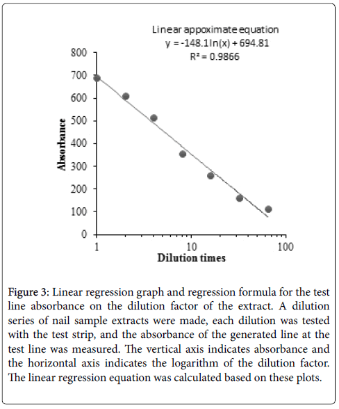
Figure 3: Linear regression graph and regression formula for the test line absorbance on the dilution factor of the extract. A dilution series of nail sample extracts were made, each dilution was tested with the test strip, and the absorbance of the generated line at the test line was measured. The vertical axis indicates absorbance and the horizontal axis indicates the logarithm of the dilution factor. The linear regression equation was calculated based on these plots.
Using a number of clinical specimens, Tsunemi et al. showed that this test strip indicated good detection capacity and it provides a reliable, convenient, and quick method to test tinea unguium [4,5]. On the other hand, they also showed that the test strip was not suitable for tinea pedis, because less antigen in the scale caused false negative results and contamination with non-trichophyton caused false positive results [8]. Since it had become clear that such clinical application for the detection of Trichophyton in the nail specimen is suitable, it was an urgent priority to clarify the usage of the test strip in tinea unguium to improve its diagnostic accuracy, and to provide accurate and easy-touse in vitro diagnostic products.
According to our results in “Measurement range” and “Reactivity with various fungi and bacteria,” the test strip reacts to some non- Trichophyton fungi. Although these fungi are not residing in the nails and skin of healthy people, due to their presence in the environment, the antigen component adhering to the nail surface may cause a positive reaction on the test strip. Therefore, it is suggested that the accuracy of diagnosis will increase by comprehensive judgment combining findings from other diagnostic methods, such as microscopic examination, in addition to the avoidance of influence of substances attached to nail samples by cleaning the surface of the nail with alcohol cotton and scraping away the nail surface to the site of fungal growth in accordance with the guidelines [9]. Since the test strip does not cover an exhaustive list of filamentous fungi besides Trichophyton, diagnosis of non-dermatophyte nail mycosis cannot be made. It appears to be limited as a diagnostic agent for tinea unguium.
Based on the results of the extraction conditions of the nail and the required minimum amount, 1 mg of a nail specimen removed from the affected area is necessary and sufficient to determine if this test strip provides a positive result. Our results suggest that a range of 150 μl to 850 μl extraction buffer was allowed for 0.5 mg of positive and negative standard nail samples in the test. It is preferable to use as small amount of the extraction buffer as possible from the point of view of sensitivity, but if the volume of the extraction buffer is within a narrow range, it is difficult for users to control the volume by dripping to the test tubes. Since the tick marks on test tubes are in positions of 250 μl and 500 μl, it was decided to use a range of 250 to 500 μl extraction buffer.
When a nail sample was stirred with a plastic rod included in the kit, it seemed to be well extracted by agitation of 10 times, but in anticipation of the consistency of the results, it was decided to agitate it more than 20 times. Taken together with the results of “Reaction time and reaction temperature? and “Effects of resting time and resting temperature after removal of the test strip from the test tube on the outcome,? it was reasonable to consider that the result of the test strip should be determined between 5 to 30 minutes after the test strip has been placed in the test tube within a range of 0 to 37°C.
Since the NC showed a false positive result, the test strip should be used as soon as possible after opening (within 5 minutes) for testing. Long-term storage stability was secured for 22 months at 30°C, if the aluminum bag is sealed.
The concentration inside the nail of itraconazole and its metabolite, itraconazole hydroxide, which has comparable antibacterial activity in the body, was up to 450 ng/g [10]. If 100 mg of nail is collected and 250 μl of the extraction buffer is added, the itraconazole concentration becomes 180 ng/ml.
Since this concentration is much lower than the itraconazole concentration of 100 μg/ml used in the current study, it is considered that itraconazole in clinical nail samples will not affect the test results of the test strip. Similarly, since it has been reported that the concentration of terbinafine penetrated to the nail is 0.78 ng/mg [11] corresponding to 0.312 μg/ml per 100 mg of collected nail, which is lower than the concentration of 0.5 μg/ml of terbinafine used in the current study. It was concluded that the test strip results are not affected by co-existing antifungal agents in the nails during the treatment with commonly used antifungal drugs.
We established the usage of a detection device for dermatophyte with clarification of its bases, and secured reproducibility and 22 month stability of the device. The detection method for dermatophyte using this device is regarded as valid and reasonably applicable to clinical practice, and a method useful as a supplemental method to diagnose tinea unguium.