Biochemistry & Pharmacology: Open Access
Open Access
ISSN: 2167-0501
ISSN: 2167-0501
Research Article - (2015) Volume 4, Issue 6
Traditional self-assembling peptide can form nanofiber scaffolds to meet the challenges of advance biomaterial, cell culture, tissue engineering and regeneration. L-amino acids have been widely used instead of D-amino acids to design nanomaterial since some D-amino acids have toxicity of cells. Here we report that using D-amino acids to design a new D-form self-assembling peptide DSAP-2 and the circular dichroism, atomic force microscopy and scanning electron microscopy show that the peptide can form nanofiber scaffold as well. Furthermore, cell inhibition assay confirmed this D-form peptide show no toxicity of cells that can support cell growth. Fluorescence microscopy results show that cells had less cell apoptosis in the 3D environment and displayed a fast proliferation after cultured for 7days. Peptide’s hydrogel not only formed nano-scaffolds surrounded by cells in a 3-D cell culture, but achieved rapid hemostasis in a rabbit liver wound model. Our study suggests this peptide could be used in the wound and beyond in the future. This work could also inspire us to design more novel D-form self-assembling peptide in biomaterials and biomedical areas.
<Keywords: Self-assembling peptide; D-Amino acid; Nanofiber; 3D Cell culture; Rapid hemostasis
At present, self-assembling peptides have been extensively studied in biomaterials and biomedical areas. More and more recent designs have shown their surprising results in structures, properties, functions and even in applications [1]. They can form well-ordered nanostructures such as nanofiber, nanotube and nanovesicle. They have multiple advantages, including stability, safety, efficiency, nonbiological, biocompatibility, biodegradability and non-immunogenicity [2]. And they are widely applied to 3D cell culture [3-5], rapid hemostasis [6,7], wound-healing [8,9], drug control release [9-11], membrane proteins stabilization [12-16] and solar cell devices [17- 21]. Compared with the topical cell culture media such as engineered substrates, animal-derived extracellular matrix or synthetic mimics, self-assembling peptides are competitive for helping the understanding and manipulation of 3-dimensional (3D) microenvironment cellular processes to proliferation, differentiation, migration, cell-cell contact interactions and apoptosis in tissue engineering and regenerative medicine [1]. Partially to 3D print bioink for soft-tissue, it is necessary to develop a kind of biomaterials which prevents body enzymes from quick degradation, where every single ingredient is known to achieve the fine-tuning and control, or suit for basic individual needs of studies and applications [22]. Peptides and proteins made of D-amino acids are more stable in the body environment since proteases can readily degrade L-form peptide bonds, but difficult to degrade D-form peptide bonds [23-26]. Proteins of animals are all synthesized by L-amino acids and some D-amino acid like D-Ala and D-Asp have toxicity of cells [23,26-29]. Therefore, most of them were designed by L-amino acid and a few by D-amino acids [1,2]. Up till now, there have been a few reports that D-form self-assembling peptide did not obtain toxicity to cell stains and was also resistant to protease digestion, as a 3D biological matrix for cell culture [6,30-32]. Amino acids and theirs isomers are a good option of nano-biomaterials for biotechnology and biomedical areas [33-36]. Here we utilized two D-amino acids, D-Ala and D-Asp, which were reported to have toxicity of cells, and designed a new chiral self-assembly peptide DSAP-2. We got D-form self-assembling peptide hydrogel, investigated its physicochemical properties by circular dichroism spectroscopy, and examined microstructures by AFM and SEM. We also assessed the situations of cell behaviors in 3D cell culture, and some of its simple applications. Our study could prompt more designers working in basic medical and clinical studies to find more similar peptides.
Peptides synthesis and purification
The sequence of peptide DSAP-2 is Ac-(DArg-DAla-DAsp-DAla)4- CONH2, and the peptide was commercially custom-synthesized by solid-phase peptide synthesis (Chengdu CP Biochem Co., Ltd., Chengdu, China). The peptide was purified to 95.27% by HPLC and characterized by mass spectroscopy. The lyophilized white powder was stored at 4°C. Solutions of the peptides were prepared at mass concentration of 1.0% in water (18.2 KΩ/cm2, Millipore Milli-Q system) and stored at 4°C before use.
Molecular modeling
Design of molecular models of these peptides was based on the principle of minimum energy and models were constructed using free modeling software from China (Hyperchem professional version 7.5, http://www.hyper.com). The software package was run on a PC machine.
DSAP-2 hydrogel
1.0 ml of the stock solution of DSAP-2 peptide (10 mg/ml) was added to 1.0 ml of phosphate-buffered saline (0.058 M NaH2PO4, 0.017 M NaH2PO4, 0.069 M NaCl, 0.002 M Mg2+, pH=7.2) which induced DSAP-2 self-assembling to form hydrogel, then the secondary structure of DSAP-2 was test by CD spectroscopy and the stable hydrogel was stained with Congo red at a glass slide after cultured for 24 h in room temperature. In vitro physiological environment: d-DARA16 solution (1%) was mixed with culture medium (DMEM or MEM or RPMI- 1640) (Gibco) which include 8-10% fetal calf serums, added the same volume (100 μl) solution into 96 well-plates, cultured it for 24 h in the incubator (37°C, 5% CO2), and dyed by Congo red in a glass slide.
Atomic force microscopy
500-1000 mmol/l peptide solution was prepared and cultured in different pH regulated by HCl, PBS and NaOH over 12 h. Then 5-10 μl peptide solution was applied onto a freshly cleaved mica surface. Each aliquot was left on mica for 30-60 s, washed with 1000 ml deionized water at least three times, dried in the air and imaged immediately. The images were obtained to scan the mica surface in the air in the Tapping Mode of AFM (Hitachi SPM400, Japan). It was important to minimize the tip tapping force to image soft biopolymer with AFM at resolution. AFM images were taken at 512 × 512 pixels resolution and displayed topographic images of the samples, in which the brightness of features increases as a function of height.
Cell cultures preparation
Human hepatoma cells SMMC-7721 and normal human hepatocyte cell L-02 (Transplantation engineering and transplantation immunity laboratory, Sichuan University) were dissolved rapidly in 37°C water bath, suspended in RPMI-1640 (Gibco Corp), added into 25 cm2 culture vessels and cultured in the incubator (37°C, 5% CO2). RPMI-1640 (Gibco Corp) medium contained 1% double-antibody solution (mycillin) and 8-10% (volume concentration) fetal bovine serum (Gibco Corp).
Cell inhibition assay
Two liver cell strains, normal cell line L-02 cells and cancer cell line SMMC-7721 cells, were seeded into 96 well plates with 10% FBS RPMI-1640 medium at a certain concentration of 1.2 × 10⁴ cells/well (L-02 cells) and 1.5 × 10⁴ cells/well (SMMC-7721 cells) respectively and incubated for overnight in 5% CO2 at 37°C. Here we set four control groups: blank control (C); before Medication control (T0); experimental groups (Te(1-5)) and positive control groups(Tp(1-5)). Cell growth rate=(T-T0/(C-T0) × 100, the relative Growth Rate (GR) was calculated by OD value as follows: GR=(T-T0)/(C-T0) × 100 if T ≥ T0, GR=(T-T0)/T0 × 100 if T
Morphology of cells in 3D cell culture
Here L-02 cells were cultured as a model for 3D cell culture. When cells spread over the whole bottom of culture bottle, removed medium and trypsinized by 1 ml 0.25% trypsin to dissociate adherent cells. 1-2 ml RPMI-1640 is added into culture bottle to terminate trypsin action. Centrifuged cells for 8 min in 1000 turn/min, discarded supernatant, resuspended culture cell by RPMI-1640 and subcultured in culture bottles, mixed the cells with peptide solution for 3D culture. More detail processes can be seen in the reference [32]. The procedures of L-02 cell 3D culture in copper wire were mentioned above, and the ingrowth of cells in the pore of copper wire mesh can be clearly observed by phase contrast microscope.
Scanning electron microscopy
L-02 cells overlap on copper wire mesh and was detected by scanning electron microscope after being cultivated for 2-3 days. At the temperature of 4°C, used 5% (volume ratio) glutaraldehyde and fixed the hydrogel and the cells for 30-60 min. The cells were dehydrated with 20%, 50%, 70%, 90% and 100% ethanol gradient and dried for 2-4 hours in critical CO2 liquid services. Metal spraying the sample after vacuum drying, then observed it by scanning electron microscope(JSM-5900, JEOL, Japan).
Fluorescence microscopy
L-20 cells were 3D cultured in 96 well plates, the positive control was added cis-platin (2.5 mg/kg) and the negative control is 3D culture system. Cells were washed twice by PBS, mixed with 500 μl binding buffer which included 2 μl Annexin V-FITC and 5 μl Propidium Iodide and shielded from light, reacted for 5 min at room temperature. When Annexin V was used in combination with PI, PI were excluded from living cells (Annexin V- / PI-) and early apoptotic cells (Annexin V+ / PI-), while late apoptotic cells and necrotic cells are simultaneously FITC and PI staining combined showed double positive (Annexin V+ / PI+). Cell apoptosis observed by using inverted Fluorescence Microscopy with double color filter (FITC and rhodamine).
The hemostasis of rabbit liver mode
In the rabbit liver transverse hemostasis experiments, 10 white New Zealand rabbits (1-3 kg, random distribution of male and female) (animal experimental center, West China hospital, Sichuan university) were chosen. The animals were anesthetized with an intraperitoneal injection of sodium pentobarbital (50 mg/kg), and then chose 3 bigger livers in each rabbit and made a sagittal liver cut about 1.5 cm long, 0.1- 0.2 cm wide and 0.2-0.4 cm deep. Applied 200 μl of 10 mg/ml DSAP-2 peptide solution into the wound and recorded the hemostasis time. In addition, we chose two rabbits as a negative control that treated with saline, and found the hemostasis time were 90-120 s. Experiment was repeated at least 10 times on different rabbits and the average time for hemostasis was recorded. Here we focus on the chiral peptide as a catalyst to hemostasis and do not estimate the clinical relevance of our findings, so some positive controls (current treatment protocol, other hydrogels) are not taken into consideration here. All our studies followed the rule of animal ethics and were approved by the Animal Ethics Committee of the Sichuan University or Chongqing Medical University.
The structure of DSAP-2
We present the molecular model of DSAP-2 (Figure 1A), and we had an interest in what structure this peptide would adopt. CD spectroscopy revealed it indeed has an inverted spectrum with an ellipticity at 222 nm and a minimum ellipticity at 208 nm (Figure 1B). The CD profile suggests the secondary structure of this D-form peptide is dominated, none-typical β-sheet, but the α-helix in aqueous solution.
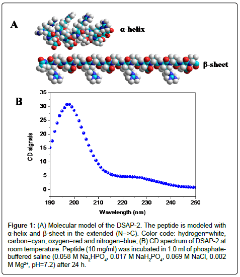
Figure 1: (A) Molecular model of the DSAP-2. The peptide is modeled with α-helix and β-sheet in the extended (N–>C). Color code: hydrogen=white, carbon=cyan, oxygen=red and nitrogen=blue; (B) CD spectrum of DSAP-2 at room temperature. Peptide (10 mg/ml) was incubated in 1.0 ml of phosphatebuffered saline (0.058 M Na2HPO4, 0.017 M NaH2PO4, 0.069 M NaCl, 0.002 M Mg2+, pH=7.2) after 24 h.
The nanostructure of DSAP-2 cultured in various pH value solution
The nanostructures of chiral peptide were obtained by AFM. Cultured in acidic solution (pH=3), DSAP-2 can form some nanofibers and nanoparticles (Figure 2A). In the neutral environment (pH=7), it can self-assembly to a large number of well-ordered nanofibers, with a diameter between 10-20 nm and a length of 1000 to 5000 nm and more (Figure 2B). In the alkaline solution (pH=10), only nanoparticles were obtained (Figure 2C). The results in the process of D-form peptide forming nanostructures were significantly affected by the pH environment. Furthermore, the self-assembly processes of this peptide were much sensitive to pH change. The results suggested that we can easily control the pH value and get the target nanostructure in our study.
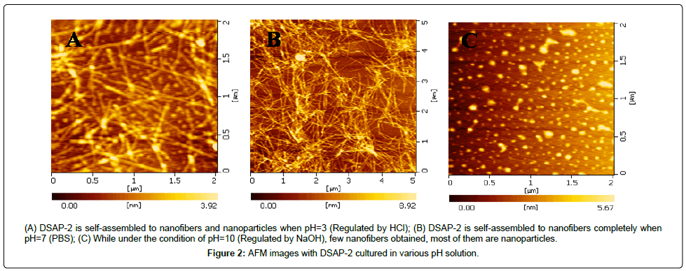
Figure 2: AFM images with DSAP-2 cultured in various pH solution.
Cell Inhibition and cell apoptosis in DSAP-2 solution
Some D-amino acids like D-Ala and D-Asp have toxicity of cells, but it is not clear whether the D-form peptide is toxic for cell strains or not [32,38]. We added peptide solutions with different concentrations into cell culture medium and tested OD value in each group compared with positive control. The data showed that, in different concentration peptide solutions, there was no significant difference in relative growth rate between normal cell line L-02 and cancer cell line SMMC-7721, and all of the growth rates were higher than 90% (Figure 3B and 3D). In the positive control drug paclitaxel group, cell inhibition increased significantly in both cell lines when the concentration was greater than 0.1 mg/ml (Figure 3A and 3C). The results confirmed DSAP-2 had no inhibited cells growth, and it was not proved this D-form peptide had toxicity of cell strains. Furthermore, Annexin V-FITC and Propidium Iodide were used to evaluate the cell apoptosis of DSAP-2 by fluorescence microscope. We found that L02 in DSAP-2 grew well and few cell apoptosis (Figure 4A-4C), and some cell apoptosis in positive control (Figure 4D-4F). When cultured for 7 days, cell proliferation is very fast, forming cell mass (Figure 5A-5D). The results confirmed that this D-form peptide can support cell growth and do not induce cell apoptosis or death.
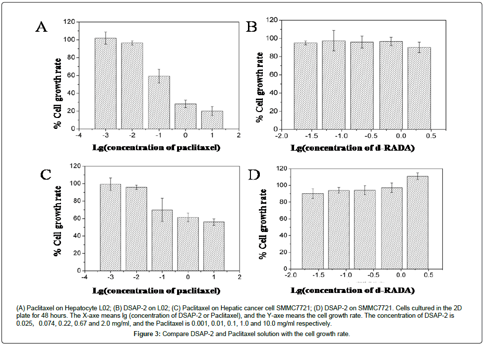
Figure 3: Compare DSAP-2 and Paclitaxel solution with the cell growth rate.
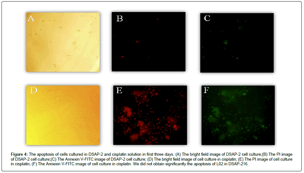
Figure 4: The apoptosis of cells cultured in DSAP-2 and cisplatin solution in first three days. (A) The bright field image of DSAP-2 cell culture;(B) The PI image of DSAP-2 cell culture;(C) The Annexin V-FITC image of DSAP-2 cell culture; (D) The bright field image of cell culture in cisplatin; (E) The PI image of cell culture in cisplatin; (F) The Annexin V-FITC image of cell culture in cisplatin. We did not obtain significantly the apoptosis of L02 in DSAP-216.
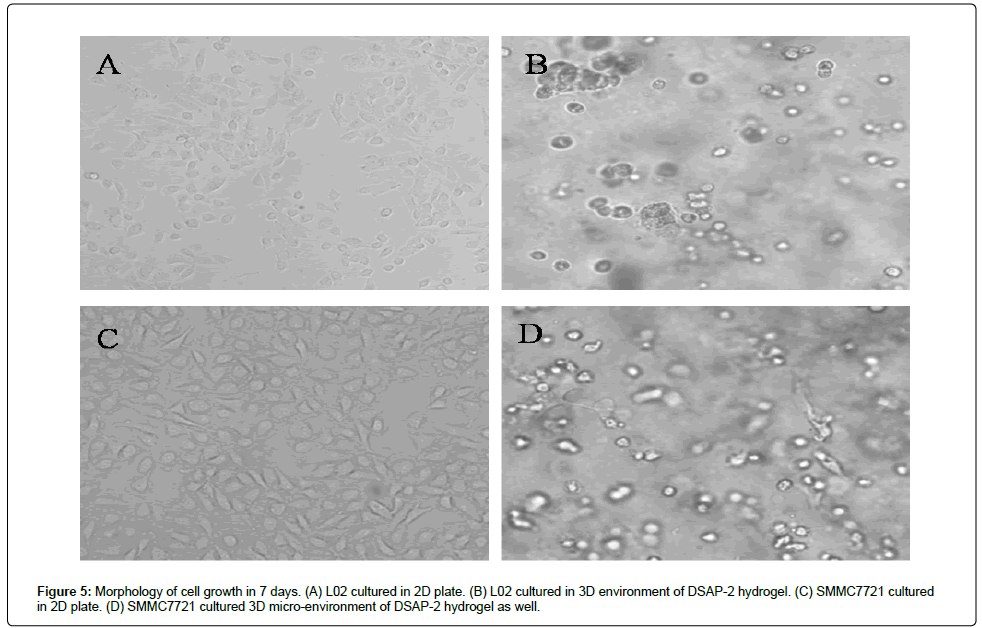
Figure 5: Morphology of cell growth in 7 days. (A) L02 cultured in 2D plate. (B) L02 cultured in 3D environment of DSAP-2 hydrogel. (C) SMMC7721 cultured in 2D plate. (D) SMMC7721 cultured 3D micro-environment of DSAP-2 hydrogel as well.
Hydrogel of DSAP-2
Stable hydrogel can be formed by DSAP-2, with >99% water content, not only in solution with ions but also in vitro physiological environment (RPMI-1640, Gibco) and the hydrogel was transparent in the naked eye. We can observe the morphology of the hydrogel by the light microscope after stained by Congo red (Figure 6A). The picture suggested that the hydrogel had a capability of moisture-keeping, providing the environment for cells growth. DSAP-2 peptides can self-assemble to ordered-nanofibers scaffolds, form the stable hydrogel scaffold in physiological environment, support cell growth without cell inhabitation or cell apoptosis, which suggests it, could be used as nanobiomaterial for three-dimensional (3D) cell culture.
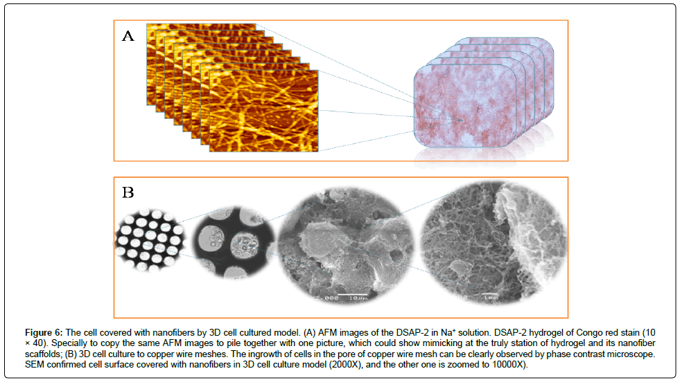
Figure 6: The cell covered with nanofibers by 3D cell cultured model. (A) AFM images of the DSAP-2 in Na+ solution. DSAP-2 hydrogel of Congo red stain (10 × 40). Specially to copy the same AFM images to pile together with one picture, which could show mimicking at the truly station of hydrogel and its nanofiber scaffolds; (B) 3D cell culture to copper wire meshes. The ingrowth of cells in the pore of copper wire mesh can be clearly observed by phase contrast microscope. SEM confirmed cell surface covered with nanofibers in 3D cell culture model (2000X), and the other one is zoomed to 10000X).
Three-dimensional cell culture microenvironment of DSAP-2
Having learned about the self-assembly property of DSAP-2, we obtained different morphology of cells (L-02 and SMMC-7721) in the two-dimensional (2D) and 3D cultured in DSAP-2 peptide solution (Figure 5A-5D). To check the 3D morphology of cells microenvironment, we cultured cells in copper wire meshes and confirmed by SEM. The nanofibers diameter was ~ 10-30 nm, and the pores between nanofibers were about 10-100nm. L-02 cells can adhere, grow and proliferate in the scaffolds interspace. The cell surface was covered with piled-nanofibers which provided 3D scaffolds support like extracellular matrix (Figure 6B). The interspaces of peptide scaffolds retained a little water and the small molecular bioactive substances can be interchanged through these interspaces. The truly nano-scaffolds microenvironment proved this D-form self-assembling peptide DSAP- 2 can be applied to 3D cell culture as the matrix material, and support cell growth, proliferation and beyond.
The rapid hemostasis function of DSAP-2
In our previous report, we found the chiral peptide d-EAK16 had an amazingly rapid hemostasis effect [6]. Here is the transverse wound model of rabbit liver. We wiped oozing bleeding 2-4 times with clean gauze on the liver surface of rabbit before adding DSAP-2 solution (Figure 7A). The weight of bleeding was 448mg on average (Figure 7C). After applying 200 μl 10 mg/ml peptide solution to the wound of rabbit liver (Figure 7B), we found that a layer of hydrogel was formed rapidly on the wound and bleeding was stopped. Hemostatic time was 18.3 ± 5.1 s (Figure 7D) compared with 106.1 ± 10.4 s (P<0.05) when treated with saline. The results confirmed that DSAP-2 has a satisfying hemostatic effect. DSAP-2 can not only support cell growth but also achieve rapid hemostasis.
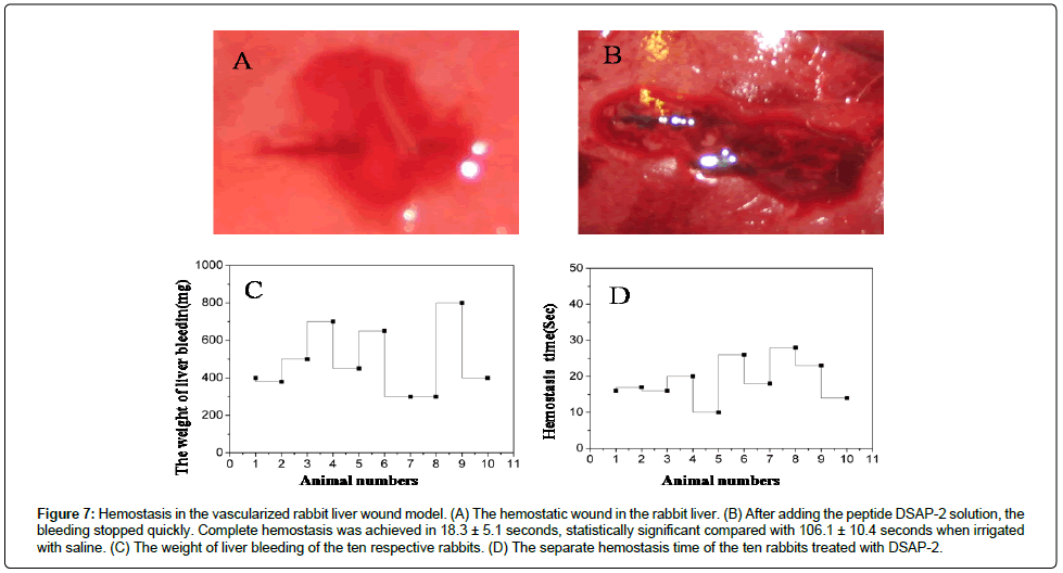
Figure 7: Hemostasis in the vascularized rabbit liver wound model. (A) The hemostatic wound in the rabbit liver. (B) After adding the peptide DSAP-2 solution, the bleeding stopped quickly. Complete hemostasis was achieved in 18.3 ± 5.1 seconds, statistically significant compared with 106.1 ± 10.4 seconds when irrigated with saline. (C) The weight of liver bleeding of the ten respective rabbits. (D) The separate hemostasis time of the ten rabbits treated with DSAP-2.
The advantage of DSAP-2 for 3D cell culture
The self-assembling processes also require some induced factors such as peptide concentration, pH, ions and temperature. They can easily meet these factors in the body physiological condition, and be widely used in the biomaterial area and medical areas. In general, cell cycle, cell differentiation and cell communication are determined by molecular gradient [39,40]. The interactions of protein-protein or protein-receptor, the cellular membrane structure, the extracellular matrix (ECM) and ligand play a vital role in cellular activities and metabolism in cell culture. 2D cell culture system can’t establish a dedicated 3D molecular gradient and thus alter these cells’ metabolism or gene expression [4,41]. DSAP-2 can self-assemble to well-ordered nanofibers then aggregate to 3D scaffolds under a certain condition like other self-assembly peptides. It has no cell growth inhibition and no cell apoptosis (Figure 5 and 6), and its nanofiber scaffolds are similar to extracellular matrix surrounded the cells (Figure 2B). These kinds of 3D tissue cell cultures systems could provide a more realistic biophysical microenvironment that can enhance cell proliferation, migration, differentiation and performing their biological function [42-44], and suggest this peptide has a potential extracellular matrix to be used in 3D tissue culture system or 3D print soft-ink.
Rapid hemostasis of DSAP-2 and its proposed plausible model
We had reported the nanofiber scaffold can stop bleeding quickly, which is associated with the nano-mechanically force [32]. Here DSAP-2 can stop bleeding quickly as well. A plausible hypothesis of rapid hemostasis of DSAP-2 is the ions induced peptide molecules self-assembling to nanofibers, and these nanofibers by forming tight nano-seals to prevent leakage of liquid and cells (Figure 8). The mechanism of hemostasis could be a complicated process like promoting/resisting platelet activation, coagulation, fibrinolysis, cell interaction and so on [45,46]. When wound bleeds without this peptide hydrogel solution, coagulation factors normally distribute in decentralized spaces and simultaneously cascade reaction center was not centralized, generating longer coagulation time. Covered the hydrogel to the wound bleeding, diversified factors could be captured in nanofibers web to fleetly form a relatively stable central bed of coagulation activation which may accelerate the hemostasis process [45,47]. More detailed evidence still needs to be found in the future.
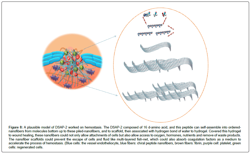
Figure 8: A plausible model of DSAP-2 worked on hemostasis. The DSAP-2 composed of 16 d-amino acid, and this peptide can self-assemble into orderednanofibers from molecules bottom up to these piled-nanofibers, and to scaffold, then associated with hydrogen bond of water to hydrogel. Covered this hydrogel to wound healing, these nanofibers could not only allow attachments of cells but also allow access to oxygen, hormones, nutrients and remove of waste products. The nanofiber scaffolds could prevent the escape of cells and fluid like multi-layered fish-net, which could also absorb coagulation factors as a medium to accelerate the process of hemostasis. (Blue cells: the vessel endotheliocyte, blue fibers: chiral peptide nanofibers, brown fibers: fibrin, purple cell: platelet, green cells: regenerated cells.
Here we designed a D form self-assembling peptide DSAP-2, obtained its nanostructure, which can be affected by pH value, and got its nanofiber hygrogel in vitro physiological environment. We also confirmed this D-form peptide can support cell growth, and nanoscaffolds surrounded by cells can use it as a 3D cell culture matrix material to support cell growth, proliferation and beyond. DSAP-2 can also achieve rapid hemostasis as well. Our study suggests this peptide could work in the wound healing in the future.
Z.L. and M.M. designed research; all members of the team contributed new reagents, analytic tools and finished the experiment; Z.L., M.M., X.F., W.R. and L.L. analyzed data; Z.L., M.M., X.F. and L.L. wrote the paper.
ZL was supported by Nature Science Foundation Project of CQ (CSTC) (cstc2015jcyjB0162), and the grant from the National Nature Science Foundation of China (NSFC 31540019), Antibiotic Research and Re-evaluation of Sichuan Provincial Key Laboratory Topics (ARRLKF14-01), Chongqing Medical University Basic Medical Science Support Foundation (JC201514), and Chongqing Medical University Research Cultivate Foundation (Natural Science) (201417).