Anatomy & Physiology: Current Research
Open Access
ISSN: 2161-0940
ISSN: 2161-0940
Research Article - (2018) Volume 8, Issue 3
This research work was aimed at studying the impact of chronic lead exposure on healing of experimentally induced Colitis in Wistar rats. The results willincrease our understanding of the roles played by heavy metals toxicity in colitis disease initiation, progression and remission. Cases of environmental lead exposure and its effects on the health of humans and animals are becoming more noticeable globally. However, there is paucity of information on how chronic lead exposure interferes with healing of chemically-induced colitis in rats. Sixty male rats (80-100 g) were randomly and equally divided into control (tap water), low dose and high dose (Lead acetate, 100 ppm and 5,000 ppm respectively, p.o.) groups. Twenty weeks post- treatment, colitis was induced in the colon of the animals by injection of 6% Acetic acid solution via a modified Teflon cannula into the colon of the animals. Colitis (stool) scoring was done betweenin all groups. Histological slides were prepared on selected days. The results showed that in control animals, coltis healing was complete on day 12 compared to day 22 in treated animals. Inconclusion, exposure of rats to Lead delayed colitis healing, buttressing the fact that people with inflammatory bowel diseaseshould avoid Lead exposure.
Keywords: Lead; Colitis; Acetic acid; Colon; Rats
The Gastrointestinal tract (GIT) is a very important system in the body, being the only natural route through which nutrients and other biological and non-biological molecules (condiments, drugs, etc.) needed for growth and development are ingested. These substances enter the stomach from the mouth via esophagus where they are digested and/or absorbed. After leaving the stomach, partially digested food, digestive enzymes, water and a myriad of other substances move to the small intestine and on to the large intestine or colon where absorption of significant quantities of water, minerals, vitamins and other substances takes place [1]. The reception of products of digestion as well as the absorption that occurs in the colon makes it to be susceptible to normal “wear and tear” processes. However, there are some mechanisms found in the colon that protect it from these assaults and minimize the damage that would otherwise have occurred. Several disorders affect the GIT such as ulcers, gastritis, ulcerative colitis, Crohn’s disease, etc. Ulcerative colitis and Crohn’s disease are amongst the most challenging human illnesses globally however it is more common in the developed world and difficult to cure pharmacologically [2-4]. Colitis is defined as an inflammation of the colon. Ulcerative colitis is a form of colitis, a disease of the colon (large intestine), that includes characteristic ulcers, or open sores. Chronic and extensive ulcerative colitis predisposes to colorectal cancer, therefore colitis should be seen as a serious health condition worthy of detailed research if only to minimize the possiblility of it degenerating into more critical conditions [5]. Current literature suggests that several immune, genetic and environmental factors influence both the initiation and continuation of the disease [6]. The main symptom of active disease is usually constant diarrhea mixed with blood and usually of gradual onset.
Healing of any gastrointestinal ulceration and/or inflammation involves some or all of the following processes: granulation tissue formation, re-epithelialization, tissue remodeling, cell proliferation, cell division, cell hypertrophy, fibroblast migration, collagen deposition, mucosal regeneration, angiogenesis, etc [7,8]. Certain substances serve as markers for quantifying mucosal healing within the GIT such as: Angiopoietins, Prostaglandin E-2, Interleukins, Tissue necrotic Factor (TNF-Alpha), Serum Response Factor (SRF), Endothelium Derived Growth Factor (EDGF) etc [8]. Although not hitherto given much attention, it is becoming clear that environmental toxicants (e.g. heavy metals) are fast being recognized as major sources of assault on the gastrointestinal tract. Examples of some heavy metals that are found in the environment are lead, cadmium, mercury, chromium, vanadium etc. These environmental toxicants come from heavy industries such as oil refinery, lead smelter plants, battery manufacturing/recycling plants, tannery, agricultural production, fertilizer production, metal mining industries, small scale industries such as artisanal welding, joinery, automechanic workshops and from natural processes such as volcanic eruptions.
Lead (Latin: Plumbum ) has been used by man since earliest times. Today, lead is used in battery grids and terminals, counterweights, plumbing components, and type metal. Lead is one of the most hazardous trace elements that contaminate the environment where we live [9]. In humans, routes of exposure to lead include the diet and by inhalation [10]. Substantial exposure of individuals to lead also occurs in the workplace for people who work in lead-prone environment [11]. Lead poisoning is a medical condition caused by increased levels of lead in the body. Symptoms of lead poisoning (in the GIT) include abdominal pain constipation, colic, diarrhea, poor appetite, etc. Environmental lead contamination is widespread in both industrialized and rural societies and poses a number of health hazards [12]. Concern about lead exposure as a public health problem has increased as evidence has mounted regarding adverse health effects at successively lower levels. This issue is complicated by the fact that there is no demonstrated biological function of lead in humans [13,14].
Results of experiments done previously in our laboratories indicate that exposure of animals to lead enhanced the formation of experimentally induced ulcers [15]. Also there is information available on effects of lead exposure on some haematologic and biochemical parameters, however there is paucity of information on effects of chronic lead exposure on colitis healing in wistar rats. That is the focus of the present study [16,17].
Animal grouping
The rats were randomly divided into three groups: control (drank tap water for 20 weeks), low-dose (100 parts per million (ppm) of Lead Acetate dissolved in drinking water for 20 weeks) and high-dose (5,000 ppm of Lead Acetate dissolved in drinking water for 20 weeks) groups [18]. Each group has twenty animals distributed evenly among four subgroups. The four subgroups were animals sacrificed on days 6, 10, 21 and 25 respectively after colitis induction in all groups.
Measurement of weekly body weight changes
The weekly body weight changes in the rats were measured using a digital scale (Citizen Model MP2000) throughout the duration of the study and recorded.
Colitis (Stool) scoring
Colitis was scored according to the pattern of Masonobi et al. as follows: normal stool-0, normal stool with blood-1, loose stool without blood-2, loose stool with visible blood-3 and, bloody diarrhea-4. The rats were inspected and scored for the presence of diarrhea according to the scale developed by Masonobiet al.[19].
Assay for plasma lead levels
Plasma lead level in all groups was assayed using spectrophotometric techniques at the Multidisciplinary Central research Laboratories, University of Ibadan, Ibadan, Nigeria.
Histopathological studies
To process for histopathological studies, colonic specimens were fixed in 10% formalin in phosphate buffered saline, embedded in paraffin and cut into 4 micrometer sections. Paraffin sections were deparrafinized with xylene, hydrated and stained with haematoxylin and eosin. The stained sections were assessed for any inflammatory changes including infiltration of cells, necrosis or damage to nucleus or tissue structures.
Superoxide Dismutase (SOD) activity
The levels of SOD activity was determined by the method of Misra and Fridovich [20]. This involves inhibition of epinephrine autoxidation, in an alkaline medium at 480 nm in a UV vial spectrophotometer. For the determination of specific activity of SOD in homogenate sample of gastric tissue, the rate of autoxidation of epinephrine was noted at 30seconds intervals in all groups. The enzyme activity was expressed in arbitrary units considering inhibition of autoxidation as 1unit of SOD specific activity.
Catalase assay
Homogenized sample of gastric tissue (0.5ml) was mixed with equal volume of 30M of Hydrogen peroxide, 1ml of H2SO4 and 7ml of 0.01M of potassium permanganate. Absorbance was read at 480nm within 30 to 60 seconds against distilled water. The result was expressed in μmol/mg protein [21].
Malondialdehyde assay (Lipid peroxidation)
Lipid peroxidation was estimated in terms of thiobarbituric acid (TBA), using malondialdehyde (MDA) concentration. Two ml of TBA reagent and 1 ml of Trichloroacetic acid (TCA) were mixed with 2mls of gastric tissue. The mixture was heated at 60°C for 20 minutes. It was then cooled and centrifuged at 400rpm for 10 minutes. The absorbance of the supernatant was read at 540 nm.
Statistical analysis
The data obtained were expressed as mean and standard error of mean (Mean ± SEM). The difference between the means was determined using independent sample Students t-test. P values less than or equal to 0.05 were considered significant.
Shown in Figure 1 is the effect of 20 weeks lead exposure on plasma lead levels in control and lead treated rats. The plasma lead level in the low lead (low dose) group increased more than five folds (560% of the control value). A ten-fold increase (990% of control values) was observed in the high dose group compared to control (**P<0.01).
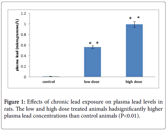
Figure 1: Effects of chronic lead exposure on plasma lead levels in rats. The low and high dose treated animals hadsignificantly higher plasma lead concentrations than control animals (P<0.01).
There was significant difference in blood lead levels in both low and high dose groups compared to control group. This result correlates with similar work by Olaleye et al. and Vahedian, in which they reported that lead exposed animals had higher plasma lead levels than controls [15,22].
Lead exposure caused significant reduction in percentage weight gain compared to control animals. This is an established fact that lead causes reduced body weight due to the compromised metabolism in lead affected rats (Figure 2). The results further show that colitis healing (as measured by stool consistency scoring pattern) was faster in control than in the lead treated groups. The delayed healing of colitis seen in this study may be as a result of the fact that lead exposure causes increased oxidative stress in the gastrointestinal tract of rats. In conclusion, lead exposure caused a significant increase in colitis scores at both low and high doses compare to control (Figure 3). Lead treatment also caused increased frequency and severity of relapse of colitis in exposed animals. Lipid peroxidation (measured as the amount of TBA reactants in the colonic mucosa) was increased in lead treated animals on days 7 and 14 only. Exposure of rats to low and high lead levels significantly decreased SOD activity compared to the control throughout the duration of the study. Similarly, lead exposure increased colonic mucosal catalase activity compared to the control.
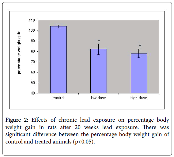
Figure 2: Effects of chronic lead exposure on percentage body weight gain in rats after 20 weeks lead exposure. There was significant difference between the percentage body weight gain of control and treated animals (p<0.05).
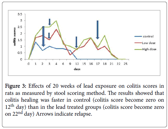
Figure 3: Effects of 20 weeks of lead exposure on colitis scores in rats as measured by stool scoring method. The results showed that colitis healing was faster in control (colitis score become zero on 12th day) than in the lead treated groups (colitis score become zero on 22nd day) Arrows indicate relapse.
The present study investigated the role of oxidative stress in the delayed healing of chemically-induced colitis seen in the colonic mucosa of lead exposure in rats. The results show that colitis healing (as measured by stool consistency scoring pattern) was faster in control than in the lead treated groups.
In histology, Plates 1-5 shows effects of chronic lead exposure on goblet cell mass, lymphoid distribution, epithelial erosions and other characteristics. On day 6 in all groups, the goblet cells were engorged and hyperplastic, there was epithelial and glandular necrosis and pronounced mucosal lymphoid migration. However, by days 22-25 there wasgoblet cell restoration, normal lymphoid distribution and no visible lesions were seen in all groups indicating that healing was complete by this time in the Lead exposed animals.
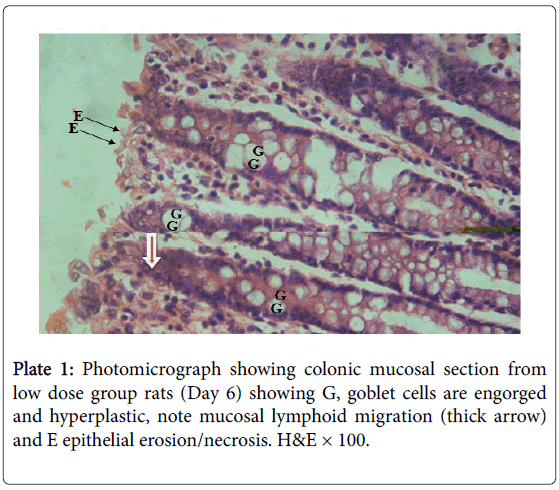
Plate 1: Photomicrograph showing colonic mucosal section from low dose group rats (Day 6) showing G, goblet cells are engorged and hyperplastic, note mucosal lymphoid migration (thick arrow) and E epithelial erosion/necrosis. H&E × 100.
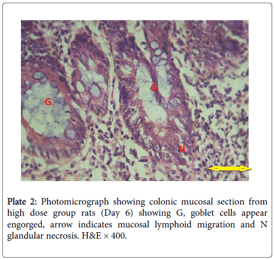
Plate 2: Photomicrograph showing colonic mucosal section from high dose group rats (Day 6) showing G, goblet cells appear engorged, arrow indicates mucosal lymphoid migration and N glandular necrosis. H&E × 400.
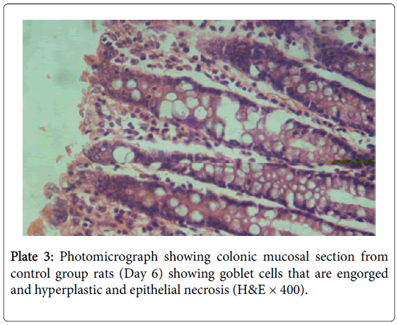
Plate 3: Photomicrograph showing colonic mucosal section from control group rats (Day 6) showing goblet cells that are engorged and hyperplastic and epithelial necrosis (H&E × 400).
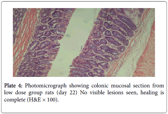
Plate 4: Photomicrograph showing colonic mucosal section from low dose group rats (day 22) No visible lesions seen, healing is complete (H&E × 100).
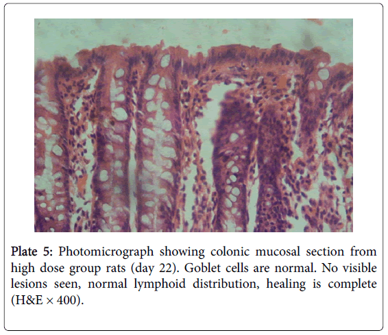
Plate 5: Photomicrograph showing colonic mucosal section from high dose group rats (day 22). Goblet cells are normal. No visible lesions seen, normal lymphoid distribution, healing is complete (H&E × 400).
Lipid peroxidation has been implicated in the etiology of damage to sub-cellular membranes and injury to the cell. In the present study, lipid peroxidation was increased by lead exposure. The implication of this is that lead causes an increase in the formation of free radicals, which, if not mopped up by free radical scavengers as SOD, CAT, or glutathione, will expose the colon to inflammation. In gastrointestinal protection, the first line of antioxidative enzyme defense is SOD which catalyses the dismutation of superoxide radical anion (O2¯) into less noxious hydrogen peroxide (H2O2). H2O2 is then inactivated by the degradation into water by Catalase [23]. Depletion of SOD levels, as evident by the results of this study, therefore predisposes the colon to a greater impact of the free radicals produced via increased lipid peroxidation, hence increased delayed colitis healing following lead exposure (Figures 4-6). The result shows that there was higher incidence and severity of colitis in treated rats compared to control (maximum colitis score of 3.0 and 1.3 respectively). Also the result further shows a higher number of relapses in treated animals (3 relapses each) compared to control (1 relapse). Finally, by day 12, colitis score was zero in control animals (an indication of complete healing of colitis), while in low and high dose groups, colitis score reached zero on day 22. This result implies that chronic lead exposure aggravate the symptoms of inflammatory bowel disease and people suffering from this disease should avoid exposure to lead as much as possible.
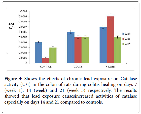
Figure 4: Shows the effects of chronic lead exposure on Catalase activity (U/I) in the colon of rats during colitis healing on days 7 (week 1), 14 (week) and 21 (week 3) respectively. The results showed that lead exposure causesincreased activities of catalase especially on days 14 and 21 compared to controls.
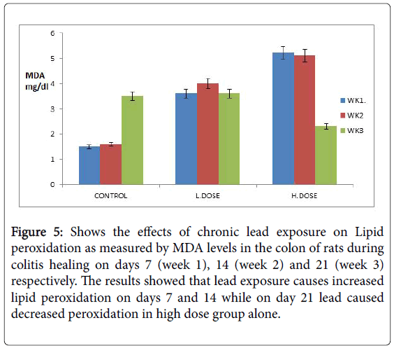
Figure 5: Shows the effects of chronic lead exposure on Lipid peroxidation as measured by MDA levels in the colon of rats during colitis healing on days 7 (week 1), 14 (week 2) and 21 (week 3) respectively. The results showed that lead exposure causes increased lipid peroxidation on days 7 and 14 while on day 21 lead caused decreased peroxidation in high dose group alone.
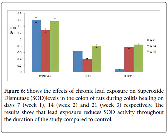
Figure 6: Shows the effects of chronic lead exposure on Superoxide Dismutase (SOD)levels in the colon of rats during colitis healing on days 7 (week 1), 14 (week 2) and 21 (week 3) respectively. The results show that lead exposure reduces SOD activity throughout the duration of the study compared to control.
The Authors gratefully acknowledge the sponsorship of this research work by the 2014-2015 Merged TETFUND (Nigeria) Research Grant released March 2017.
This work was carried out in collaboration between all authors. Authors AGS and OOA designed the study, wrote the protocol and checked the manuscript. Author OO managed literature searches, designed the model and carryout analyses of the data. Authors AAO and ABS corrected the manuscript. All authors read and approved the final manuscript.