Journal of Cell Science & Therapy
Open Access
ISSN: 2157-7013
ISSN: 2157-7013
Research Article - (2015) Volume 6, Issue 5
Background: In this article we described the influence of transplantation of pregnant rat BMC to pregnant rats of the same date of pregnancy to study the influence on fetus’s development.
Methods: To study we made a single intravenous transplantation of BMC of pregnant rats of 4-5, 7-8 or 11, 12 days to pregnant rats of the same date of pregnancy. The analyses were made at 18th day of pregnancy.
Results: The transplantation of rat BMC at 4, 5 pregnant days increases pre- and post-implantation death of fetuses without change of their weight and weight of placentas. The transplantation of BMC after implantation at 7, 8, 9 days of pregnancy during gastrulation increased the weight of 18th day fetuses and of placentas in comparison with the same parameters of usual normal fetuses. The survival of fetuses was not disturbed. In case of BMC transplantation during placentation at 11, 12 days of pregnancy the weight of fetuses and survival of fetuses was significantly decreased and the weight of placentas was increased.
Conclusions: Results of BMC transplantation depends on the stage of rat pregnancy. The increase of the weight of fetuses after BMC transplantation within gastrulation may be explained by positive paracrine effects of allogenic transplanted cells on the size of decidua and in turn on the growth of fetuses and placentas or by transplanted cells itself. The transplantation of BMC at 11, 12 day of pregnancy during placentation decrease the weight of fetuses at 18th day of pregnancy and increase the weight of placentas. The possibility to regulate a weight of fetuses by allogenic BMC transplantation at gastrulation is important results for stem cell therapy of fetuses.
Keywords: Pregnancy; Bone marrow transplantation; Gastrulation; Placentation; Fetuses
Decidualization of the endometrium is an obligatory condition for fetus development for some mammalians. Decidual cells (DC) controls the growth of fetuses and pregnancy progress. Bone marrow origin of rat large decidual cells of antimesometrial part of decidua (LDC) and of endometrial granulated cells were shown [1-4]. Rat LDC and human parietalis DC are characterized by high level of turnover. In accordance with flow cytometry data the proliferative activity of human decidua parietalis at first pregnant trimester is near 3.5 ± 0.3%. Before birth the proliferative activity decreases up to 1.6 ± 0.2%. In case of severe preeclampsia the level of proliferating cells decreases up to 0.45 ± 0.2%. Severe preeclampsia is also characterized by abnormality of DC DNA content and by decrease of decidua thickness. It means that human DC loss is not compensated by precursors or by stem cells proliferation [5]. We suggested that in case of severe preeclampsia or in case of recurrent abortion it is possible to compensate loss of DC by stem cells therapy. Previously we used the decidua development of the pseudopregnant rats as a model to control the capacity of human endometrial stem cells and of rat bone marrow cells to influence on decidua formation after transplantation to uterus of pseudopregnant [6]. To maintain the point of view for bone marrow stem cells as source of precursors for DC we studied an influence of rat bone marrow cells (BMC) transplantation on rat fetus development.
We used for the experiments outbred white rats produced at specialized animal farm “Rappolovo” (St. Petersburg, Russua) with weight 150-200g. The day of observation of spermatozoid in the morning vaginal smear was marked as first day of pregnancy [7]. Bone marrow cells (BMC) were prepared from long bones of pregnant rat. After 63% Percoll fractionation mononuclear cells were collected and injected intravenously (v. jugularis) in volume 1 ml PBS to rat of the same date of pregnancy. The quantity of mononuclear cells for every transplantation were (135 ± 12) 106. The animals were opened at 18th of pregnancy with help of diethyl ether narcosis. The fetusis and placentas were fixed at Bouin solution and weighted at next day. The level of preimplantation death was counted as discrepancy of quantity of yellow bodies in ovary and quantity of implantation sites at uterus; the level of post implantation death was counted as discrepancy between quantity of implantation sites and quantity of alive fetuses.
Aspirated bone marrow was further processed in order to isolate the mononuclear fraction, a heterogeneous population containing differentially matured B-cells, T-cells and monocytes, as rare progenitor cells such as hematopoietic stem cells, mesenchymal stromal cells, endothelial progenitor cells and very small embryonic-like cells. In accordance with multiple studies such cell mixture promotes distinct angiogenic properties, mediates vascular repair, express several cytoprotective growth factors and cytokines [8]. It was shown that transplantation of BMC of pregnant rats into pregnant rats of the same date of pregnancy is not provided by teratogenic effects after superficial examination (Figure 1). At the same time there are significant difference of fetuses weight in case of BMC transplantation during implantation (7-8; 9 days of pregnancy) in compare with weight of control fetuses of 18th day of pregnancy (Table 1 and Figure 1). There was retardation in the fetuses weight in case of BMC transplantation during placentation (11-12 days of pregnancy). Growth of fetuses after BMC transplantation at 7-8th and 9th was accompanied by placenta growth. The retardation of weight of fetuses after transplantation of BMC at 11-12 pregnant days was accompanied by an increase of placenta weight. Transplantation of BMC from nonpregnant animals at diestrus stage of estrus cycle increased the weight of fetuses in low level and did not change the weight of placentas. The peaks of curves of BMC transplantation from pregnant and nonpregnant rats coincides (Figure 2). It means about common nature of BMC activities of pregnant and nonpregnant rats.
| Experiments | Weight of fetuses, mg | Weight of placentas, mg |
|---|---|---|
| Normal rats 18th days (11 families) | 720 ± 20 | 303 ± 14 |
| 4-5th days of transplantation of BMC, (4 families) | 760 ± 20 | -- |
| 7-8th days of transplantation of BMC (8 families) | 815 ± 16 | 343 ± 12 |
| 7-8th days (diestrus) of transplantation of BMC (3 families) | 787 ± 7 | 309 ± 17 |
| 9th day of BMC transplantation (7 families) | 838 ± 60 | 334 ± 18 |
| 11-12 days of BMC transplantation (3 families) | 587 ± 5 | 375 ± 80 |
Table 1: Influence of bone marrow cells (BMC) transplantation at early stages of rat development for growth of fetuses at 18th day of pregnancy.
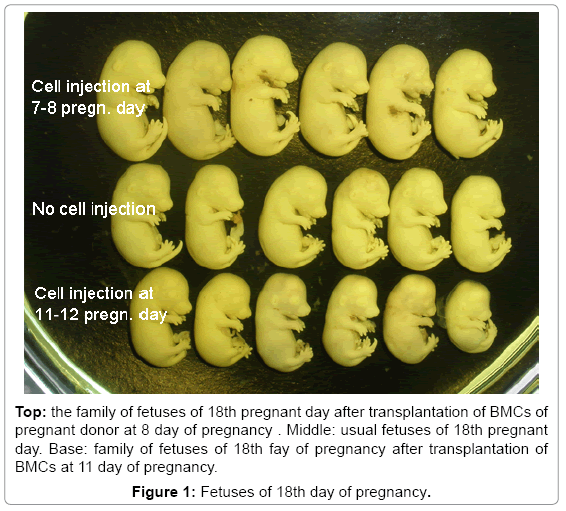
Figure 1: Fetuses of 18th day of pregnancy.
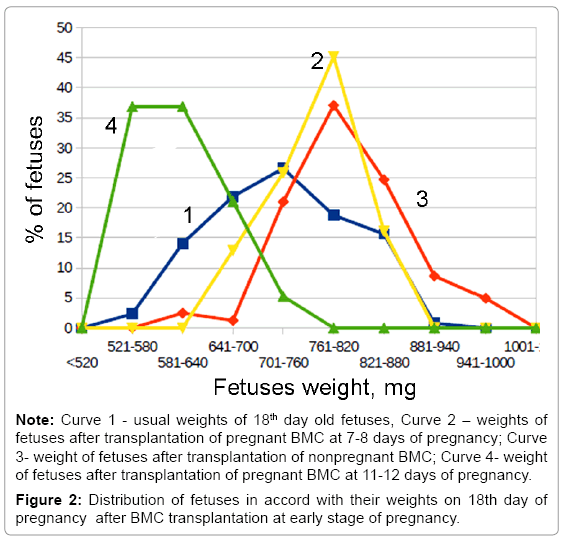
Figure 2: Distribution of fetuses in accord with their weights on 18th day of pregnancy after BMC transplantation at early stage of pregnancy.
BMC transplantation before implantation at 4-5 days of pregnancy was characterized by an increased level of pre- and post-implantation death (Table 2). The weight of fetuses did not change. At this time the blastocysts are in the lumen of uterus. The high level of pre- and post-implantation death may be explained by the disturbance of implantation because of excesses of transplanted BMC. Probably an excess of BMC violates the coordination of numerous molecular and cellular mechanisms that regulate the interaction of blastocysts and uterine during implantation [9]. The normal weight of alive fetuses at 18th day of pregnancy points to the absence of the direct influence of BMC on blastocysts.
| Pregnant days of BM cells transplantation | Preimplantaion embryo survival, % | Postimplantation embryo survival, % |
|---|---|---|
| without transplantation of BMC | 94.2 ± 2.4 | 94.8 ± 2.7 |
| transplantation of BMC at 4-5 day of pregnancy | 72.3 ±8.5 | 45.0 ± 15.4 |
| transplantation of BMC at 7-8 day of pregnancy | 96.6 ± 2.1 | 97.8 ± 2.4 |
| transplantation of BMC at 11-12 day of pregnancy | 89.3 ± 6.7 | 71.7 ± 11.8 |
Table 2: Pre- and post implantation survival of rat’s fetusis of 18th day of pregnancy after transplantation of mononuclear bone marrow cells (BMC) during early stages of pregnancy (%).
The increase of fetuses weight after BMC transplantation during gastrulation may be explained by positive paracrine effects of transplanted allogenic BMC for growth of fetuses as the consequence of increased decidua growth. Early we had observed that human endometrial stem cells and rat BMC local transplantation to pseudopregnant rats increased the sizes of rat decidual tissue [6]. It is very important that gastrulation in rat and mice takes place in the environment of large decidual cells (LDC). Specific morphology of LDC determines the specificity of decidual tissue [10]. Such morphogenetic events as formation of mesoderma and endoderma, determination of body axes, beginning of heart and of brain formation, high level of embryonal cells proliferation are content of gastrulation [11-13]. Development of blastocysts in vitro or in ectopic sites without decidual tissue in vivo have been stopped at stage of gastrulation [14]. The gastrulation claim special cover against oxidize radicals. Our results for stimulation of decidual growth by transplantation of BMC was without disturbance of structure of decidua [6] and growth of fetuses with placentas (Table 1) show for general positive influence of BMC transplantation for embryonal growth. We suggest that main target of BMC action during gastrulation is decidual influence for embryo cell proliferation. Absence of teratogenic effects of BMC transplantation confirms the influence of BMC transplantation for growth of embryos and of ectoplacental cone as general action to increase proliferation of embryo cells. The LDC of antimesometrial part of decidua as of rat as of mouse disappear during 11-12 pregnant days [5].
At 11th day of pregnancy ectoplacental cone begin to form placenta. Transplantation of BMC during placentation (11-12 days of pregnancy) induces the retardation of fetuses’ growth and the increase of placenta weight. Retardation of growth may be explained by appearance in BM during this stage of pregnancy of the precursor of granulated metrial gland cells with NK activity (uNK cells) [2,13,15,16]. In accord with results of Podporina metrial gland granulated cells are characterized by uNK activity at beginning of differentiation and by immunosuppressor activity at the end of differentiation. These capacities of granulated cells explain the function of rat granulated cells as regulator of immunological situation in placenta [17].
The results show that early stages of rat development have own characteristics of relationships between bone marrow and embryo development. The dates confirm the reality of participation of bone marrow cells in the regulation of embryo development. Attract attention the possibility to increase growth of fetuses by transplantation of allogenic bone marrow mononuclear cells. From another side the dates caution against direct interference with stem therapy in the regulation of pregnancy without tentative study in another experimental models of regulation of pregnancy.
The work was supported by Grant RFBR # 14-04-00259-a and by Grant RNF # 14-50-00068.