Clinical & Experimental Cardiology
Open Access
ISSN: 2155-9880
ISSN: 2155-9880
Mini Review - (2023)Volume 14, Issue 4
We ever received the case of a middle-aged man with Aortic valve Stenosis (AS) and severe mid- and apical-ventricular septal hypertrophy in our department. We tried to manage both concerns in one operation. Due to severe AS, Aortic Root Enlargement (ARE) is commonly used for aortic valve replacement to prevent Patient-Prosthetic Mismatch (PPM). Konno-Rastan’s (K-R’s) operation is a common procedure of ARE which can be used to resolve the two complications of AS and obstructive Hypertrophic Cardiomyopathy (HCM). Morrow’s procedure is also a general standard operation for HCM. However, they both only can resolve upper-ventricular septal hypertrophy, which are not the best options for the patient. aortoventriculoplasty, differs from K-R’s and Morrow’s procedure, since it mainly involves dissection of the aorta and pulmonary artery to expand the operation field to the apical-ventricular septum. Until now, few doctors have previously preferred and reported it and we also have less experiences. We successfully relieved the rare obstruction, and completed the AV replacement. The patient was followed up for twenty-two months and he had an uneventful recovery. This was an important attempt of the relatively unfamiliar operation for us. We now believe that aortoventriculoplasty could be considered in similar patients with hypertrophy in the middle and lower ventricular septum regardless of valve lesions.
Aortoventriculoplasty; Aortic valve stenosis; Obstructive hypertrophic cardiomyopathy
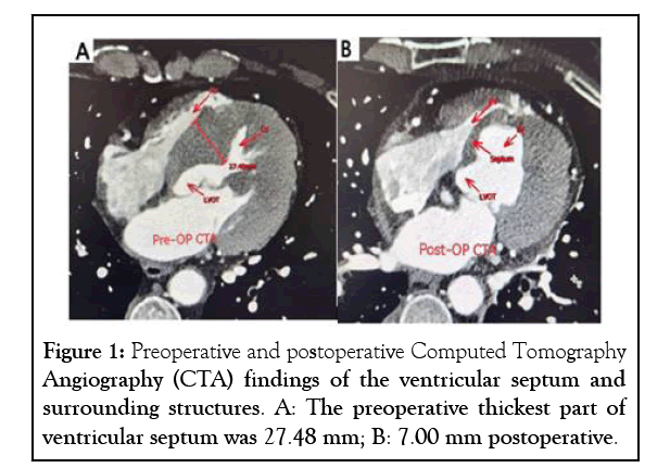
The patient had two obvious complications including severe AS with a valve area of 0.8 cm2, along with severe HCM in the midand apical-ventricular septum measuring 27.48 mm preoperatively in the Colour Doppler Echocardiography scan (Table 1).
| Preoperative | Postoperative | |
|---|---|---|
| Thickness of ventricular septum (mm) | 27.48 | 7 |
| Peak aortic velocity (m/s) | 5.9 | 1.86 |
| Mean gradient (mmhg) | 75 | 14 |
| Aortic regurgitation velocity (m/s) | 4.3 | 1.6 |
| Aortic valve annulus diameter (mm) | 19 | 25 |
| Pulmonary valve regurgitation velocity (cm/s) | 116 | 85 |
| EDV* (ml) | 63 | 131 |
| ESV* (ml) | 28 | 64 |
Note: EDV: End-diastolic volume; ESV: End-systolic volume
Table 1: Preoperative and postoperative Colour Doppler Echocardiography scan findings of the ventricular septum and surrounding structures.
Preoperative End-Diastolic Volume (EDV) of the left ventricular was 63 ml. In order to Prevent Patient-prosthetic Mismatch (PPM), Aortic Root Enlargement (ARE) is used to facilitate the implementation of artificial valves of appropriate size for AS. There are four main techniques used to perform ARE, namely, Nicks, Manouguian, Nunez (modified Manouguian), and the Konno-Rastan’s (K-R’s) procedure [1,2]. The K-R’s operation is among the most common and traditional techniques, which also can be used for some types of obstructive Hypertrophic CardioMyopathy (HCM). Morrow's procedure, that is generally accepted as the standard surgical treatment for HCM. It passes through the patient's aortic valve orifice and try to remove the upper hypertrophic ventricular septum [2]. Morrow’s as well as K-R’s surgery are difficult to perform an adequate septal myectomy for mid-ventricular and apical HCM [2]. Moreover, we attempted to manage both concerns in a single operation, a type of traditional aortoventriculoplasty, is another method for ARE [3,4].
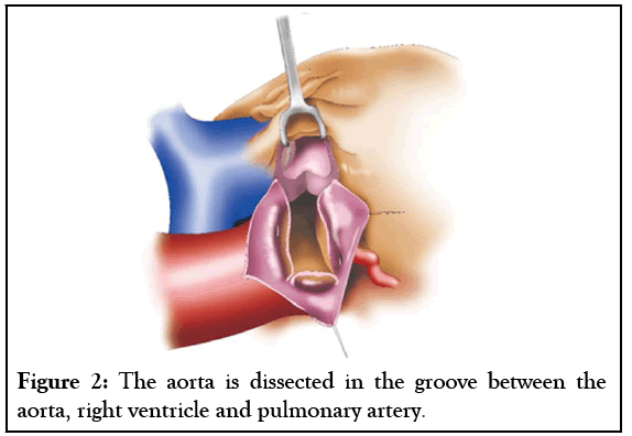
In a common procedure, the aortic incision is extended into the commissure between the left and right coronary cusps of the aortic valve into the interleaflet triangle and the outflow chamber of the right ventricle is opened transversely. Thus, the incision into the right ventricle is made in the outlet portion and into the infundibular septum. This offers the option of preserving the aortic valve if it is normal or repairable. The thickness of the hypertrophied ventricular septum is reduced by resecting the left ventricular side of the septum to relieve the left ventricular outflow tract obstruction [4]. operation can significantly expand the visual field and accurately control the width and thickness of the resected ventricular septal muscle. The surgeon can clearly see how much muscle is excised, moreover, it is more important that the appropriate remainder of the septum can be guaranteed. Not only can the Left Ventricular Outflow Tract Obstruction (LVOTO) be completely eliminated, but also the left ventricular volume can be significantly enlarged. Hence, there were fewer opportunity of complication such as prevention of iatrogenic interventricular septum perforation. Herein, the aortoventriculoplasty was performed. After operation, the patient’s symptoms such as chest tightness and shortness of breath disappeared, the murmur in aortic valve area was not heard, an ultrasound scan showed that the ventricular septal thickness had reduced to 7 mm whereas, and the cardiac hemodynamic parameters were close to the normal level (Figures 1 and 2).

Figure 1: Preoperative and postoperative Computed Tomography Angiography (CTA) findings of the ventricular septum and surrounding structures. A: The preoperative thickest part of ventricular septum was 27.48 mm; B: 7.00 mm postoperative.

Figure 2: The aorta is dissected in the groove between the aorta, right ventricle and pulmonary artery.
At the time of this reporting this case, twenty-two months have passed since his surgery without any complications.
We ever think the surgery is the better choice for the patients with hypertrophy in the middle and lower ventricular septum without AS. But now we believe, regardless of valve lesions, the aortoventriculoplasty will be a good choice as long as the hypertrophy of patients is in the mid or, and apical-ventricular septum because of the successful patient. We think the aortoventriculoplasty may be an option for the patients with obvious distal ventricular septal hypertrophy and obstructive HCM with or without valve lesions.
Citation: Xiao C, Wang J, Chi H (2023) Aortoventriculoplasty for Obstructive Hypertrophic Cardiomyopathy along with Aortic Stenosis. J Clin Exp Cardiolog. 14:788.
Received: 10-May-2023, Manuscript No. JCEC-23-24016; Editor assigned: 12-May-2023, Pre QC No. JCEC-23-24016 (PQ); Reviewed: 31-May-2023, QC No. JCEC-23-24016; Revised: 08-Jun-2023, Manuscript No. JCEC-23-24016 (R); Published: 16-Jun-2023 , DOI: 10.35248/2155-9880.23.14.788
Copyright: © 2023 Xiao C, et al. This is an open-access article distributed under the terms of the Creative Commons Attribution License, which permits unrestricted use, distribution, and reproduction in any medium, provided the original author and source are credited.