Biochemistry & Pharmacology: Open Access
Open Access
ISSN: 2167-0501
ISSN: 2167-0501
Research Article - (2014) Volume 3, Issue 3
A bioassay was developed to provide a process for the synthesis of cyanine dye labeled base modified nucleoside triphosphates analogues as a novel labeling agent for biomolecules. It is also to provide a low cost process and more specifically relates to a process for the synthesis of cyanine fluorophore - base modified nucleoside triphosphates analogues as well as for efficient and robust labeling of RNA and cDNA as hybridization probe in microarray detection and analysis.
<Keywords: Cyanine fluorophore; Fluorescent dyes; Labeling agent; Microarray detection
Cyanine dyes, used as fluorescent dyes belong to the polymethine group. Cy3 and Cy5 are reactive water-soluble fluorescent dyes of the cyanine dye family. They are usually synthesized with reactive groups on either one or both of the nitrogen side chains so that they can be chemically linked to either nucleic acids or protein molecules. Cyanine dyes are introduced as new fluorescent reagents for covalently labeling proteins and other biomolecules [1]. Labeling is done for visualization and quantification purposes.
Cyanine 3- and cyanine 5-N-hydroxysuccinimidyl esters (NHS esters) are high quality, reactive fluorescent dyes optimized for amine labeling. The cyanine 3 dye provides bright orange signal (~ 550 nm excitation, ~ 570 nm emission), while the cyanine 5 dye provides bright red fluorescence signal (~ 650 nm excitation, ~ 670 nm emission). The reactive groups allow the dyes to be chemically linked to either aminoallyl-modified nucleic acids or directly to proteins and peptides. The dyes are suitable for a wide variety of biological applications including comparative genomic hybridization and expression array profiling, as commonly employed in transcriptomics [2]. They are also suitable for labeling proteins and nucleic acids for a variety of other applications relating to proteomics and genomics [3]. The dyes display good aqueous solubility and low non-specific binding, allowing convenient labeling and assay set-up.
The synthesis of two new cyanine dye-labeled dUTP analogs, Cy3-dUTP and Cy5-dUTP has been reported [4]. They are efficient substrates for DNA polymerases and can be incorporated into DNA probes by standard nick translation, random priming and polymerase chain reactions. Optimal labeling conditions have been identified which yield probes with 20–40 dyes per kilobase. The directly labeled DNA probes obtained with these analogs offer a simple approach for multicolor multisequence analysis that requires no secondary detection reagents and steps.
In another method for increasing the fluorescent signal intensities, amino-allyl reverse transcription (AA-RT) can be used for probe preparation. In this process cDNA is synthesized from total RNA or mRNA in the presence of amino-allyl dUTP (AA-dUTP, Sigma, St. Louis, MO, USA) instead of Cy3- or Cy5-dUTP. The AA-dUTPs incorporated into the synthesized cDNA are coupled with Cy3 or Cy5 monofunctional dye (Amersham Pharmacia Biotech, Inc.).
Before the two labeled samples are pooled, AA-dUTP is quenched by the addition of hydroxylamine. This technique have shown an increase in fluorescent signal intensities compared with the direct fluorescent dye incorporation method, It is suspected that the enhancement of sensitivity by AA-RT is due to an increase in the reverse transcription rate compared with reverse transcription in the presence of Cy-dUTP [5].
In the present study, a process is provided for the synthesis of fluorescent cyanine dye labeled AA-dUTP analogues.
Process for the synthesis of fluorescent cyanine dye labeled AA-dUTP analogues from allyl-amine dUTP and Cy3-NHydroxysuccinimide ester
Anhydrous dimethyl formamide (DMF) was prepared by combining 250ml of fresh DMF with 50gm of diatomaceous earth and was incubated at room temperature for overnight. 10mg of AA-dUTP was then dissolved in 4ml of 0.1M of sodium borate buffer of pH 8.5. 1.0mg of monofunctional N-Hydroxysuccinimide (NHS)-ester-Cy3 dye (Amersham Pharmacia Biotech, Catalogue No. Q-13108) was resuspended in 0.61ml of incubated anhydrous dimethyl formamide (DMF) solution and 2ml of the solution thus prepared was mixed with 2ml of AA-dUTP solution for incubation for 4hrs at room temperature to obtain AA-dUTP-cy3. Both the products obtained were then purified by HPLC.
Process for the purification of synthesized AA-dUTP-cy3 by HPLC
The Products AA-dUTP-cy3 were purified through HPLC at 550nm respectively on Nucleogen Deae 60-7 column of 4mm X 125mm with a flow rate of 1ml/min. The detector used for the HPLC was UV detector at 1 absorbance units full scale (AUFS) and the solvent used was a low salt buffer consisting of 25 mM of triethylammonium acetate (TEAA) of pH 8.0 in 10% Acetonitrile and a high salt buffer consisting of 1M of triethylammonium acetate (TEAA) of pH 8.0 in 10% Acetonitrile.
Assay for the activity of AA-dUTP-Cy3 as labeling agent for RNA and cDNA in microarray experiments
Activity of AA-dUTP-Cy3 was assayed through the synthesis of dsDNA from ssDNA by reverse transcription containing AA-dUTPCy3. For annealing 0.25μl of ssDNA of 0.65 μg/μl concentration and 0.375 μl of T3 primer of 0.5 μg/μl concentration were incubated at a temperature of 70.degree.C for 10minutes and then this reaction mixture was chilled on ice for 10 minutes. For dsDNA synthesis the reagents including 1.0 μl of 5X1st Strand Buffer (500ml of 5X1st Strand Buffer comprising 250mM Tris-HCL pH 8.3, 375mM Potassium Chloride,15mM Magnesium Chloride, 50mM DTT. 500mL, Sterile), 0.5μl of 0.1M dithiothreitol (DTT), 0.5μl of Super ScriptII Reverse Transcriptase, 0.5μl of 2mM of deoxynucleoside triphosphates (dNTPs) excluding dttp and dUTP, 5.0μl of dH2O, AA-dUTP-Cy3 in the volume as needed for 0.2mM final concentration were added to the reaction mixture and incubated at 42°C for 2hours. The DNA was then separated from unincorporated dUTP/Cy dye on G-50 sephadex column by centrifugation at 2000 rpm for 5minutes and the flow was discarded. G-50 Sephadex column was prepared by combining Sephadex and TE in the ratio of 1:1. 50 μl DNA solution obtained after separation from unincorporated dUTP/Cy dye on G-50 sephadex column was mixed onto the resin bed and centrifuged at 2000 rpm for 5 minutes. The eluent was saved. The DNA solution comprised of the following reagent in a total volume of 50 μl (DNA solution: 5.0μl; EDTA:10 μl; Yeast RNA carrier (4 mg/ml) 2.5 μl; dH2O 32.5 μl). 50μl of DNA solution comprising of above reagents, two volumes of 100% ETOH and 15μl of 3M NaOAc were mixed. The reaction mixture was incubated at -80°C for a time period of more than 1 hour to concentrate the volume of the reaction mixture to 5 μl. The reaction mixture was then centrifuged at 4°C for 25 minutes and was air dried for 3 to 5 minutes. 5μl 3X SSC was added to each tube to resuspend the concentrate. 1ml of reconstituted DNA solution and 1ml of 1:10 dilution in 3X SSC was spotted onto a poly-L-lysine-coated slide. The slide was scanned in microarray. The HPLC elution profile of synthesized AA-dUTP-Cy3 is 27.79min which is lower than the elution profile of AA-dutp-Cy3 dye synthesized and marketed by Amersham, USA which is 27.82 as shown in Figure 1-3 respectively.
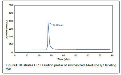
Figure 1: Illustrates HPLC elution profile of synthesized AA-dutp-Cy3 labeling dye
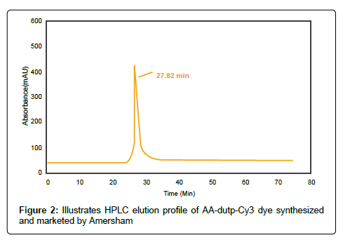
Figure 2: Illustrates HPLC elution profile of AA-dutp-Cy3 dye synthesized and marketed by Amersham
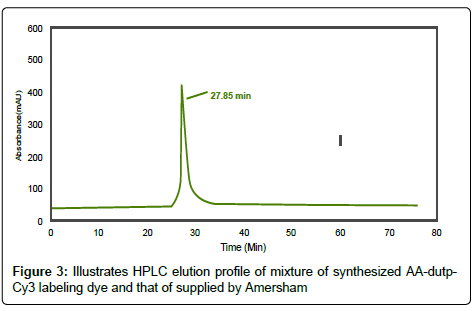
Figure 3: Illustrates HPLC elution profile of mixture of synthesized AA-dutp- Cy3 labeling dye and that of supplied by Amersham
In the present study the activity of AA-dUTP-Cy3 as labeling agent is assayed through synthesis of dsDNA from ssDNA by in vitro reverse transcription and thus labeled RNA and cDNA functions as hybridization probe in microarray detection and analysis. The labeling technique, mainly two-step processes is very much consistent and versatile because of the efficient insertion of primary amines and also the chemical labeling reaction can be done with a variety of amino dyes. In this procedure using aminoallyl-dUTP is gaining popularity due to the increased labeling efficiency and reduction in dye bias and cost. In this two-step procedure, primary aliphatic amino groups are first incorporated during cDNA synthesis. In the second step, the monofunctional N-hydroxyl succinimide-activated fluo-rescent dye (Cy3) is coupled to cDNA by chemical reaction with the amino functional groups.
These amino reactive dyes required for nucleic acid labeling are hydrophilic in nature which is advantageous for stabilization when conjugated to DNA [4]. Fluorescence-based nucleic acid hybridization assays are much important in various studies like gene expression. The cDNA probes for array hybridization are synthesized at first from total RNA by reverse transcriptase and then labeled with radioisotopes (such as P32 orthophosphate) or fluorescent markers using random or specific primers. The amount of probe used for hybridization depends on the array format and labeling method [6]. In a typical hybridization reaction, equal amounts of Cy3- labeled probes based on the incorporated dye concentration are combined [7-16].
The present work provides a simple and cost effective process for the synthesis of cyanine labeled fluorescent analogues as a novel labeling agent for the nucleotides. The product is also a low cost fluorescent labeling agent for the labeling of RNA and cDNA as hybridization probe in microarray detection.
The author acknowledges the financial assistance from Amity Institute of Biotechnology, Amity University, Noida, UP and Council of Scientific and Industrial Research, CSIR, Govt. of India.