Anatomy & Physiology: Current Research
Open Access
ISSN: 2161-0940
ISSN: 2161-0940
Research Article - (2019)Volume 9, Issue 2
Background: Dissecting the vertebral column and studying the features of individual vertebrae is an integral part of the curriculum in anatomy. The vertebral column consists of 33 vertebrae which are stacked one upon the other and joined to each other by the intervertebral joints and kept in a position of by various ligaments, chief of which includes anterior and posterior longitudinal ligaments. Disease processes involving these ligaments leads to various spectrums of aliments extending from backache to deformities and nerve entrapments. Aims and Objectives: To study the Anterior and Posterior longitudinal ligament and note the incidence of different types of ossification and the other associated features in various levels of the vertebral column. Material and Methods: The study was conducted on 25 disarticulated sets of bones and 30 cadavers used in dissection at GMC and ESICH, CBE. Results: It was observed that out of 25 dry sets of vertebrae examined, only 5 sets showed evidence of ossified ligaments at various levels. While no ossification of ligaments was observed in cadaveric specimens. Conclusion: Ossification of Anterior ligaments of the spine is rare. If the present cause should be identified and corrected to improve, preserve flexibility, prevent the development of deformities and neurological deficits.
Anterior longitudinal ligament; Intervertebral joints; Vertebral spine
The human vertebral column consists of a series of vertebrae firmly connected to each other by joints and ligaments. Among the ligaments, Anterior and Posterior longitudinal ligaments are integral in keeping the bodies of vertebrae in alignment with each other and check the anterior and posterior displacement of the vertebra over each other. The anterior longitudinal ligament is a flat strong band extending from the second cervical vertebra (axis) to sacrum along the anterior surface of vertebral bodies. It is thickest in the thoracic region. The posterior longitudinal ligament is extending from the axis, at the level axis is continuous as membrane tectoria to sacrum along the posterior surface of vertebral bodies. When these ligaments undergo ossification or calcification the normal alignment of the vertebrae is deranged leading to disturbance in the posture and gait.
The present study was conducted on 25 dry bony sets and 30 human cadavers under dissection available in our institution. In the cadavers, Anterior and Posterior longitudinal ligaments were visualized and palpated for change in texture to find out any evidence of ossification. In the dry bones, the margins of the body of the vertebra and margins of the lamina were examined to find ossified remnants of the ligaments.
All the spinal ligaments inspected in the cadaveric specimens looked normal and there was no evidence of ossification or calcification on inspection and deep palpation.
In the dry bone sets, we observed no ossification of ligaments in cervical vertebrae. While 3 sets of vertebrae showed ossified ligaments in thoracic level (all types) i.e. segmental, continuous, mixed and 2 sets showed ossified ligaments at lumbar level (segmental and mixed). In Segmental ossification of ligaments, either partial or entire vertebral body was involved while an intervertebral disc space was intact, whereas in continuous-type the ossification involved disc space also. Mixed type of ossification showed the presence of both segmental and continuous type in different parts of the same specimen. Apart from ossified anterior longitudinal ligament, these segments showed the fusion of costovertebral and costotransverse joints, narrowing of intervertebral foramen and ossification of Supraspinous ligaments in some regions. The observations were noted and tabulated as in the figure.
DISH was reported and recognized as an entity in recent decades. It is described in the literature with various names. Meyer et al. coined the term “Moniliform Hyperostosis” to describe the thoracic spine hyperostosis and calcification [1]. Forestier et al. used a similar term called “Senile Ankylosing Vertebral Hyperostosis” or “Forestier’s disease”, but it has to be differentiated from other spinal diseases [2].
Resnick et al. introduced the term DISH (Diffuse Idiopathic Skeletal Hyperostosis) explains the condition more clearly stating that ossification of Anterior Longitudinal Ligament with normal disc space [3]. But it has to be differentiated from the degenerative disease [4].
By nature, anterior longitudinal fibres are adhered strongly to the intervertebral disc, hyaline cartilage end plates and margins of adjacent vertebrae and loosely attached to the intermediate level of bodies. Ligamentous fibres blend with subjacent periosteum, perichondrium and periphery of annulus fibrosus in various levels [5].
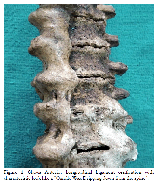
Hyperostosis is a reparative reaction to mechanical stress of tendons and ligaments. In this condition, osteoblasts from mesenchymal precursor cells migrate into the intact periosteum and lay down new bones formation in tendons and ligaments lying close to the bone which is subjected to stress [6]. In case of DISH (diffuse idiopathic skeletal hyperostosis) has a tendency for ossification of ligaments, tendons and joint capsule insertion, but most common region to be affected is the spine, particularly thoracic [7]. The classical description of the appearance of ossified Anterior Longitudinal Ligament is “Candle Wax Dripping down the Spine”. The same picture was found in our study as shown in Figure 1 [8].

Figure 1. Shows Anterior Longitudinal Ligament ossification with characteristic look like a “Candle Wax Dripping down from the spine”.
The prevalence of DISH in European and Caucasian population is reported as 27.3% by Pappone N et al. [9], while in Korean population is 2.9% by Kim SK et al. [10]. No percentage has been quoted for the Indian population by Vivek Singh Malik et al. [11]. In the current study, the prevalence rate is 9%. Westerveld LA et al. stated that DISH has male preponderance compared to females [12].
Khozaim N and Grays observed that incidence of the disease is 6%-12% in both sexes [13]. In the current study, there was no ossification observed in the cadaveric specimens. Ossification was observed in the dry bones but sex determination could not be done.
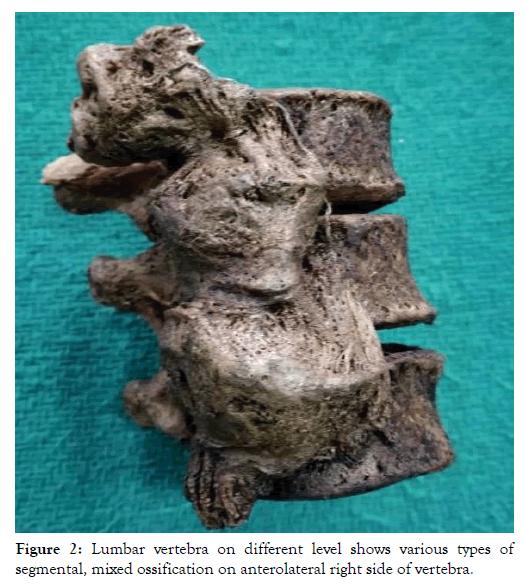
Vivek Singh Malik et al. observed the commonest site involved is thoracic vertebrae. In the current study, the same was observed and two specimens showed lumbar vertebral involvement shown in Figure 2 [11].

Figure 2. Lumbar vertebra on different level shows various types of segmental, mixed ossification on anterolateral right side of vertebra.
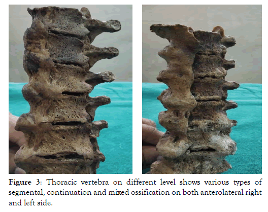
Resnick and Niwayama found T7 to T10 level involvement in both sides [14] and Talijanovic et al. observed and stated that most commonly involved region is thoracic vertebrae, in that particularly 7th thoracic to 11th thoracic vertebrae are involved more frequently than any other [15]. In the current study bilateral ossification was seen in the thoracic level in 2 specimens. In one specimen right side extent was T7 to T12 and on the left extension from T9 to T12 and in another specimen right side extent was T7 to T10 and on the left extension from T8 to T10 was noted shown in Figure 3 and tabulated in Table 1.

Figure 3. Thoracic vertebra on different level shows various types of segmental, continuation and mixed ossification on both anterolateral right and left side.
| S. No. | Vertebral level | Ossification noted |
|---|---|---|
| 1. | Cervical | No ossification |
| 2. | Thoracic | Thoracic Specimen no 1: Ossification T7 to T12 Thoracic Specimen no 2: Ossification T7 to T12 Thoracic Specimen no 3: Ossification T7 to T10 |
| 3. | Lumbar | Lumbar Specimen no 1: Ossification L2 to L4 Lumbar Specimen no 2: Ossification L1 to L4 |
Table 1: Study of bilateral ossification seen in the thoracic level in 2 specimens.
The exact reason for this not implicated but the probable role of descending thoracic aorta on the left preventing the onset of ossification was suspected this was explained by Nagaraj Mallashetty et al. [16]. Very rarely, DISH was also observed in pediatric cases reported by Coakely et al. [17].
Belanger T. A et al. observed extensive ossification on the anterior or right anterolateral aspect of contiguous vertebrae [18]. Mamata et al. reported ossification in the right anterolateral surface of vertebral bodies in 56 cases and in the left anterolateral surface of vertebral bodies in 3 cases [7].
In the current study, we found all types of ossifications segmental, continuous, mixed in thoracic and lumbar segments in anterior and right and left anterolateral regions. Similar findings have reported already by Kalyan Chakravarthi Kosuri et al. and also he stated that such extensive ossification leads to restriction of active and passive respiratory movements and leads to the development of pulmonary diseases [19].
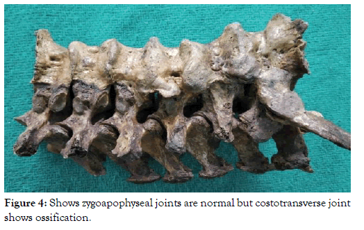
Regarding the type of ossification, Kalyan Chakravarthi Kosuri et al. noted the maximum incidence of segmental type around (17.89%) while yet another study showed an incidence of 4% segmental type. In the current study maximum incidence of segmental ossification (14.5%) was seen followed by mixed type (12.7%) and at least was continuous type. In the current study, simultaneous ossification of Supraspinous ligament was seen in all thoracic vertebral sets extend from T7 to T12. The fusion of Costotransverse and Costovertebral joints were seen in two thoracic vertebral sets extend from T11 to T12 was shown in Figure 4. Stenosis of intervertebral foramen was seen in the level of T12 in one of the thoracic vertebral sets were tabulated in detail in Table 2.

Figure 4. Shows zygoapophyseal joints are normal but costotransverse joint shows ossification.
| Parameters | Cervical vertebra | Thoracic vertebral set no:1 shows ossification from T7 to T12 | Thoracic vertebral set no:2 shows ossification from T7 to T12 | Thoracic vertebra set no:3 shows ossification from T7 to T10 | Lumbar vertebra set no:1 shows ossification from L2 to L4 | Lumbar vertebra set no:2 shows ossification from L1 to L4 |
|---|---|---|---|---|---|---|
| Segmental ossification of ALL | - | 1.T7 to T8 (A,ALRS) 2.T8 to T9 (A,ALRS) |
1.T7 to T8 (A,ALRS) 2.T8 to T9 (A,ALRS) |
1.T7 to T8 (A,ALRS) | 1.L2 to L3 (A,ALRS) 2. L3 to L4 (A,ALRS) |
1.L1 to L2 (A,ALRS) |
| Continuous ossification of ALL | - | 1.T9 to T10 (A,ALRS) 2.T10 to T11 (A,ALRS) |
1.T9 to T10 (A,ALRS) 2.T10 to T11 (A,ALRS) 3.T11 to T12 (A, ALLS) |
1.T8 to T9 (A,ALLS) 2.T9 to T10 (A,ALLS) |
- | |
| Mixed type | - | 1.T11 to T12 (A,ALRS) | T11 to T12 (A,ALRS) T10 to T11 (A,ALLS) |
1.T8 to T9 (A,ALRS) 2.T9 to T10 (A,ALRS) |
- | 1.L2 to L3 (A,ALRS) 2.L3 to L4 (A,ALRS) |
| Side of ossification | - | Anterior (A) and Anterolateral right side (ALRS) | Anterior (A) and Anterolateral right and left side (ALRS and ALLS) | Anterior (A) and Anterolateral right and left side (ALRS and ALLS) | Anterior (A) and Anterolateral right side (ALRS) | Anterior (A) and Anterolateral right side (ALRS) |
| Supraspinous ligament ossification present or absent and its nature | - | + Complete from T7 to T12 | + Incomplete T7 to T12 | + Complete T8 to T10 | - | - |
| Fusion of Costotransverse joint and level involved | - | Present T11 to T12 |
Present T11 to T12 |
- | - | - |
| Fusion of Costovertebral joint and vertebral level involved | - | Present T11 to T12 | Present T11 to T12 | - | - | - |
| Intervertebral foramen narrowing | - | Yes at T12 | Normal | Normal | Normal | Normal |
| Zygoapophyseal joint normal or fused | - | Normal | Normal | Normal | Normal | Normal |
Table 2: Detail study of stenosis of intervertebral foramen seen in the level of T12 in one of the thoracic vertebral sets.
Etiopathogenesis of diffuse skeletal hyperostosis of the spine with regional ossification of fibres of Anterior Longitudinal Ligament is attributed to hypercalcemia and disorders of pyrophosphate metabolism [20].
Ossification of posterior longitudinal ligament has been reported in the Japanese population involving cervical spine and stenosis of the intervertebral foramen. In some cases, patients were asymptomatic, but others presented with radiculopathy and spastic palsy of extremities [21].
DISH has often been misdiagnosed as Ankylosing spondylitis. DISH has no genetic transmission and organ involvement while ankylosing spondylitis has both [22].
The normal pattern of arrangement of vertebrae in the human spine confers springiness, flexibility, and strength to the vertebral column; this is possible because of the various ligaments and joints binding the vertebra together. Hence ossification of ligaments deranges the alignment of the vertebra and also affects the flexibility of entire spine movements. Ossification in the anterior longitudinal ligament is one component of diffuse idiopathic skeletal hyperostosis (DISH) or Forestier's disease. Posterior longitudinal ligament, Supraspinous ligament, costotransverse and Costovertebral joint are not usually involved. Such ossifications are associated with low backache, dysphagia, odynophagia, compression of the brachial plexus end up in nerve weakness or damage, recurrent laryngeal nerve palsy, aphonia, and immobility or mucosal thickening of the larynx, sometimes reduced movements of vertebral column flexibility predisposes to acute spinal fracture known as “chalk stick fracture”. DISH is a rare entity usually not encountered by clinical examination because of vague non-specific symptoms and diagnosis is usually made through investigating modalities like X-ray, CT or MRI.
Nil
None
Citation: Bently J. D, Rieyaz H. A (2019) A Study on Anterior Longitudinal Ligament Ossification in Vertebral Spine in Southern Tamilnadu PopulationProspective Study. Anat Physiol 9: 315. doi: 10.24105/2161-0940.9.315
Received: 01-May-2019 Accepted: 17-May-2019 Published: 27-May-2019 , DOI: 10.35248/2161-0940.19.9.315
Copyright: © 2019 Bently J.D. et al. This is an open-access article distributed under the terms of the Creative Commons Attribution License, which permits unrestricted use, distribution, and reproduction in any medium, provided the original author and source are credited.