Advanced Techniques in Biology & Medicine
Open Access
ISSN: 2379-1764
ISSN: 2379-1764
Review Article - (2015) Volume 3, Issue 2
Peptide Nucleic Acid (PNA) is a nucleobase oligomer in which the whole backbone is mainly replaced by N-(2- aminoethyl) glycine units. PNA is considered as DNA with a neutral peptide backbone due to negative charged sugar–phosphate backbone. It is chemically stable and resistant to hydrolytic cleavage. PNA can be categorized specific sequences of DNA and RNA according to Watson–Crick hydrogen bonding structure. Hybridization process showed high thermal stability and unique ionic strength effects. It is formed a stable PNA/DNA/PNA triplex with a looped-out DNA strand. PNA hybridization technology is promptly developed within in situ hybridization. In our review paper was elaborated the PNA superior hybridization characteristics, importance’s of PNA and major applications of PNA in the diagnostic and pharmaceutical fields. And also PNA could be replaced DNA in uses as a probe for many investigation purposes. PNAs antisense activities have found in nerve cells and even in rats upon injection into the brain, and in Escherichia coli.
Keywords: Peptide nucleic acid, Hybridization characteristics, Deoxyribonucleic acid DNA/ Ribonucleic acid duplex, Antisense
DNA: Deoxyribonucleic Acid; RNA: Ribonucleic Acid; PNA: Peptide Nucleic Acid; WC: Watson-Crick; HG: Hoongsteen; NMM: N-Methyl Morpholine; DSC: Differential Scanning Calorimetry; RP-HPLC: Reverse Phase High Performance Liquid Chromatography; DsRed: Red Fluorescent Protein; SNPs: Single Nucleotide Polymorphisms; ACE: Affinity Capillary Electrophoresis; PNA-FISH: PNA Fluorescence In Situ Hybridization; NDV: Newcastle Disease Virus; HS-PNA: Thiolated Pyrrolidinyl Peptide Nucleic Acid; SPR: Surface Plasmon Resonance; RAIRS: Reflective Absorption Infrared Spectroscopy; APTES: 3-Aminopropyltriethoxysilane; FRET: Fluorescence Resonance Energy Transfer; ERGO: Electrochemical Reduced Graphene Oxide; LSAW: Leaky Surface Acoustic Wave
The double helix DNA is functioning as storage, recovering and communicating the genetic information’s of a living organism. There are various important characteristics of DNA allowing them to perform these functions. It has formed the double helix to be unwound and/ or then rewound in the same configuration. With the advancement in the field of DNA/RNA synthesis, synthetic oligonucleotides are played a key role in molecular biology, genetic diagnostics and medicine [1].
Peptide Nucleic Acid (PNA) are single stranded which potentially mimic DNA capable of hybridizing to complementary DNA, RNA or PNA [2,3]. PNAs have high chemical stability and specificity [4,5]. Several types of nucleotides with peptide backbone have been synthesized to improve the limitation of solubility and the structural flexibility of nucleic acid recognition [3]. In PNA, a neutral and achiral polyamide backbone has comprised of N-(2-amino-ethyl) glycyl (AEG) units linked by peptide bonds replacing the charged sugar-phosphate backbone of DNA. The nucleobases (A/G/C/T) are attached to the backbone through tertiary acetamide linker. When PNA binding with the target DNA/RNA sequences, it has showed high sequence specificity and affinity [6,7]. In Figure 1 shows the comparison of structures between PNA and DNA in their backbone.
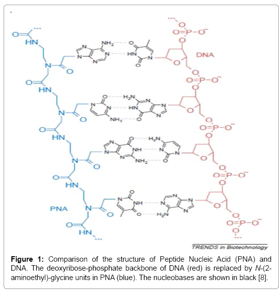
Figure 1: Comparison of the structure of Peptide Nucleic Acid (PNA) and DNA. The deoxyribose-phosphate backbone of DNA (red) is replaced by N-(2- aminoethyl)-glycine units in PNA (blue). The nucleobases are shown in black [8].
PNA binding with complementary DNA and/ RNA on the basis of Watson-Crick (WC) base pairs then formed duplexes. The combinations of WC and Hoongsteen (HG) hydrogen bonding have formed triplexes due to their natural backbone [8-10]. The PNA probe has offered greater affinity in binding to complementary DNA. PNA is an invaluable tool in molecular biology due to its biological stability, the excellent nucleic acids binding properties and the appreciable chemical simplicity, and improved distinction between closely-related sequences (including single-base mismatches) [11]. Such mismatch discrimination has a particular importance in the detection of diseaserelated mutations [12]. In spite of the radical difference in the chemical composition of the backbone, PNA is not only retained but also improved the hybridization characteristics of RNA and DNA [13].
Natural oligonucleotides are attributed the lack of electrostatic repulsion comparing to the stronger binding properties of PNA [14]. However, under low-salt concentration, the neutral amide backbone is enabled PNA to hybridize with DNA molecules because of no positive ions are necessary for counteracting the inter-strand repulsion that hampers duplex formation between two negatively charged nucleic acids [15]. This review paper is overcome these disadvantages, focused on the hybridization characteristics of PNA conjugates and it importance’s of PNA, and applications of PNA in the diagnostic and pharmaceutical fields.
PNA usually synthesized via solid phase techniques, either by manual or automated [14,16]. In large scale (>20 μM) of PNA is more efficient way synthesized through manual way synthesis. PNA synthesis is accomplished using monomers protected by either t-Boc or Fmoc protecting groups. Manual synthesis is mainly time consuming [17]. On the other hand, automated synthesis can be produced long sequences in few hours but required substantial investment in instrumentation [18]. There is a main problem encountered during synthesis of PNA, which is the tendency of the growing PNA oligomer to fold over itself (aggregate). A low solubility of PNA is occurred in organic solvents, the complementary recognition and binding of the nucleobases on the chain. Poletti and Giannini have developed a new technique using ionic liquid as reaction media rather than classical organic solvents for PNA synthesis in order to overcome the chain aggregation; the reactivity of coupling reaction increases the final yield [16]. Ionic liquids have simply been used to replace organic solvents, mainly due to their lack of vapor pressure; they have often led to improve process. The advantages of using ionic liquids over the use of organic solvents as reaction medium for biocatalysis include enhancement of enzyme activity, stability and selectivity [19].
The protocols of manual synthesis are often adapted in an automated synthesis. The synthesis by Fmoc chemistry of PNA oligomers protocol has developed by Egholm and Casale for the automated synthesis of PNA oligomers using Fmoc/Bhoc protected PNA monomers [20]. The manual coupling of Fmoc/Bhoc has efficiently done the synthesis using activators, HOBT/HBTU, in the presence of N-Methyl Morpholine (NMM)/pyrimidine as bases [21]. Thus, large amounts of pyrimidine is higher the yield of oligomers because of high contents of purine. PNA’s analysis and purifications have used by RP-HPLC [22].
PNAs showed several attractive features such as high chemical and thermal stability, resistance to enzymatic degradation, and stable binding to their RNA or DNA targets in a sequence-specific manner. However, the main hindrance to the effective use of PNAs is their poor uptake by cells as well as the difficult and laborious chemical synthesis. Joshi et al. have developed the automated protocol as well as cost-effective semi-automated synthesis of PNAs and PNA-peptide constructs on an automated peptide synthesizer [23]. The facile synthesis of PNAs can helpful in generating PNA libraries usable, e.g. for high-throughput screening in biomolecular studies. Efficient synthetic schemes, the automated procedure, the reduced consumption of costly reagents, and the high purity of the products are attractive features of the procedure.
Modifications of PNA (α-, β- and γ- position) are showed superior properties such as water solubility, high binding affinity and specificity [14]. Comparison of the effects of substituents at α- position with those of the γ-position has found that γ-modification is more effectively improved than the DNA binding ability. The β-position of the PNA backbone corresponds to the C4’ of the deoxyribose moiety of DNA and the C4’ is a chiral carbon atom. The introduction of a chiral center at the β- position may significantly affect the conformation and the DNA binding ability of PNA oligomers [10]. Sugiyama et al. have discussed a new PNA monomer possessing a methyl group of S-configuration at the β- position of the PNA backbone [10]. In chiral amino acid based PNAs, the molecules are characterized in the presence of one or more chiral monomers substituted in the position 2, 5 or both with amino acid derived side chain. The modifications of these side chains have increased the complementarities with the nucleic acid sequences and improved the specificity of mismatch recognition. New class of chiral PNAs, namely arginine-based PNA (Arg-PNAs) has synthesized by inserting an arginine in position 2 or 5 or both of the backbone. The binding performance of the PNA is evaluated the melting temperature of perfect match/ mismatch PNA-DNA/PNA-RNA hybrids and never observable effect on the binding affinity and selectivity toward DNA [24].
Inherently cationic and chiral Cγ–aminopropylene Peptide Nucleic Acid (amp-PNA) have been synthesized with three carbon spacer chain from the backbone [25,26]. The modified amp-PNAs have showed to stabilize (DTm) the PNA: DNA duplexes better than that of corresponding PNA: RNA duplexes. However, the PNA: RNA duplexes have higher Thermal stability (Tm) compared to PNA: DNA duplexes for both modified and unmodified PNAs.
Important properties of PNA
Thermal stability (Tm) of PNA and its hybrid complexes: The stability of the hybridization has formed between the two strands can be assessed with different biophysical techniques. Thermodynamics of the thermal dissociation transitions of 10 bp PNA/DNA duplexes and their corresponding DNA/DNA duplexes in 10 mM sodium phosphate buffer (pH 7.0) are determined from Differential Scanning Calorimetry (DSC) measurements [23,24]. The PNA/DNA transition temperatures ranged from 329 to 343 K and the calorimetric transition enthalpies ranged from 209 +/- 6 to 283 +/- 37 kJmol-1. The corresponding DNA/ DNA transition temperatures were 7-20 K lower and the transition enthalpies ranged from 72 +/- 29 to 236 +/- 24 kJmol-1. Agreement between the DSC and UV Monitored Melting (UVM) determined transition enthalpies validated analyzing the UVM transitions in terms of a two-state transition model. PNA (in contrast to DNA) duplexes show almost unaffected stability in up to 70% Dimethylformamide (DMF) or dioxane, and extrapolation of the data to conditions of 100% organic solvents indicates only minor (or no) destabilization of the PNA duplexes [27,28]. The differences in behaviour between the PNA and the DNA duplexes are attributed to the differences in hydration and counter ion release rather than to the differences in nucleobase interaction. These results support the possibility of having stable nucleobase paired double helices in organic solvents.
Therefore, Ahn et al. have developed a Peptide Nucleic Acid (PNA)- based fluorescence melting curve analysis (FMCA) method. PNA oligonucleotides have a much higher melting Temperature (Tm) value than DNA [29]. Short PNA probes have adequate Tm values for FMCA, and short probes have higher specificity and accuracy in FMCA. Moreover, dual-labelled PNA probes have self-quenching ability via single-strand base stacking, which makes PNA more favorable.
Stronger binding independent of salt concentration: The greater affinity of PNA/DNA or PNA/RNA complexes is reflected in their higher thermal stability. PNA sequences showed that the melting Temperatures (Tm) are higher for PNA hybrids than for either DNA/DNA or DNA/ RNA [30]. On average, the Tm of a PNA/DNA duplex is 1ºC higher for base pair compared to that of the corresponding DNA/DNA or DNA/ RNA duplex. The thermal stability of a PNA/DNA duplex is essentially independent of the salt concentration in the hybridization solution [31]. Unlike DNA, PNA does not need proper salt concentration for hybridization. The Tm of a 15-mer duplex decrease by only 5ºC as the NaCl concentration is raised from 10 mM to 1 M. PNA can act as a good probe to detect target sequence of secondary structure which is not stable in low salt concentration [32].
Strand invasion: Peptide Nucleic Acids (PNAs) are neutral and are commonly appended with positively charged lysine residues to enhance their solubility and binding affinity for nucleic acid targets. Thus obtained cationic PNAs very effectively interact with the designated duplex DNA targets in a sequence-specific manner forming strandinvasion complexes. Abibi et al. have reported that a typical range of salt concentrations used when working with strand-invading PNAs (10–20 mM NaCl) the PNA binding rates essentially do not depend on the presence of non-target DNA in the reaction mixture [33]. However, at lower salt concentrations (<10 mM NaCl), the rates of PNA binding to DNA targets are significantly slowed down by the excess of unrelated DNA. This effect of non-target DNA arises from depleting the concentration of free PNA capable of interacting with DNA target due to adhesion of positively charged PNA molecules on the negatively charged DNA duplex.
Homopyrimidine PNA oligomers are usually formed triplex structure of PNA/DNA/PNA. This triplex formation is able to form double-stranded DNA targets by a mechanism that involving the pyrimidine DNA strand. This triplex is formed by Watson-Crick basepairing (first PNA) and the other PNA held by Hoogsteen hydrogen base-pairing which produced the higher stability of triplexes, whereby, the “strand invasion” of DNA/DNA. As Figure 2 shows the two homopyrimidine PNA oligomers within the P-loop formed a (PNA) 2-DNA triplex [34]. Binding of the PNA resulting in the formation of a P-loop in the double-stranded DNA, where the PNA is displaced one of the DNA strands [15,35].
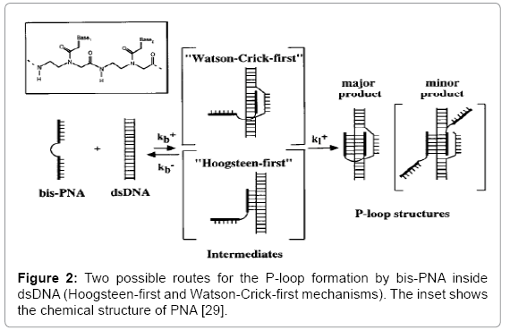
Figure 2: Two possible routes for the P-loop formation by bis-PNA inside dsDNA (Hoogsteen-first and Watson-Crick-first mechanisms). The inset shows the chemical structure of PNA [29].
Xiao et al. have developed very simple approaches to increase the efficiency of strand invasion [36]. First strategy, the use of PNA is facilitated an active primer-template formation and found that polyurine-polypyrimidine regions within PNA is promoted strand invasion when comprised the part of PNA. Yamamoto et al. are effectively used chiral PNA monomers bearing N-(aminoethyl)- D-lysine backbone, and introduced to PNAs bearing pseudocomplementary nucleobases [37]. Lysine side-chains have induced the electrostatic repulsion to suppress the PNA-PNA duplex formation, and hence, the PNA-DNA duplexes are stabilized by electrostatic repulsion as the strand invasion can be occurred. This modified PNA result is successfully invaded the target site in double-stranded DNA giving resistance to nucleases and proteases.
Peptide Nucleic Acid (PNA) is a mimic of DNA that shows a high chemical stability and can survive the enzymatic degradation of nucleases and proteases. The superior binding properties of PNA enable the formation of PNA/DNA or PNA/RNA duplex with excellent thermal stability and unique ionic strength effect [38]. The unique structure of PNA is composed of N-(2-aminoethyl) glycine units and thus gave PNAs resistance to DNAses and proteinases and higher affinity of binding to complementary ssRNA and DNA [15,39,40].
Solubility of PNA: Generally, the solubility of PNA decreases as the polymer length increases [41]. Furthermore, the synthesis, purification and use of purine-rich PNA oligomers is known to be especially problematic [41,42]. Attempts to improve the solubility of PNA oligomers have included terminal modification with charged amino acids such as L-lysine as well as modification of the aminoethylglycyl backbone [43-45]. These modifications, typically introduce reactive groups and/or chiral centers into the otherwise achiral and unreactive PNA structure. We opted to prepare N-(2-aminoethyl)-glycine (aeg) amino acids wherein the nucleobases of the standard PNA monomers were replaced with either neutral or positively charged hydrophilic moieties.
The hybridization strength is known as Thermal stability (Tm) of PNA-DNA duplexes and found more complex sequences while comparing to DNA-DNA duplexes. Targeting of double-stranded (ds) DNA with PNA has occurred at least four different binding modes which are triplex, triplex invasion, duplex invasion and double duplex invasion. PNA-DNA duplexes have higher thermal stability than that of DNA-DNA or DNA-RNA hybrids because of the PNA are neutral and are commonly appended with positively charged lysine residues to enhance their solubility and binding affinity for nucleic acid targets [7,15]. In order to the elimination of electrostatic repulsion between the two hybridized strands [46]. PNA-DNA hybridization has increased the electrochemical signal after hybridization of PNA with the ssDNA modified electrode [47].
De et al. have done the comparison study of the PNA-DNA duplex hybridization characteristics of vertically tethered and new horizontally tethered PNA probes on solid surfaces [5]. The horizontal 15-mer PNA probe is synthesized with linker molecules attached at three locations (γ-points) positioned along the PNA backbone that provided covalent attachment of the probe with the backbone aligned parallel to the surface, which is important for DNA hybridization assays that use electric field effect sensors for detection. A radioactive labelled assay and real-time Surface Plasmon Resonance (SPR) biosensor are used to assess the probe surface density, nonspecific binding, and DNA hybridization affinity, respectively, of the new PNA probe configuration.
Rashatasakhon et al. have studied the dendritic polycationic phenylene-ethylene as a FRET donor in the detection of DNA sequence via the PNA/DNA hybridization principle [37]. The fluorescin-labeled pyrolidinyl PNA (Fl-acpcPNA), the energy acceptor is showed superior specificity in the DNA hybridization comparing to the classical PNA. The complementary DNA sequence is able to detect by the sensing system at submicromolar concentration level and can distinguish it from the DNA with a single mismatch base.
In order to increase the PNA performance in the hybridization with complementary DNA, gold nanoparticle-PNA (AuNP-PNA) conjugates have produced surfactant polyoxyethylene (20) sorbitan monolaurate (Tween 20) [48,49]. The ability of AuNP-PNA conjugates have hybridized to its complementary DNA and verified using enzymelinked immunoassay. The hybridization of AuNP-PNA conjugates with its DNA complementary is showed the increase in TMB absorbance peak values due to the specific hybridization of conjugates with its DNA complementary and its ability to hybridize with its complement.
Immobilization of PNA probe onto the silica glass surface is hybridized with its complementary [50]. The development of immobilization method has introduced which commercially available reagents used, 3-Aminopropyltriethoxysilane (APTES), succinimidyl-4- (N-maleimidomethyl)cyclehexane-1-carboxy-(6-amino-caproate) (LCSMCC) and cysteinyl-PNA due to increase its hybridization properties. Various concentration of standard assay mixture, FITC-Tgt, is used to determine the absorptivity of the PNA-glass for complementary oligonucleotide and the result is found 50 pM of complementary target.
(PNA)2 -DNA triplexes: There is remarkably smaller binding constant of the PNA/DNA hetero-duplex with a single-base mismatch than that of the entirely matched one. Thus, the PNA probe is able to discriminate a single-base difference in the ssDNA analyte [51]. Binding of 2PNA/1DNA is through Watson-Crick base pairing while the second PNA strand binds by Hoongsteen base pairing [15].
Bis-PNA: Bis-PNA is contained two homopyrimidine PNA oligomers which are connected by a flexible linker. Under low salt concentration, the bis-PNA (“clamps”) is showed rapid strand invasion into both linear and supercoiled double-stranded (ds) DNA [52]. When, together uses of LNA and bisPNA have increased the specificity and stability of coupling specific functions to plasmids. The high biological stability of both PNAs and LNAs are made short LNAs as “opener”, in combination with the bisPNA as anchor molecules, an attractive method for attachment of biological functions to plasmids in a sequence-specific manner.
Applications of PNA
PNAs are a powerful molecular tool with a wide range of important applications. Due to their ability of interacting with high sequence specificity to a chosen target in a gene sequence, it’s have major interest in medicinal and biotechnological contexts [53,54]. They show promise for the development of gene therapeutic agents, diagnostic devices for genetic analysis, and as molecular tools for nucleic acid manipulations. In vitro studies indicate that PNAs could inhibit both transcription and translation of genes to which they have been targeted, which holds promise for their use for antigene and antisense therapy.
Antigene and antisense therapy: Antigene strategy using Oligonucleotides (ODNs) have showed potential against viral targets [55]. PNA has resistance towards nucleases and proteases digestion, which have a low affinity for proteins properties, thus PNAs can make an attractive agent for biological and medical applications [10]. PNA is successfully applied in antisense technology for their ability to bind a messenger RNA (mRNA). It has high biological stability and strong inhibiting translation of the target genes [56]. Antisense PNAs have higher hybridization affinity because of their natural backbones. PNAs also exhibit superior stability compared with other anti-sense agents due to nuclease resistant properties resulting from the replacement of the deoxyribose phosphate backbone with a polypeptide backbone [57,58]. Consequently, a new concept of drug design has exploited by several structural modification of oligonucleotides and to modulate the expression of genetic information by antisense and antigene oligonucleotides [3]. The antisense and antigene properties of PNAs have improved the binding affinity for DNA or RNA by suitable preorganization. Several backbone modifications of PNAs have explored under the concept of preorganization. However, only limited numbers of modifications have been improved the hybridization properties [10].
A new mRNA targeting contrast agent, compost of mRNA of a red fluorescent protein (DsRed) and an antisense PNA, which are specifically targeted to a complementary region of DsRed mRNA, which is developed and synthesized. This PNA can hybridize to its complementary mRNA in the cytosol and can provide cell specific targeting for cells expressing the respective mRNA. This new contrast agent is showed efficient cellular uptake and significant contrast enhancement at very low labelling concentrations of 0.5 μM [40].
Rajasekaran et al. have studied the inhibition of Brucella suis growth using PNA linked covalently to cell-penetrating peptides (CPPs) [2]. After 24 hour of treatment, Brucella genes are involved in DNA, RNA, cell envelope, fatty acid and protein synthesis that are inhibited the growth of B. suis in culture and macrophages. This study has revealed the potential of antisense PNA as novel therapeutic agents against intacellular Brucella.
Monoamines Oxidase (MAO) functions in catalysis of oxidative deamination of many biogenic amines. Differential expression of the MAO isoform, MAO-A gene, has linked to numerous central nervous disorders. An MAO-A mRNA imaging agent in live neuronal cells have developed and composed to a complementary Peptide Nucleic Acid (PNA). The PNA is labelled with fluorescent dye which is attached to a peptide moiety for receptor-mediated intracellular delivery. Incorporation of a receptor-targeting peptide is efficiently deliver PNA imaging agents into targeted cells. The result has showed very specific delivery of WT4879 (receptor-specific agent) to the human neuroblastoma cell line [59].
Dye-labelled PNA probe has hybridized with its complementary dsDNA reported by Baker et al. [60]. In advance biosensor, Fluorescence Resonance Energy Transfer (FRET) has utilizing water-soluble Cationic Conjugated Polymer (CCP) donors and dye-labelled PNA acceptors used as sensitive hybridization assays (Figure 3) [61]. FRET analysis has showed specific PNA/dsDNA binding by a positive (acceptor) signal with complementary dsDNA. The binding specificity between PNA and noncomplementary/ or complementary dsDNA are examined by several techniques. Hence, fluorescent dye labeled-PNA in conjugation with cationic conjugate polymer which is allowing a specific detection of dsDNA without denaturing step.
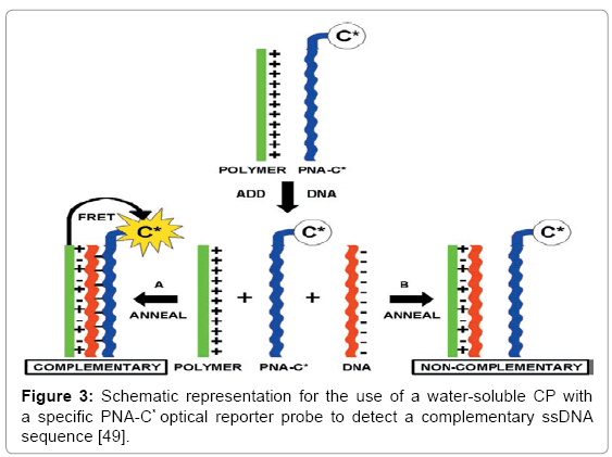
Figure 3: Schematic representation for the use of a water-soluble CP with a specific PNA-C* optical reporter probe to detect a complementary ssDNA sequence [49].
The cationic liposomes as non-viral vectors able to ionic bind to DNA molecule and deliver them in cells both in vitro and in vivo have described by Nastruzzi et al. [62]. Liposomes are composed at least a positive charged lipid which is interacted negatively charged DNA molecules generating stable complexes. PNAs never suitable delivered by cationic liposomes because of its neutral charged which cannot form electrostatic interactions with the positively charged liposomes. The binding of PNA/DNA molecules are increased the cationic liposomes efficiently complexate PNA/DNA hybrid and mediate their delivery to target cells.
Tridecamer PNAs are successfully antisense agents when targeted to the coding region (codon 12) of oncogenic Ha-ras mRNA forming stable duplexes [63]. PNA is inhibited the translation of mutated Ha-ras mRNA toward RNA processing, nucleoplasmic transport or translation, not lead to RNA degradation. The Tm value of the tridecamer PNARNA duplex has found to be high (Tm= 86 OC).
Single nucleotide polymorphisms (SNPs) detection using PNA: Single Nucleotide Polymorphisms (SNPs) are one of the major sources of genetic variation in human genome. Diseases such as cystic fibrosis, thalassemia and Azhemeir’s disease are related to SNPs. Kaijuan et al. have developed PNA-DNA films on gold electrodes with Ni2+ ions to detect single-nucleotide mismatch in genotype of apoE 4 [64]. It can be associated with lipid disorders and related to Alzheimer’s disease by Electrochemical Impedance Spectroscopy (EIS). The single-nucleotide mismatches can be detected directly from DNA fragments isolated from blood or cell lysates samples with PNA as a capture probe. Moreover, Tsukada et al. have studied the SNP allele frequencies estimation in pools of hairpin structured ssDNA analytes with secondary structures [51]. They are developed a block copolymer composed of an allelespecific PNA probe modified with polyethylene glycol, which is disrupted secondary structures of analysts and hybridized during electrophoresis. PEG has served as a good probe for Affinity Capillary Electrophoresis (ACE). The DNA-binding capability has allowed the separation of the folded analytes with a single-base difference within 20 minutes. Besides, Hyou et al. have reported successful PNA microarray based method to detect mutation in human gene, BRCA1, which is involved in over 50% of breasts and ovarian cancer [65]. This technique has several advantages on the cleavage of single-specific strand nuclease which is specific for mismatched sequences in heteroduplex DNAs formed between SSS probes and target strands.
Zhang et al. have developed a highly sensitive and selective method for protein or hormone markers assay [66]. This method takes advantage of PNA-coupled dual amplification simultaneously both in solution and on electrode interface, as well as Zr4+ mediated phosphate group’s interaction, receiving greatly increased sensitivity of the detection and efficiently monitor the insulin level for the evaluation of serum insulin in pregnant women.
PNA fluorescence in situ hybridization (PNA FISH): FISH is a molecular assay for detection of microbe. It has developed in the combination of the microscopy observation and the specificity hybridization of DNA/rRNA [67]. The detection of Lactobacillus genus by Fluorescent In Situ Hybridization (FISH) method using Lac663 PNA probe has found high probe specificity and selectivity when hybridized with all Lactobacillus collection strains rather than hybridization with other species and non-Lactobacillus strains [68]. When tested with fresh milk samples added with Lactobacillus strains at probiotic range concentrations, other taxonomically bacteria and pathogenic bacteria, the Lac663 probe exclusively detected of Lactobacillus spp. with concentration of 1 x 10⁷ CFU/mL. Xiao Feng et al. have developed fluorescein-labelled PNA probes using 16S rRNA-based FISH method to allow in situ detection of Listeria species [69]. Keefe et al. also have developed fluorescently labelled PNA-FISH probe targeting specific rRNA sequences of bacterial species consisted of gram-negative and gram-positive bacteria and yeast species for rapid detection [70]. Short PNA able to penetrate a hydrophobic cell wall and hybridized with rRNA without additional enzymatic permeabilization steps and required simpler fixation step. PNA FISH method is allowed the morphology examination in combination with specific bacterial staining provided by PNA-FISH. Thermotolerant of Campylobacter spp. (C. coli, C. jejuni and C. lari) have detection using rapid PNA FISH methods [67]. The sequence of PNA based on the 16S rRNA sequence of C. jejuni. The same sequence as PNA probe is applied to a conventional DNA probe, after that, the DNA probe has failed to hybridize with the target sequence. Because of PNA has good permeability and higher affinity to the Class 3-4 rRNA sites. Simple fluorescence microscopy analysis has able to accurate detection at the presence of 18S rRNA from the parasite Trypanosoma brucei [71]. By coating a pair of biotinylated PNA with streptavidin fluorescent beads, to which these PNA oligomers probes can be coupled to create a hybridization co-localization detection method for nucleic acids. Development of PNA probe is allowed the detection of Takayama pulchella with high specificity, sensitivity using application of FISH in combination with epifluorescence microscopy and flow cytometry [72].
A peptide nucleic acid fluorescence in situ hybridization (PNA– FISH) method has developed for specific detection of the Vibrio genus reported by Zhang et al. [73]. In silico analysis by BLAST and ProbeCheck showed that the designed PNA probe targeting the 16S rRNAs is suitable for specific identification of Vibrio. Specificity and sensitivity of the probe Vib-16S-1 are experimentally verified by its reactivity against 18 strains of 9 Vibrio species and 14 non-Vibrio strains of 14 representative species. The PNA–FISH assay has able to identify 47 Vibrio positive samples from selectively enriched cultures of 510 samples of aquatic products and environments, comparable with the results obtained by biochemical identification and real-time PCR. So they concluded that PNA–FISH can be an alternative method for rapid identification of Vibrio species in a broad spectrum of seafood or related samples.
PNA as biosensor for nucleic acid: The developments of biosensors have high sensitivity and selectivity. It has paramount importance in application of clinical diagnostics, genomics and drug discovery. Detection of mutation point in the human p53 gene using PNA biosensor has explored by Wang et al. [74]. A 17-mer PNA with a lysine residue is immobilized on the Carbon Paste Electrode (CPE) with incorporation of the Co (phen) 33+ as the label binding. The hybridization of PNA probe with its complementary p53 sequence has monitored by the high sensitive constant-current chronopotentiometry. The PNA biosensor is detected until sub-micromolar and micromolar concentration of the p53 target sequence.
Newcastle Disease Virus (NDV) RNA detection method has developed using label-free visual assay, PNA probes and gold nanoparticles [75]. In this assay, the specific agglomerative behavior of PNA with gold nanoparticles have manipulated by its complementation with NDV RNA. When the PNA is hybridized with its complement, PNA complex is retained the stability of gold nanoparticles and preventing in color changing. This phenomenon forms on the basis of label-free viral RNA detection using gene-specific PNA probes and gold nanoparticles.
Kerman et al. have developed the detection Single-Nucleotide Polymorphisms (SNPs) using electrochemical DNA biosensor, with the addition of ferrocene-conjugated Chitosan nanoparticles (Chi- Fc) as the electroactive hybridization indicator [39]. PNA probe is immobilized onto the surface of gold electrode; has hybridized with its complementary target DNA, whereby the Chi-Fc electrostatically binds to the negatively charged phosphate backbone of DNA. This attachment of Chi-Fc raises a high electrochemical oxidation signal from ferrocene. SNP in target DNA is determined by monitoring the changes in the electrical current response of Chi-Fc.
High sensitive and specific detection of single-stranded oligonucleotides using nanoxidized Silicon Nanowires (SiNWs) have found the detection limit of 10 Fm. Fluorescently labelled complementary DNA has able to the SiNWs immobilized with PNA. Strong fluorescent signals on the SiNWs are obtained. This fluorescent imaging is indicated the hybridization of target DNA with the PNA probe [76].
Electrochemical parameters are used Methylene Blue (MB) as a redox indicator on binding to DNA at Hanging Mercury Drop Electrode (HMDE), Glassy Carbon Electrode (GCE), and Carbon Paste Electrode (CPE) in the solution and at the electrode surface. MB, which interacts with the immobilized calf thymus DNA, was detected by using singlestranded DNA-modified HMDE or CPE (ssDNA-modified HMDE or CPE), bare HMDE or CPE, and double-stranded DNA-modified HMDE or CPE (dsDNA-modified HMDE or CPE) in combination with Adsorptive Transfer Stripping Voltammetry (AdTSV), Differential Pulse Voltammetry (DPV), and Alternating Current Voltammetry (ACV) techniques [77]. The structural conformation of DNA and hybridization between synthetic Peptide Nucleic Acid (PNA) and DNA oligonucleotides are determined by the changes in the voltammetric peak of MB. The PNA and DNA probes are also challenged with excessive and equal amount of non-complementary DNA and a mixture that contained one-base mismatched and target DNA. The partition coefficient has obtained from the signal of MB with probe, hybrid, and ssDNA-modified GCEs. These results demonstrated that MB could be used as an effective electroactive hybridization indicator for DNA biosensors.
The increase in the signal of MB at the ssDNA-modified electrode has showed the interaction of MB with guanine bases. Therefore, when comparing between DNA and PNA probe, the selectivity of the DNA probe for mismatched DNA target sequence has found to be poorer than that of the PNA probe. Thus, the MB resembled signal has obtained from the DNA hybrid-modified CPE. Danxin et al. have fabricated electrochemical DNA biosensor based on electrochemical reduced graphene oxide (ERGO) on the glassy carbon electrode [47]. A 1-Pyrenebutanoic Acid Succinimidyl Ester (PASE) is used as a linker to immobilize the PNA probe onto the surface of the ERGO. The PNA biosensor is detected with different concentration of target DNA in range of 1.0 × 10⁻⁷ to 1.0 × 10⁻¹² mol L-1 with a detection limit of 5.45 × 10⁻¹³ mol L-1.
Thiolated pyrrolidinyl - Peptide Nucleic Acid (HS-PNA) is bearing D-prolyl-2-Aminocyclopentanecarboxylix Acid (ACPC) backbone. Thermodynamics stability of (2’R,4’R)-acpcPNA.DNA showed higher than (2’R,4’S )-acpcPNA.DNA [78]. The analysis of HS-PNA is assessed by Surface Plasmon Resonance (SPR), an alternative for non-labeling technique. The immobilization of HS-PNA on gold-coated glass is through the formation of Self-Assembled Monolayer (SAM). The effects of spacer and blocking thiol are showed the increases in sensor surface for improving PNA/DNA hybridization reaction. The developed biosensor method can be detected at lower target DNA concentration of 2 μM which equivalent to 10 pM that could differentiate between completely complementary and single-mismatched hybridization. In similar study done by Ananthanawat et al. using aegPNA and acpcPNA /or pyrrolidinyl PNA to interact with dsDNA [79]. The binding of PNA to dsDNA has resulting in Triplex-Forming Oligonucleotides (TFOs). TFOs have bright potential in gene-targeted therapeutic, can inhibit gene expression or induce target mutagenesis. In this study has found that the aegPNA able to bind with dsDNA to form triplexes. On the other hand, the conformationally rigid acpcPNA is not bind with dsDNA in the SPR measurement. Due to the lower conformational flexibility of the acpcPNA backbone, may decrease its ability to find the complementary region in the center of the immobilized dsDNA. Another studied of 16S rRNA detection of E. coli using SPR biosensor system has developed using PNA probe by Hyou et al. [65]. 16S rRNA is hybridized on the PNA probe-immobilized SPR sensor chip, with ionic interaction with gold nanoparticles. The detection limit of E. coli rRNA is 58.2 ±1.37 pg mL-1. The developed SPR detection system is highly sensitive in identification of bacteria.
SAM of thiol-derivatized PNA is successfully again developed by Martí et al. and mentioned that PNA with the terminal cysteine has provided a thiol group that allows binding to gold surface and detected a low concentration of PNA (1 μM) of target DNA [80]. Reflective Absorption Infrared Spectroscopy (RAIRS) analysis is showed the PNA probe successfully adsorbed on the gold surface. A label-free electrochemical detection method using PNA probes have been reported by Bin et al [81]. In their approach, 20-mer PNA probe is immobilized onto a gold electrode transducer, and the formation of the PNA/DNA hybrids is detected using ferrocene-containing cationic polythiophene. This ferrocene-containing cationic polythiophene has adsorbed on the DNA backbone, resulting clear hybridization detection signal. The detection limit is calculated at 1.0 × 10⁻¹¹ mol L-1. The same approach also used by Raymond et al. using fluorescent cationic polythiophenes, which is eliminated the labeling step of the probe [82]. The cationic polymer bind strongly with the negatively-charged backbone of the complementary oligonucletides bound to PNA probes. Properties of PNA and LNA capture probes are investigated in the development of an electrochemical hybridization assay of enzymatic product.
Laschi et al. used streptavidin-coated paramagnetic micro-beads as a solid support to immobilize biotinylated DNA, PNA and LNA [83]. These paramagnetic micro-beads are well known versatile solid support for electrochemical nucleic acid biosensing. The electrochemical detection of biotinylated DNA as well as RNA sequences has performed on the surface of a disposable electrode. The detection limits have found to be 152, 118 and 91 pM for DNA, PNA and LNA probes, respectively, with their target DNA. For DNA target, the detection limits are 51, 60 and 78 pM for DNA, PNA and LNA probes, respectively.
PNA based biosensor is immobilized with probe PNA on gold surfaces via a thiol linker (DTSP, 3, 3’-dithio-bis(propionic acid N-hydroxysuccinimide ester)) in two different ways; namely onestep- immobilization and two-step-immobilization [84]. In one-stepimmobilization, an equal amount of PNA probes and DTSP are dropped onto the surface of gold surface. For two-step-immobilization, the gold surface is modified with DTSP following the immobilization of PNA on the modified gold layer. These two different methods of immobilization are analyzed using TOF-SIMS measurements. The binding of PNA is significantly longer than 24 h of DTSP layer attached to the surface. This result showed that the two-step-immobilization method is more preferable to the first one.
Ali et al. have explored the PNA-modified nanofluidic device for the uses high selective sequence of a short ssDNA [85]. An amineterminated PNA probe is covalently immobilized on the inner wall of a single conical nanochannel through carbodiimide coupling method. The immobilization and hybridization of PNA/DNA duplex on the inner walls are monitored through the change in the rectified ionic flux and perm-selectivity is displayed by the single conical nanochannels.
PNA-modified Ion-Sensitive Field-Effect Transistor (IS-FET)-based biosensor is used for direct detection of DNA hybridization without any reporter molecules and labeling [86]. IS-FET-based biosensors are very attractive because of their smaller in size and have light weight, high reliability, fast response and low cost of mass production. PNA has immobilized on a silicon nitride gate insulator. On the immobilization surface, the succinimide group of N-(6-maleimideocaproyl-oxy) sucinimide is reacted with the thiol group of the terminal cysteine in PNA. This study has showed a good correlation between the calculated dissociation equilibrium constant (KD) and the measure Tm from those PNA-DNA duplexes.
Cysteine-labeled PNA can efficiently immobilize onto crystalgold surface at 200ºC [87]. A 15-mer cysteine labeled-PNA sequence is attached with gold surface on top of a quartz crystal using ethylene glycol linker. Application of physical transducer of a quartz crystal microbalance has offered an in-situ detection of nucleic acid hybridization events. This concept is able to detect a single mismatch in a 15-mer p53 tumor suppresser gene.
Label-free electrochemical DNA biosensor incorporation with cationic ruthenium complexes [Ru(NH3)6]3+ is successfully applied in the detection of 23S rRNA gene of Helicobacter pylori by using PNA probes immobilized onto the gold electrode surface [88]. Helicobacter pylori are a gram negative, slow growing, and spiral in shaped and flagellated bacteria causing ulcers. The lack of the negative charges of PNA is allowed strong binding with ssRNA. After the hybridization of PNA with DNA form, [Ru(NH3)6]3+ is interacted with the negatively charged phosphate groups and adsorbed on the DNA backbone which is giving a clear hybridization detection signal. The biosensor has able to detect of H. pylori gene with a good selectivity and sensitivity.
According to new Leaky Surface Acoustic Wave (LSAW) bis-PNA probe is constructed electrochemical biosensor to detect Human Papiloma Virus (HPV) sequences more effective with high specificity [89]. LSAW is confined strongly on the surface of the device in the range of the acoustic wave. Bis-PNA probe is immobilized onto the gold electrode surface of LSAW via thiol linker where target DNA sequence in the range from 1 pg/L to 100 μg/L of HPV detection. Despite using synthetic oligonucleotides target, the detection of HPV in the clinical samples are successfully performed and the detection limit is calculated of 1.21 pg/L to 0.78 μg/L. Rapid detection method of HPV is successfully employed without PCR amplification.
Another study employing PNA as capture probe in DNA biosensor is done by Pita et al. [90]. They are synthesized gold-covered by ferrite nanoparticles, functionalized with amino and thiol groups to facilitate the electrostatic attraction and further chemisorpton of the gold nanoparticles. The thiolated-PNA onto gold-magnetic nanoparticles is immobilized via SAM on gold surfaces. The hybridization signal of PNA with target DNA is monitored by the detection of fluorescent dye marker, Rhodamine 6G, which intercalates into the hybrids.
Detection of HER2 oncogene, which encodes a 185 kDa tyrosine kinase in the family of human epidermal growth factor receptors have been reported by Metaferia et al. [91]. Over-expression of HER2 oncogene constitutes 20–30% in breast cancer patient. HER2 PNA probe is designated to detect HER2 mRNA. PNA probe has amine terminated mini-PEG linkers for efficient conjugation to the carboxymodified beads, glass slides and gold chips. The PNA probes have able to discriminate between the wild type HER2 target and the mutant target.
PNA has high binding affinity towards complementary DNA sequence when comparing to DNA probe in label-free potentiometric biosensor [92]. Hybridization event of probe and target DNA have changed the charge density at the solution/electrode interface on the SAM-forms microelectrodes. This charge density directly transformed into potentiometric signals. The length of probe has influenced their sensitivity in biosensor performance.
Accordingly antisense PNA is highly inhibited Duck Hepatitis B Virus (DHBV) Reverse Transcription (RT) and has found PNA targeting the bulge and upper stem of HBV encapsidation signal, ε RNA, appeared as more efficient RT inhibitor than the PNA targeting only the bulge [55]. However, the PNA has high affinity towards RNA; the ε RNA sequence has highly conserved and reduced the opportunities to escape inhibition by mutation in this region.
Two types of PNA-PEG-functionalized manolayers on alkylphosphonates and alkenylsiloxanes on silicon oxide-coated silicon surfaces have been synthesized [93]. Phopsphonate SAM is formed with PNA using a maleimido-terminated PEG45 meanwhile siloxanes monolayers are functionalized with PNA using a maleimido-terminated PEG45 spacer and modified with a shorter methoxy-terminated PEG12. They have found both hybridization of complementary cDNA and noncDNA using phosphonate and siloxane biofunctional surfaces are effective with minimal nonspecific adsorption of analyte.
Peptide nucleic acid is a novel class of compound which is applied various applications in the field of biology. It has very specific interactions with DNA and RNA and their chemical and biological stability make them promising in wide applications. The main advantage of PNA is combination of many features that are very similar to those of natural DNA with additional characteristics that are rather different. Development of PNA technology is gaining momentum, and several applications exploiting the unique properties of PNA, the ones that actual distinguish it from DNA, are being taken advantage. PNA has found more exciting new and very powerful detection methods, such as ‘sensors’ are being employed. The chemical and biological consequences of hybrid structure; PNA has a unique position compared with many other nucleic acid derivatives and could make an impact particularly in the fields of antigene and antisense therapy and probe-based assays. There is very prospect in the near future, with the collaboration of biologists and chemists, PNA can comprehend in entirely its massive practical potential.
This work was supported by grants from the Ministry of Science, Technology and Innovation, MOSTI (No. SCF0090- IND- 2013).