Journal of Research and Development
Open Access
ISSN: 2311-3278
ISSN: 2311-3278
Review Article - (2023)Volume 11, Issue 3
Brain is the nonsupervisory unit in mortal body. It controls the places similar as mind, unreality, hail, knowledge, personality, case working, etc. Sometime brain faces monumental case like excrescence and can’t do its work duly. Brain excrescence occurs owing to unbridled and rapid-fire excrescency of cells. Brain excrescence is considered as one of the ambitious conditions, among children and grown-ups. Brain excrescences grow veritably presto and if not treated well, the survival chances of the case are veritably less. Currently, brain excrescence discovery has turned up as a general reason in the demesne of health care. The usual system to descry brain excrescence is glamorous Resonance Imaging (MRI) reviews. From the MRI images information about the anomalous towel excrescency in the brain is linked. There are colorful ways to descry brain tumor. This paper uses colorful ways for discovery like Artificial Neural Network (ANN), Complication Neural Network (CNN), Visual Figure Group (VGG 16), (VGG19), DenseNet-121, Resnet-50, LeNet to descry the excrescence grounded on the MRI images. The most competent and operative algorithms are bandied in this paper after studying a number of applicable algorithm. In the testing data, the algorithms outperformed the current usual approaches for detecting brain excrescences and achieved an excellent delicacy of CNN 96%, VGG-16%-96.5%, VGG-19%-99.20%, DenseNet-121-95, Resnet-50 75.50%, LeNet 98%. Experimental effects have shown off that VGG-19 gives better delicacy as assimilated (VGG 16) DenseNet-121, Resnet-50, LeNet.
(VGG 16); (VGG19); DenseNet-121; Resnet-50; LeNet; MRI
The operation of information technology and machine literacy in drug has gained significance in moment’s world. Artificial intelligence is a scientific field concerned with developing a machine that can learn on its own without mortal intervention to prepare itself for dealing with implicit cases on its own. Operation of this wisdom finds high applicability in developing interventions for brain excrescences, as the excrescence cells show largely uncertain geste that's too complex to be controlled through conventional drug. The brain is a most important organ in the mortal body which controls the entire functionality of other organs and helps in decision timber. It's primarily the control center of the central nervous system and is responsible for performing the diurnal voluntary and involuntary conditioning in the mortal body [1]. The excrescence is a stringy mesh of unwanted towel growth inside our brain that proliferates in an unconstrained way. On this time at age of 15 about, 540 children get diagnosed with a brain excrescence. The right way of understanding of brain excrescence and its stages is an important task to help and to carry out the way in curing the illness. To do so, Glamorous Resonance Imaging (MRI) is extensively used by radiologists to dissect brain excrescences. The result of the analysis carried out in this paper reveals whether the brain is normal one or diseased bone by applying the deep literacy ways. At present, opinion and operation are grounded upon clinical and radiological information. Glamorous Resonance Imaging (MRI) is the dependence for the assessment of cases with brain excrescences, although conventional imaging has significant limitations in assessing excrescence extent, prognosticating grade, and assessing treatment response. New accession ways are under development to ameliorate lesion characterization, remedy assessment and operation; still, new approaches to image analysis have been gaining traction thanks to the wealth of information held by radiological images. Automatic disfigurement recognition in medical imaging has surfaced as a promising field for colorful medical individual procedures. The discovery and shadowing of excrescences in glamorous Resonance Imaging (MRI) is pivotal because it offers details about abnormal, demanded for remedial interventions. MRI brain excrescence discovery is a complicated task, due to the complications and different forms of excrescences. Collecting, organizing, and assaying medical images has come digitized in moment’s digital realm. In this paper the performance of colorful ways are analysis and we get what's the stylish algorithm for discovery a tumor.
A group of irregular brain cells make a brain excrescence. Nebulous evolution in the synapses characterizes brain excrescences. The high hardness and strictness of the cranium makes it tricky to contain implicit expansion in measure. The jolt may be endured in the shape of hindrance with mortal capacities along with swelling in to other body corridor [2]. Over 130 non identical brain, and intermediary anxious, diseases categorize from benign to nasty, exceedingly delicate to nicely common or garden. Still, each of the 130 brain excrescences is codified as either primary or secondary. Meiyan Huang, et al., presents utilizing the LIPC (original independent protuberancegrounded bracket) system for categorizing the voxel of the brain. Also utilizing this system, path point is uprooted. Unequivocal regularization need not be performed in LIPC. Low delicacy is achieved. BjoernH, Menze, et al., presents new brain excrescence segmentation also appertained to as multimodal brain excrescence segmentation gambit. Colorful segmentation algorithm are being connected to gain better interpretation in discrepancy to the being system. Though, still it depicts high complication. Shamsul Huda, et al., presents cold-blooded point election utilizing ensemble bracket for per forming brain excrescence opinion. For acquiring of resolution regulations, resolution tree, GANNIGMAC, Bagging C grounded wrapper path are espoused and the resolution regulations are simplified by making use of mongrel point election that merges (Decision Tree+MRMR C+GANNIGMAC+Bagging C. Sergio Pereira, et al., presents automated styles for brain excrescences relating and type listing by exercising MRI images of brain birthright from the original time when one could essay to overlook and freight medical images in the computer system. On the negative, NN (Neural Networks) and SVM (Brace Vector Machine) being the generally espoused styles recently as they extend better interpretation [3].
Data set
The dataset is taken from Kaggle. This dataset contains MRI images of brain excrescence. There are two flyers one represents the normal brain image and the other represents the excrescence images. Completely there are 2065 images in both these flyers. Figure 1 shows the sample normal and brain excrescence image. Completely 1085 tumorous and 980 non-tumorous images are taken. The images are of different shapes (e.g. 630X, 225 X225) and these images are resized to 256 × 256.1672 images for training, 186 images for confirmation and 207 images for testing is taken [4]. Out of 1672 training image, 877 images are excrescence image and 795 images are non-excrescence image. 92 excrescence and 94 non excrescence images taken from 186 confirmation images. Among 207 testing images, 116 excrescence images and 91 non excrescence images (Figure 1).
Figure 1: Normal brain and brain with tumor preprocessing.
Preprocessing
Before feeding the MRI images to the proposed structure preprocessing step is performed on the images. In the image preprocessing part first image data generator system of Keras library is used to induce batches of tensor image data with real time data addition so that our model gets different types of images. Also the preprocess input parameter of the image data generator system of Keras library is set to acceptable MRI image to the format the model requires preprocessing. All the necessary steps of including cropping of image, gyration of image, defining brilliance range, flipping the image and chancing the 4 blockish boundaries equals of the cropped image is done inside this image data generator system of Keras library [5].
Data modelling
After we've standardized our entire dataset we've slitted our dataset into training set as well as test set. 80 data is used for training and 20 data is used for testing. Also using image data generator, images are spitted into test and training sets.
Proposed method
In this paper we proffered Convolution Neural Network (CNN), Visual Figure Group (VGG 16), (VGG19), DenseNet-121, Resnet-50, LeNet to descry the excrescence grounded on the MRI images. Then we also study and descry the styles delicacy and loss. These styles contain the following pre-processing in the MR image there are several inapplicable bowwows, consequently we've to ameliorate the quality of the MR images by preprocessing styles. The main task in pre-processing is to ameliorate the single bruit rate, enhancing the visual appearance of the MR image, removing the bruit and uninvited corridor in the ground, smoothing the inner corridor and conserve the bite skull disrobing skull disrobing is an important process in biomedical image dissection with the help of this process we can examination of brain excrescence from MR images. By this process it's practicable to remove fresh napkins similar as redundancy, face, and cranium in the brain image. There are numerous ways for cranium stripping and other popular ways like spontaneous cranium disrobing utilizing image figure, cranium disrobing grounded on segmentation and morphological operation, and cranium disrobing grounded on histogram dissection or a threshold value. Morphological operation the morphological operation is exercised for the excerpt the boundary areas from the brain images [6]. This operation is only rearranging the relative order of pixel value, not fine value, so it's able for only double images. Dilation and corrosion is introductory operation of morphology. Dilation adds pixels to the boundary region of the object, while corrosion removes the pixel from the boundary region of the object. Point birth point birth is the process of collect advanced position information about image like shape, texture, color and discrepancy. Texture dissection is an important parameter of the mortal visual perception and engine literacy system. Texture finding and dissection can ameliorate opinion at non identical stage of the excrescence discovery. It's exercised for effectively ameliorate the delicacy of opinion system by opting statistic features like mean, discrepancy, dynamism, entropy, standardissue divagation, skewness, kurtosis etc.
Complication Neural Network (CNN)
Convolutional neural networks are a technical type of artificial neural networks that use a fine operation called complication in place of general matrix addition in at least one of their layers. They're specifically designed to reuse pixel data and are used in image recognition and processing. Convolutional neural networks are distinguished from other neural networks by their superior performance with image, speech, or audio signal inputs. They've three main types of layers, which are:
• Convolutional sub caste.
• Pooling sub caste.
• Completely connected (FC) sub caste.
The convolutional sub caste is the first sub caste of a convolutional network. While convolutional layers can be followed by fresh convolutional layers or pooling layers, the completely connected sub caste is the final sub caste. With each sub caste, the CNN increases in its complexity, relating lesser portions of the image. Before layers concentrate on simple features, similar as colors and edges. As the image data progresses through the layers of the CNN, it starts to fete larger rudiments or shapes of the object until it eventually identifies the willed object (Figure 2).
Figure 2: Input into a single node within a convolutional neural network.
Visual figure group (VGG 16)
VGG is a notorious CNN armature that first showed it was possible to do accurate image recognition with a deep network and small convolutional pollutants. It proved this to the world scoring largely in the image net challenge. VGG-16 is a convolutional neural network that's 16 layers deep. You can load a pre trained interpretation of the network trained on further than a million images from the ImageNet database. VGG model is generated for brain excrescence discovery. The training is followed by testing and confirmation which dissect the model performance and delicacy. The mode provides us with probabilistic affair which helps us to descry the MRI has brain excrescence (Figure 3).
Figure 3: Block diagram of brain tumor detection system using VGG-16 architecture.
VGG-19
VGG stands for visual figure group; it's a standard deep Convolutional Neural Network (CNN) armature with multiple layers. The “deep” refers to the number of layers with VGG-16 or VGG-19 conforming of 16 and 19 convolutional layers. The VGG armature is the base of ground breaking object recognition models (Figure 4).
Figure 4: VGG-19.
DenseNet-121
In a traditional feed forward Convolutional Neural Network (CNN), each convolutional sub caste except the first one (which takes in the input), receives the affair of the former convolutional sub caste and produces an affair point chart that's also passed on to the coming convolutional sub caste. Thus, for 'L' layers, there are 'L' direct connections; one between each sub caste and the coming sub caste. Still, as the number of layers in the CNN boost, i.e. as they get deeper, the' evaporating grade' case arises. This means that as the path for information from the input to the affair layers increases, it can beget certain information to' evaporate' or get lost which reduces the capability of the network to train effectively [7]. Factors of DenseNet carry:
• Connectivity.
• Dense blocks.
• Excrescency rate.
• Tailback layers.
DenseNets resolve this case by modifying the standard-issue CNN armature and simplifying the connectivity pattern between layers. In a DenseNet armature, each sub caste is connected directly with every other sub caste, hence the name densely connected convolutional network. For 'L' layers, there are L (L1)/2 direct connections (Figure 5).
Figure 5: DenseNet-121.
ResNet-50
ResNet has numerous variants that run on the same conception but have different Figures of layers. Resnet-50 is used to denote the variant that can work with 50 neural network layers. While the Resnet-50 armature is grounded on the below model, there's one major difference. In this case, the structure block was modified into a tailback design due to enterprises over the time taken to train the layers. This used a mound of 3 layers rather of the before. Thus, each of the 2-subcaste blocks in Resnet was replaced with a 3-subcaste tailback block, forming the Resnet 50 armature. This has much advanced delicacy than the 34-subcaste ResNet model. The 50-sub caste ResNet achieves a performance of 3.8 bn duds (Figure 6).
Figure 6: ResNet-50.
LeNet
In general, LeNet refers to LeNet-5 and is a simple convolutional neural network. Convolutional neural networks are a kind of feed-forward neural network whose artificial neurons can respond to a part of the girding cells in the content range and perform well in large-scale image processing. At a high position, LeNet (LeNet-5) consists of two corridors:
• A convolutional encoder conforming of two convolutional layers.
• A thick block conforming of three completely connected layers.
The introductory units in each convolutional block are a convolutional sub caste, a sigmoid activation function, and a posterior normal pooling operation (Figure 7).
Figure 7: LeNet.
Note that while ReLUs and maximum pooling work more, these discoveries hadn't yet been made at the time. Each convolutional sub caste uses a 5 × 5 kernel and a sigmoid activation function. These layers collude spatially arranged inputs to a number of two-dimensional point charts, generally adding the number of channels [8]. The first convolutional sub caste has 6 affair channels, while the second has 16. Each 2 × 2 pooling operation (stride 2) reduces dimensionality by a factor of 4 via spatial down sampling. The convolutional block emits an affair with shape given by (batch size, number of channel, height, range) [9].
In this section the result is displayed, here the result is given all of the models. We get that every model satisfied the dataset properly and we the accuracy of all the models to our dataset. From our dataset we detect the brain is healthy or not health. The unhealthy brain is called tumor detected brain. Here we get of CNN 96%, VGG-16, 96.5%, VGG-19, 99.20%, DenseNet-121%-95%, ResNet-50 75.50%, LeNet 98%. For this result we take a decision that VGG-19 is the best model to detect brain tumor (Figures 8-13) [10].
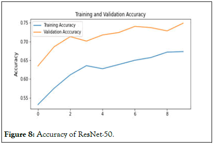
Figure 8: Accuracy of ResNet-50.
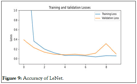
Figure 9: Accuracy of LeNet.
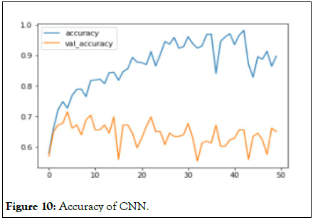
Figure 10: Accuracy of CNN.
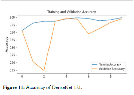
Figure 11: Accuracy of DenseNet-121.
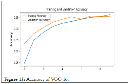
Figure 12: Accuracy of VGG-16.
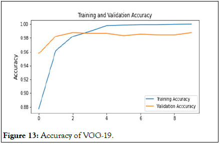
Figure 13: Accuracy of VGG-19.
Early detection and classification of brain tumors is an important research domain in the field of medical imaging and accordingly helps in selecting the most convenient treatment method to save patients life therefore [9,10]. We are gonna use many methods to predict the type of a brain tumor base on MRI pictures (Figure 14).
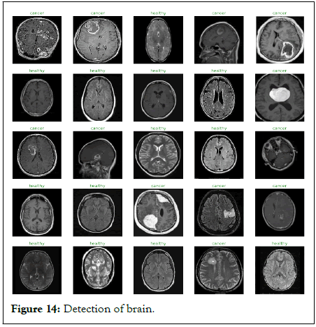
Figure 14: Detection of brain.
In this paper, check on colorful ways for brain excrescence discovery is done. Different ways are used by colorful experimenters to descry the brain excrescence from MRI images are bandied. This paper addresses about overview of the brain, brain excrescence and also about MRI images. Some segmentation ways and conventional system are also bandied. According to this review we find that robotization of brain excrescence discovery and segmentation using CNN from MRI images is most effective and helpful exploration area. Discovery of brain excrescences has a veritably significant part to play in medical interventions. It plays an integral part in the processing of medical images, as medical images have different variations. With the automatic brain excrescence discovery system, opinion isn't only intuitive but also strengthens tremendously the possibility of the case's survival.
[Crossref] [Google Scholar] [PubMed]
[Crossref] [Google Scholar] [PubMed]
[Crossref] [Google Scholar] [PubMed]
[Crossref] [Google Scholar] [PubMed]
[Crossref] [Google Scholar] [PubMed]
[Crossref] [Google Scholar] [PubMed]
[Crossref] [Google Scholar] [PubMed]
Citation: Rana M, Moon AM, Hossain S, Rahman N (2023) A Comparative Study on Brain Tumor Detection from MRI Images in Various Machine Learning Algorithm. J Res Dev. 11:225.
Received: 06-Jan-2023, Manuscript No. JRD-23-21293; Editor assigned: 09-Jan-2023, Pre QC No. JRD-23-21293 (PQ); Reviewed: 23-Jan-2023, QC No. JRD-23-21293; Revised: 20-Apr-2023, Manuscript No. JRD-23-21293 (R); Published: 27-Apr-2023 , DOI: 10.35248/2311-3278.23.11.225
Copyright: © 2023 Rana M, et al. This is an open-access article distributed under the terms of the Creative Commons Attribution License, which permits unrestricted use, distribution, and reproduction in any medium, provided the original author and source are credited.