PMC/PubMed Indexed Articles

Indexed In
- Online Access to Research in the Environment (OARE)
- Open J Gate
- Genamics JournalSeek
- JournalTOCs
- Scimago
- Ulrich's Periodicals Directory
- Access to Global Online Research in Agriculture (AGORA)
- Electronic Journals Library
- Centre for Agriculture and Biosciences International (CABI)
- RefSeek
- Directory of Research Journal Indexing (DRJI)
- Hamdard University
- EBSCO A-Z
- OCLC- WorldCat
- Scholarsteer
- SWB online catalog
- Virtual Library of Biology (vifabio)
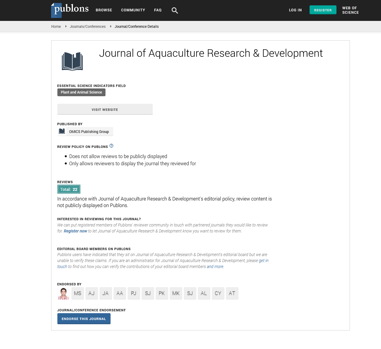
- Publons
- MIAR
- University Grants Commission
- Euro Pub
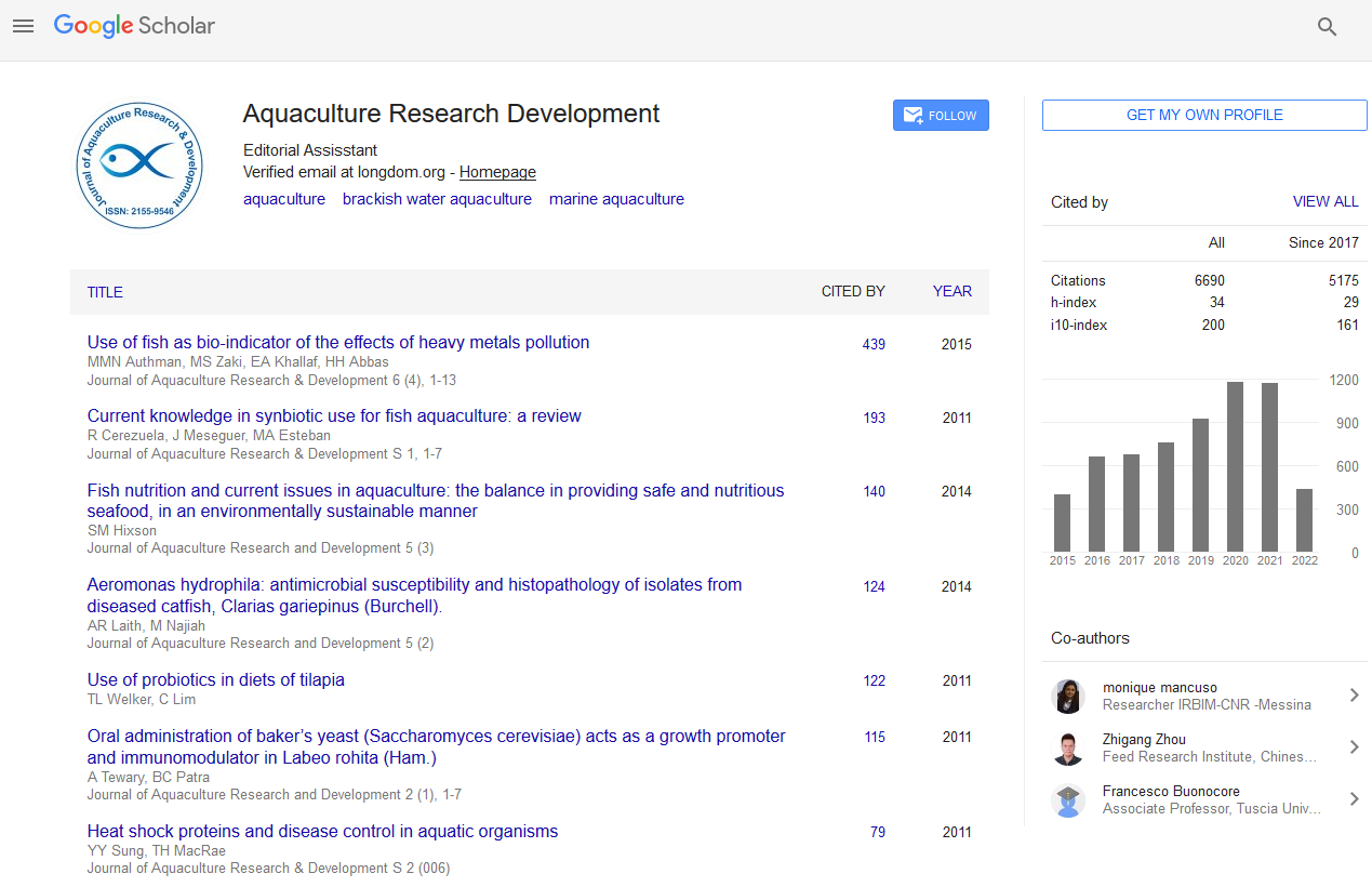
- Google Scholar
Useful Links
Share This Page
Journal Flyer
Open Access Journals
- Agri and Aquaculture
- Biochemistry
- Bioinformatics & Systems Biology
- Business & Management
- Chemistry
- Clinical Sciences
- Engineering
- Food & Nutrition
- General Science
- Genetics & Molecular Biology
- Immunology & Microbiology
- Medical Sciences
- Neuroscience & Psychology
- Nursing & Health Care
- Pharmaceutical Sciences
Effect of starvation on hepatopancreas of whiteleg shrimp (Penaeus vannamei) using computer-assisted image analysis on paraffin and frozen sections
3rd International Conference on Aquaculture & Fisheries
September 29-October 01, 2016 London, UK
Fabio Cervellione
Skretting Aquaculture Research Centre, Norway
Ghent University, Belgium
Scientific Tracks Abstracts: J Aquac Res Development
Abstract:
The hepatopancreas is the main organ of the gastro-intestinal tract in crustaceans and it is routinely assessed by pathologists for signs of disease and health monitoring because it is the site of digestion, nutrient absorption, reserve storage, detoxification, synthesis and secretion of digestive enzymes. The hepatopancreas is mainly composed of four different cell types: E-cells, B-cells, F-cells and R-cells. R-cells resemble the absorptive cells of the vertebrate intestine and store mainly lipids and glycogen. Many decapods can survive weeks or even months of total starvation. In the past, a few studies focused on the influence of feeding on hepatopancreas structure but none of them using computed-assisted image analysis. Image analysis is a fast, objective and applicable method for routine screening of high number of samples, both for diagnostic perspectives and research applications. In the present study, whiteleg shrimp (2±1 g, C inter-moult stage) were housed individually in glass tanks (27±1°C, pH at 7.8-8.1, and salinity at 20±1 gL-1). Three feeding regimes were compared over a 15 days period: fed (5% of body weight/day); starved; and re-fed after 10 days of starvation. Morphological changes caused by starvation were analysed in paraffin sections and frozen sections with computerassisted image analysis software (Visiopharm®). Hepatopancreatic parameters measured were: total tissue area, lumen area, lipid droplets, F-cells, and infiltration of haemocytes. Effect of starvation on the ultrastructure of the hepatopancreas was also studied by Transmission electron microscopy (TEM). Application of image analysis on a routine basis will permit health monitoring of the nutritional status in farmed decapods.
Biography :
Fabio Cervellione graduated as DVM at Milan University, and completed his MSc in Aquatic Veterinary Studies at Stirling University. He worked for 9 years as a Diagnostic Veterinarian for fresh water farmed species for Skretting, which is a leading shrimp and fish feed company in the aquaculture sector. He works now for Skretting Aquaculture Research Centre in Norway. He is a PhD candidate at Ghent University, focusing on semi-quantitative histology of the gastro intestinal tract of whiteleg shrimp (Penaeus vannamei) with Prof. Wim Van den Broeck, Professor of Cell Biology and Histology.