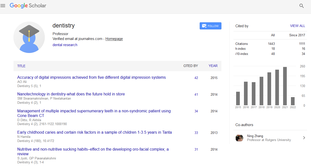
Citations : 1817
Dentistry received 1817 citations as per Google Scholar report
Indexed In
- Genamics JournalSeek
- JournalTOCs
- CiteFactor
- Ulrich's Periodicals Directory
- RefSeek
- Hamdard University
- EBSCO A-Z
- Directory of Abstract Indexing for Journals
- OCLC- WorldCat
- Publons
- Geneva Foundation for Medical Education and Research
- Euro Pub
- Google Scholar
Useful Links
Share This Page
Journal Flyer

Open Access Journals
- Agri and Aquaculture
- Biochemistry
- Bioinformatics & Systems Biology
- Business & Management
- Chemistry
- Clinical Sciences
- Engineering
- Food & Nutrition
- General Science
- Genetics & Molecular Biology
- Immunology & Microbiology
- Medical Sciences
- Neuroscience & Psychology
- Nursing & Health Care
- Pharmaceutical Sciences
Effect of low intensity pulsed ultrasound on the tooth slice organ culture of Ame1x knockout mice
30th International Conference on Dental Science & Advanced Dentistry
May 22-23, 2017 Las Vegas, USA
Krittika Bali, Harmanpreet Kaur, Mac Dederich and Tarek El-Bialy
University of Alberta, Canada
Posters & Accepted Abstracts: Dentistry
Abstract:
Orthodontically induced root resorption (OIRR) is one of the adverse outcomes of orthodontic treatment and dental replantation/transplantation which may lead to permanent loss of the root structure. Several treatment attempts have been done in the past with little clinical success. Low intensity pulsed ultrasound (LIPUS) is a form of mechanical energy that is transmitted through and into living tissue as acoustic pressure waves where this energy is absorbed at a rate proportional to the density of the tissues in which it passes through. The micromechanical strains produced by these pressure waves in biological tissues were assumed to initiate biochemical events that affect hard and soft tissue. Previous studies have shown stimulatory effect of LIPUS on different cell types like fibroblasts, osteoblasts, odontoblasts and chondrocytes. In this study our aim was to investigate the effect of LIPUS on the tissue organ culture extracted from C57BL/6 Ame1x knockout mice to prevent root resorption. Mandibles were dissected from male and female mice and sliced into 2.0 mm thickness slices. The slices were cultured in medium containing DMEM, 10% fetal bovine serum and 1% penicillin-streptomycin at 37 �?�C and 5% CO2 in humidified incubator. Slices were randomly divided into eight groups (n=6). The groups were as follows: Control (wild type with no treatment), Ame1x knockout mice (positive control); LIPUS; Ame1x+ tooth movement (TM); LIPUS+TM; Ame1x+Emdogain (EMD); EMD+LIPUS; and EMD+LIPUS+TM. In tooth movement groups, 50 gm force was applied on tooth slice by custom-made springs prior to LIPUS application. EMD solution was prepared with a 1.0 L concentration and was added to the culture media. LIPUS was applied for 10 minutes per day for 5 consecutive days before histological and histomorphometric analysis. After treatment, we looked for histological sections of the tooth slice for the area of root resorption. Based on our in vivo experiment, we expect that LIPUS treated groups to show an increase in the thickness of cementum and dentine and decrease in the area of resorption lacunae. These data may support that LIPUS may influence remodeling of the dentine-pulp complex and associated tissues during orthodontic force application ex vivo.
Biography :
Email: krittik@ualberta.ca


