PMC/PubMed Indexed Articles
Indexed In
- Open J Gate
- Genamics JournalSeek
- Academic Keys
- JournalTOCs
- ResearchBible
- Ulrich's Periodicals Directory
- Access to Global Online Research in Agriculture (AGORA)
- Electronic Journals Library
- RefSeek
- Hamdard University
- EBSCO A-Z
- OCLC- WorldCat
- SWB online catalog
- Virtual Library of Biology (vifabio)
- Publons
- MIAR
- Geneva Foundation for Medical Education and Research
- Euro Pub
- Google Scholar
Useful Links
Share This Page
Journal Flyer

Open Access Journals
- Agri and Aquaculture
- Biochemistry
- Bioinformatics & Systems Biology
- Business & Management
- Chemistry
- Clinical Sciences
- Engineering
- Food & Nutrition
- General Science
- Genetics & Molecular Biology
- Immunology & Microbiology
- Medical Sciences
- Neuroscience & Psychology
- Nursing & Health Care
- Pharmaceutical Sciences
Review Article - (2021) Volume 0, Issue 0
The SARS-Cov-2 Novel Coronavirus: A Reflection about Enhancement of High-Resolution Microscopic Images
Roberto Rodríguez1*, Brian A. Mondeja2, Leandro D. Lau1, Ananayla Vizcaino2, Emilio F. Acosta2, Yorexis González2 and Angelina Díaz22Center for Advanced Studies of Cuba (CEA), Valle Grande, Cuba
Received: 11-May-2021 Published: 31-May-2021, DOI: 10.35248/2155-9597.21.s10.004
Abstract
After a year of hard battling with the novel coronavirus SARS Cov-2, the COVID-19 pandemic continues had a catastrophic effect on society and health worldwide. This pandemic has changed labor and economic relations in almost every country in the world, and the investment that has been made in the development of new treatment protocols and the creation of vaccines has been enormous. Important laboratories, hospitals and research centers around the world have been fighting against SARS-Cov-2, and within these researches computer vision has played a prominent role. The main aim of this work is to carry out a reflection on the enhancement of the microscopic images of the novel coronavirus SARS-Cov-2 from the results obtained and published. We will analyze the effectiveness of the algorithms proposed to highlight the S-spikes, and we will detail why deep learning, despite the popularity achieved, in this case was not beneficial.
Keywords
Image enhancement; Segmentation; Algorithms; Coronavirus; SARS-CoV-2; Microscopy
Introduction
The world has been battling the novel SARS-Cov-2 coronavirus for more than a year and, however, the COVID-19 pandemic continues had a catastrophic effect on society and health everywhere. This pandemic has changed labor and economic relations in almost every country in the world, and the investment that has been made in the development of new treatment protocols and the creation of vaccines has been enormous. Important laboratories, hospitals and research centers around the world have been fighting against SARS- Cov-2, and within these researches computer vision has played a prominent role [1-5].
Indeed, Computer Vision (CV) has had an outstanding impact in various areas of knowledge; and in the field of medicine, and particularly in bio-images, due to their non-invasive nature, the use of these techniques is already essential. However, it is striking that when a new problem arises in the world of bio-images, many researchers want to solve it with the most current state of the art in CV, as is now the case of Deep Learning (DL), and many times, researchers apply DL without taking into account its advantages and disadvantages [6].
Electron Microscopy (EM) is a widely used technique in viral analysis, which allows fast morphological identification and a diagnosis of infectious elements contained in a specimen without the need for special considerations and reagents, and many times, it is often sufficient for the physician recognizes an unknown infectious agent [7].
However, as was pointed out in [1, 2], microscopic images present some blurring, due to physical problems inherent in electron microscopes, which usually occur, even, in high performance microscopes and high-resolution images. Some of these phenomenons are, scattering, out-of-focus, imperceptible vibrations, voltage disturbances, among others, which causes certain blurring in the microscopic images, and often makes it difficult to analyze correctly to them [8,9].
In this case, of the novel coronavirus SARS-Cov-2, we need to highlight the S-spikes and visualizing those areas that show a high density, which are related to active zones of viral germination and major spread of the virus. In addition, the S-spikes are high frequency information, and have a random spatial distribution; that is, they do not have a certain pattern.
From what was expressed in the previous paragraph, one can note the importance of studying these SARS-Cov-2 high-resolution microscopy images before applying any technique or algorithm, this issue being the essence of the published [1,2]. Thus, the obtained algorithms have better performance.
The main aim of this work is to carry out a reflection on the enhancement of the microscopic images of the novel coronavirus SARS-Cov-2 from the results obtained and published [1,2]. We will analyze the effectiveness of the algorithms proposed to highlight the S-spikes, and we will detail why deep learning, despite the popularity achieved, in this case is not beneficial.
We organized the rest of paper as follows: a Section on the materials and methods is given. We proposed a Section on some algorithmic aspects and its relationship with the characteristics of microscopic images. Later, another Section contains some obtained results and discussion by using other SARS-Cov-2 micro-images. Finally, we describe our conclusions.
Literature Review
As was pointed out in, we captured the samples from nasopharyngeal swabs collected from Cuban individuals with COVID-19 symptomatic and positive for SARS-CoV-2, and we treated to them (samples) with an aldehyde solution and processed by Scanning Electron Microscopy (SEM) [1,2,10]. In, we offered a comprehensive explanation of the importance of SEM [1,2]. Here, we will present in Figures 1-3 some typical SARS-Cov-2 coronavirus images (Figures 1-3).
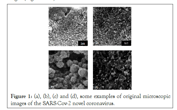
Figure 1: (a), (b), (c) and (d), some examples of original microscopic images of the SARS-Cov-2 novel coronavirus.
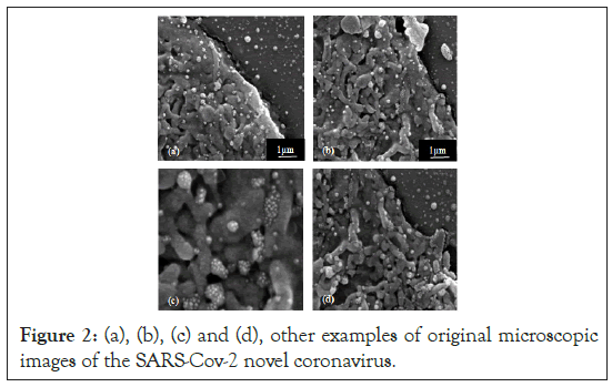
Figure 2: (a), (b), (c) and (d), other examples of original microscopic images of the SARS-Cov-2 novel coronavirus.
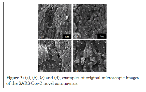
Figure 3: (a), (b), (c) and (d), examples of original microscopic images of the SARS-Cov-2 novel coronavirus.
We in carried out a study and analysis of some of the characteristics of these microscopic images of SARS-Cov-2 novel coronavirus [1]. One can observe that the most interesting in those micro-images are in their blurred appearance, where the S-spikes do not look well defined. On the other hand, these S-spikes, in addition to being information of high spatial frequencies, are hyper-dense areas (brighter points) and have a random spatial distribution (that is, S-spikes do not have a certain pattern).
In, we highlighted the importance of the study and characterization of the micro-images before applying any type of algorithm [1]. In this case, much more important when it comes to a novel coronavirus that information about the same did not exist. This helps one to develop algorithms much more adapted to the problem one want to solve, in this case, the enhancement of S-spikes or S-proteins, which allow the virus to fuse with the host cell membrane [11]. It is known that a high density of S-spikes is related to active zones of viral germination and major spread of virus [10].
Some algorithmic aspects and its relationship with the micro-images
As we pointed out, the study and characterization of the micro- images is an important step for the development of new algorithms. One must carry out this procedure before applying any algorithm, and the study must be deep and effective [12]. In image classification, Deep Learning (DL) has dominated this field due to a substantially better performance compared to traditional methods. However, as good was explained in, DL is not making traditional CV techniques obsolete, and it will not solve all the problems that arise in this mega-field [6]. In CV, there are many problems where traditional techniques with global features are a better solution [6]. This is the case when the problem to be solved is not framed in the image classification field, or the objects to be processed do not have a certain pattern. In this sense, S-spikes of the SARS-Cov-2 novel coronavirus have this behavior.
However, the current trend of many researchers is to apply DL to all the problems that arise in CV, without taking into account the advantages and disadvantages of DL [6]. Nevertheless, published works applying DL for the enhancement or segmentation of S-spikes of the SARS-Cov-2 novel coronavirus do not appear, as was reflected in Table presented in [2].
The CV works by using DL have been fundamentally applied to detect COVID-19 from chest CT radiography Digital Images [2]. This may be motivated by the fact that the chest radiographs of patients affected with COVID-19 present a certain pattern that differs from those who do not have the disease.
In Figure 4, we present two examples from chest CT radiography of patients with COVID-19 (Figure 4).
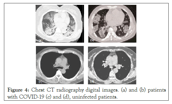
Figure 4: Chest CT radiography digital images. (a) and (b) patients with COVID-19 (c) and (d), uninfected patients.
Although nowadays, in CV the transfer learning is being used increasingly, one of the problems that persists in DL is the computational cost in the training, besides the need of big data [13]. Nevertheless, DL has proven to be sufficient and easy solution for ad-hoc problems, but once that the system is trained and optimized; the framework is a black box [14].
What we expressed in the previous paragraph means that the change of meta-parameters to adapt the system to a new problem in DL is not trivial, and a new training is necessary. Many times, while more complex a task becomes, one requires more flexibility in algorithms and a global tuning, which will be a better solution.
It is today known that the SARS-Cov-2 novel coronavirus is mutating in different countries, and this mutation can result in a change in its morphology and spatial distribution of S-spikes. Algorithms presented in are easy to adapt with simple global adjustments in case of need to enhancement the microscopic images of the novel virus strains [2]. In addition, these algorithms are simple of using to study, without need of training, micro-images of new bacteria and virus. Carrying out this task with DL is not trivial.
Therefore, as far as possible to carry out a study of characteristics of the micro-images and of their physical environment, one will be in better conditions of developing or applying algorithms that best suit the problem to be solved. This is what we name: algorithmic aspects and its relationship with the microscopic images or study images [2].
Other obtained results and discussion using the proposed algorithms
In this Section, we will carry out other enhancement of the microscopic images from Figures 1-3 by using of Algorithm No. 3 presented [2] In Figures 5-8, we show the obtained results.
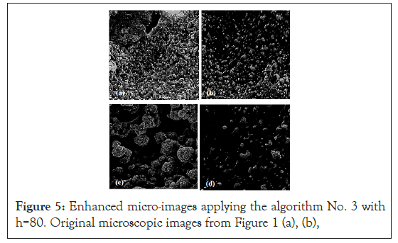
Figure 5: Enhanced micro-images applying the algorithm No. 3 with h=80. Original microscopic images from Figure 1 (a), (b),
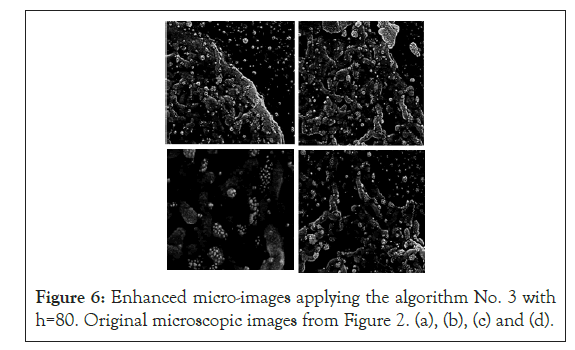
Figure 6: Enhanced micro-images applying the algorithm No. 3 with h=80. Original microscopic images from Figure 2. (a), (b), (c) and (d).
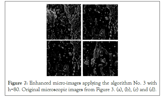
Figure 7: Enhanced micro-images applying the algorithm No. 3 with h=80. Original microscopic images from Figure 3. (a), (b), (c) and (d).
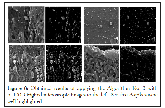
Figure 8: Obtained results of applying the Algorithm No. 3 with h=100. Original microscopic images to the left. See that S-spikes were well highlighted.
One can note in the obtained results that S-spikes were remarkably improved, and in some microscopic images the S-spikes appear to be practically segmented (very light S-spikes) on a dark background (Figures 5-8).
Discussion
The most important of these results is that we processed all the micro-images with the same value of the h parameter, which denotes the stability of this algorithm for image databases captured under the same boundary conditions (microscope, illumination, among others).
As we expressed in, the h parameter is who determines the expression of final result in the processed micro-image (improved S-spikes or segmented) [2]. The selection of one or the other result will depend of the aim of specialists (virologist, bacteriologist, biologist, or other).
Other examples of processed micro-images of patients with the SARS-Cov-2 virus appear. In this case, for one to be able to see better the result of the enhancement and visually to contrast, we will place the original micro-imagen to the left, and the improved one to right. We used a value of parameter h=100 for all processed microscopic images. One can see that S-spikes were practically isolated, and the intensity value of background was equal to zero. Of this result, one can denote the importance of this unique parameter.
Conclusion
We carried out, from the papers published in, a reflection about enhancement of high-resolution microscopic images of the SARS- Cov-2 novel coronavirus. In addition, in this work, we made a broad explanation of the importance of DL, and we analyzed their disadvantages too. We explained that in the case of enhancement of S-spikes is not convenient to use DL. Finally, we carried out an extensive experimentation with our Algorithm, and discussed the results.
REFERENCES
- Rodríguez R, Mondeja BA, Valdés O, Resik S, Vizcaino A, Acosta EF, et al. SARS-CoV-2: enhancement and segmentation of high-resolution microscopy images-Part I. Signal Image Video Process. 2021;23(1):1-9.
- Rodríguez R, Mondeja BA, Valdés O, Resik S, Vizcaino A, Acosta EF, et al. SARS-CoV-2: Theoretical analysis of the proposed algorithms to the enhancement and segmentation of high-resolution microscopy images-Part II. Signal Image Video Process. 2021;23(1):1-9.
- Yan Q, Wang B, Gong D, Luo C, Zhao W, Shen J, et al. COVID-19 chest CT image segmentation -- A deep convolutional neural network solution. ArXiv. 2020.
- Fan DP, Zhou T, Ji GP, Zhou Y, Chen G, Fu H, et al. Inf-Net: Automatic COVID-19 lung infection segmentation from CT images. IEEE Trans Med Imaging. 2007;39(8):2626-2637.
- Amyar A, Modzelewski R, Li H, Ruan S. Multi-task deep learning based CT imaging analysis for COVID-19 pneumonia: Classification and segmentation. Computers in Biol Med. 2020;126(1):104037.
- O' Mahony N, Campbell S, Carvalho A, Harapanahalli S, Velasco-Hernandez G, Krpalkova L, et al. Deep learning vs. traditional computer vision. ArXiv. 2019.
- Schramlová J, Arientová S, Hulínská D. The role of electron microscopy in the rapid diagnosis of viral infections--review. Folia Microbiol (Praha). 2010;55(1):88-101.
- Goldsmith CS, Miller SE. Modern uses of electron microscopy for detection of viruses. Clin Microbiol Rev. 2009;17(2):3-12.
- Yang SJ, Berndl M, Ando DM, Barch M, Narayanaswamy A, Christiansen E, et al. Assessing microscope image focus quality with deep learning. BMC Bioinformatics. 2018;19(1):77.
- Mondeja B, Valdes O, Resik S, Vizcaino A, Acosta E, Montalván A, et al. SARS-CoV-2: High-resolution microscopy study in human nasopharyngeal samples. Virol. 2020;19(2):565-574.
- Huang Y, Yang C, Xu XF, Xu W, Liu SW. Structural and functional properties of SARS-CoV-2 spike protein: potential antivirus drug development for COVID-19. Acta Pharmacologica Sinica. 2021;41(1):1141-1149.
- Rodríguez R, Sossa JH. In: Narayan R (eds) Mathematical techniques for biomedical image segmentation (3rd edn), Elsevier, Amsterdam, The Netherlands, 2019.
- Muñoz PL, Rodríguez R, Montalvo N. Automatic segmentation of diabetic foot ulcer from mask region-based convolutional neural networks. J Biomedical Research and Clinical Investigation. 2020;2(1):1-9.
- Camilleri D, Prescott T. Analysing the limitations of deep learning for developmental robotics. Conference on Biomimetic and Biohybrid Systems. 2017.
Citation: Rodríguez R, Mondeja BA, Lau LD, Vizcaino A, Acosta EF, González Y, et al. (2021) The SARS-Cov-2 Novel Coronavirus: A Reflection about Enhancement of High-Resolution Microscopic Images. J Bacteriol Parasitol. S10: 004
Copyright: © 2021 Rodríguez R, et al. This is an open-access article distributed under the terms of the Creative Commons Attribution License, which permits unrestricted use, distribution, and reproduction in any medium, provided the original author and source are credited.
