PMC/PubMed Indexed Articles
Indexed In
- Open J Gate
- Genamics JournalSeek
- JournalTOCs
- Ulrich's Periodicals Directory
- RefSeek
- Hamdard University
- EBSCO A-Z
- OCLC- WorldCat
- Proquest Summons
- Publons
- Geneva Foundation for Medical Education and Research
- Euro Pub
- Google Scholar
Useful Links
Share This Page
Journal Flyer

Open Access Journals
- Agri and Aquaculture
- Biochemistry
- Bioinformatics & Systems Biology
- Business & Management
- Chemistry
- Clinical Sciences
- Engineering
- Food & Nutrition
- General Science
- Genetics & Molecular Biology
- Immunology & Microbiology
- Medical Sciences
- Neuroscience & Psychology
- Nursing & Health Care
- Pharmaceutical Sciences
Case Report - (2021) Volume 12, Issue 5
Kupffer Cell SarcomaâA Rare Hepatic Sarcoma
Amaresh Aruni*Received: 20-May-2021 Published: 10-Jun-2021, DOI: 10.35248/2155-9864.21.12.465
Abstract
A 62-year male presented with pain abdomen and palpable liver mass. On CT-imaging there was a large heterogeneously enhancing mass with internal non-enhancing area in left lobe of liver. Biopsy showed the spindle cell morphology, and a final diagnosis of hepatic sarcoma was made. Patient underwent left lateral sectionectomy and sleeve resection of pyloroduodenal wall. At 3 months post op follow up imaging showed multiple right lobe liver metastasis.
Keywords
Kupffer cell; Sarcoma; Liver
Introduction
Primary hepatic sarcomas are extremely rare tumors constituting 1-2% of the primary hepatic malignant tumors, with peak incidence in adults and with male preponderance. Diagnosis usually occurs at advanced stage due to rarity of the tumor and lack of symptoms in early stages. Surgical hepatic resection with clear margins and systemic chemotherapy is the optimal management.
Case Representation
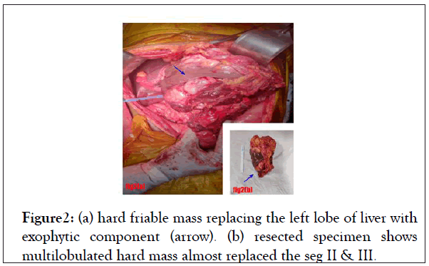
A 62-year male presented with pain in right hypochondriac region for 3 years, mass per abdomen for 3 months, with early satiety, anorexia and loss of weight. On examination, there was a hard non-tender15x15 cm liver mass extending in to right hypochondrium, epigastrium and umbilical region. Blood investigations and tumor markers were within normal limits [1]. Initial sonography findings suggested an enlarged heterogeneous mass of size 7.6 × 10.4 cm in relation to left lobe of liver and the stomach. CECT imaging revealed large heterogeneously enhancing partially exophytic mass with internal non-enhancing area in left lobe of liver without evidence of metastasis (Figures 1 and 2).
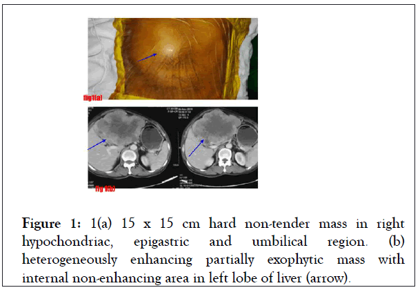
Figure 1: 1(a) 15 x 15 cm hard non-tender mass in right hypochondriac, epigastric and umbilical region. (b) heterogeneously enhancing partially exophytic mass with internal non-enhancing area in left lobe of liver (arrow).
Figure 2: (a) hard friable mass replacing the left lobe of liver with exophytic component (arrow). (b) resected specimen shows multilobulated hard mass almost replaced the seg II & III.
UGIE was done in view of occasional symptoms of GOO which revealed narrowing of D1-D2 junction due to extrinsic compression. Pre-operative tissue diagnosis revealed spindle shaped tumor cell morphology.
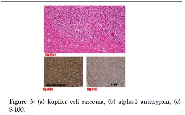
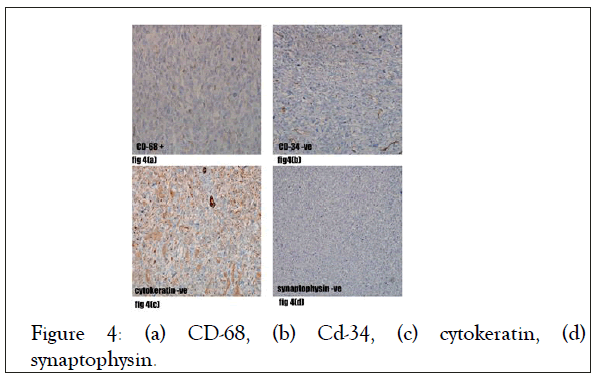
Patient underwent left lateral sectionectomy and sleeve resection of pyloro-duodenal wall (the intra-operative findings were 17 × 20 × 15 cm hard multilobulated soft tissue tumor replacing the left lobe of liver, the mass was adhered to the pyloro-duodenal wall, however duodenal mucosa was normal and there was no evidence of metastasis noted (Figures 3 and 4) [2].
Figure 3: (a) kupffer cell sarcoma, (b) alpha-1 antitrypsin, (c) S-100.
Figure 4: (a) CD-68, (b) Cd-34, (c) cytokeratin, (d) synaptophysin.
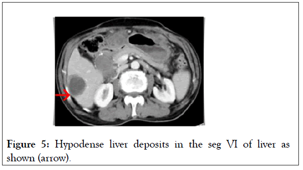
Post-operative course was uneventful. On final histopathology report, there was grossly no capsular breach by the tumor, microscopy showed infiltrating spindle shaped tumor cells arranged in sheets, fascicles, nodules. Left portal vein was infiltrated by the tumor cells. IHC was positive for alpha 1 antitrypsin (diffuse), vimentin, S-100, CD68/CD45, negative for CD34. Pancytokeratin Impression of Kupffer cell sarcoma was made (Figure 5).
Figure 5: Hypodense liver deposits in the seg VI of liver as shown (arrow).
Patient was planned for adjuvant radiotherapy but was lost to follow-up. Patient now presented at 3 months post-op with no complaints [3]. CECT imaging which was performed however sadly revealed multiple liver deposits with biopsy suggestive of metastasis. Patient is now on chemotherapy.
Clinical presentations are mainly pain abdomen followed by mass per abdomen, jaundice, chest pain, GOO due to pressure effects of the mass. Other symptoms such as fever, anorexia related to release of the inflammatory mediators from the tumor cells. Symptoms of hypoglycaemia has been associated with MFH due to secretion of the insulin-like growth factor II.Discussion
Primary mesenchymal hepatic neoplasms are extremely rare accounting for 1-2% of all primary malignant tumors of liver. Hepatic sarcomas described in literature are angiosarcoma, epitheloid hemangioendothelioma, fibrosarcoma, leiomyosarcoma, liposarcoma, fibrosarcoma, embryonal sarcoma malignant fibrous tumor and follicular dendritic cell sarcoma have also been found to arise in the liver. Angiosarcoma is most common type of primary liver sarcoma originating from vascular endothelial cells and has a recognised association with thorotrast, arsenic and vinyl chloride monomer but no etiological factors are associated with other variety of hepatic sarcomas except radiation exposure. Due to rarity of primary hepatic sarcomas, the natural history, prognostic factors and optimal management are poorly characterized. Further complicating the study of hepatic sarcoma is the diversity of subtypes. We report a rare variety of liver sarcoma with kupffer cell origin in 62-year male patient who’s a chronic smoker. These tumors occur in infants and in adults, but the peak incidence occurs in adults. Cases in infants present in the first months of life, generally as abdominal masses, and lead to death within the first 6 months of life from local pressure or cardiac failure from arteriovenous shunting in the tumor. In adults, the tumors are three times as common in males as in females [4].
Clinical presentations are mainly pain abdomen followed by mass per abdomen, jaundice, chest pain, GOO due to pressure effects of the mass. Other symptoms such as fever, anorexia related to release of the inflammatory mediators from the tumor cells. Symptoms of hypoglycaemia has been associated with MFH due to secretion of the insulin-like growth factor II. Presentation of the hepatic sarcoma occurs usually in advanced stage since liver is the largest solid organ in the body, and symptoms and signs of a tumor in the liver usually go undetected unless the tumors are large. Early suspicious of the sarcoma is unlikely due to rarity of the tumor. Initial diagnosis is difficult and usually made only after surgery or at autopsy. Recent advanced imagings can delineate the nature of the lesion and early consideration of sarcoma as a diagnosis. USG and CECT are better imaging tools in diagnosis, CECT is more accurate in diagnosis, differentiation of the tumors based on enhancement and to assess for the resectability of the tumors. Pre-operative tissue biopsy confirms the diagnosis but may be difficult with a small specimen, since appearances vary greatly in different parts of tumor.
Conclusion
Surgical curative liver resections with clear margins remains the mainstay of treatment. In advanced cases with metastasis palliative treatment can be given. The randomized trials have shown that radiotherapy decreases the local recurrence in extremity sarcoma, but the use of radiotherapy is constrained in the liver due to potential toxicity. Therefore complete resection followed by systemic chemotherapy might be the optimal treatment for these tumors.
REFERENCES
- Naik PRT, Kumar P, Kumar PV. Primary pleomorphic liposarcoma of liver: A case report and review of the literature. Case Rep Hepatol. 2013; 2013:398910.
- Weitz J, Klimstra DS, Cymes K, Jarnagin WR, D’Angelica M, La Quaglia MP, et al. Management of primary liver sarcomas. Cancer. 2007;109(7):1391–6.
- Forbes A, Portmann B, Johnson P, Williams R. Hepatic sarcomas in adults: A review of 25 cases. Gut. 1987;28(6):668–74.
- Zimmermann A. Malignant fibrous histiocytoma of the liver: A neoplasm of the undifferentiated high-grade pleomorphic sarcoma group. Tract Springer Cham. 2016;1-9.
Citation: Aruni A, (2021) Kupffer Cell Sarcoma–A Rare Hepatic Sarcoma. J Blood Disord Transfus. 12: 465.
Copyright: © 2021 Aruni A. This is an open-access article distributed under the terms of the Creative Commons Attribution License, which permits unrestricted use, distribution, and reproduction in any medium, provided the original author and source are credited.
