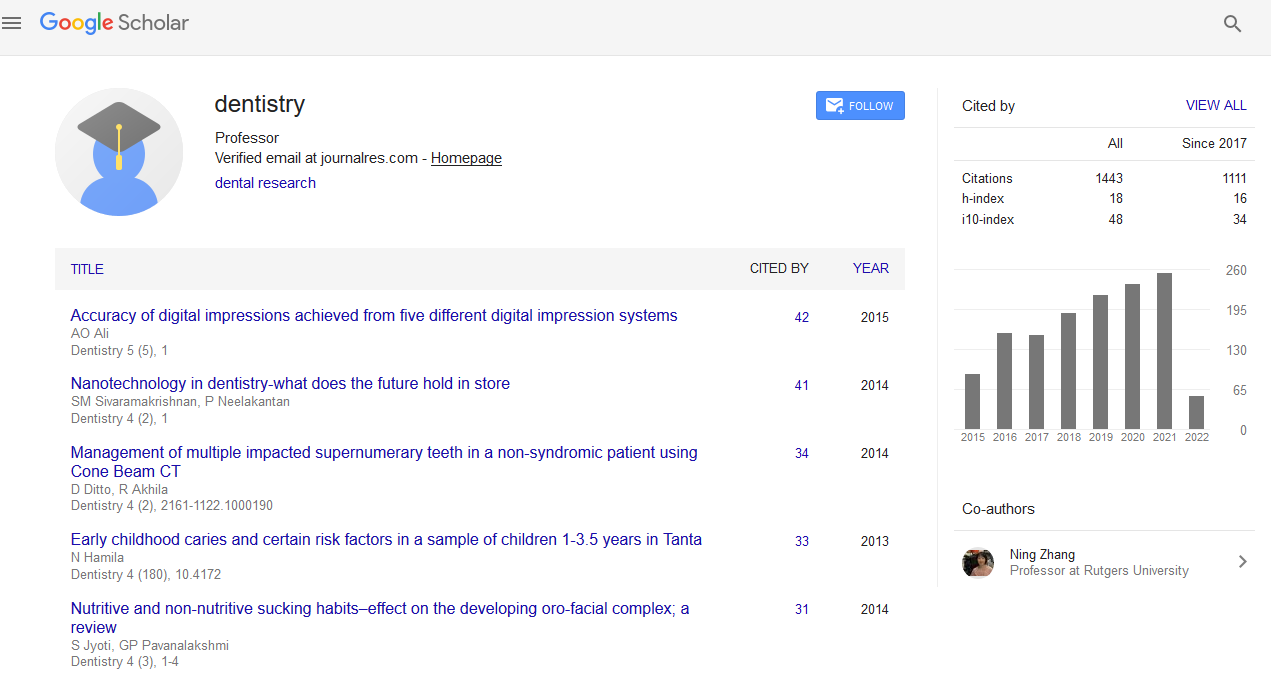
Citations : 1817
Dentistry received 1817 citations as per Google Scholar report
Indexed In
- Genamics JournalSeek
- JournalTOCs
- CiteFactor
- Ulrich's Periodicals Directory
- RefSeek
- Hamdard University
- EBSCO A-Z
- Directory of Abstract Indexing for Journals
- OCLC- WorldCat
- Publons
- Geneva Foundation for Medical Education and Research
- Euro Pub
- Google Scholar
Useful Links
Share This Page
Journal Flyer

Open Access Journals
- Agri and Aquaculture
- Biochemistry
- Bioinformatics & Systems Biology
- Business & Management
- Chemistry
- Clinical Sciences
- Engineering
- Food & Nutrition
- General Science
- Genetics & Molecular Biology
- Immunology & Microbiology
- Medical Sciences
- Neuroscience & Psychology
- Nursing & Health Care
- Pharmaceutical Sciences
Abstract
Valproic Acid Contributes to Bone Cavity Healing in Rats
Mamunur Rashid, Yosuke Akiba, Kaori Eguchi, Nami Akiba, Masaru Kaku, Masako Nagasawa and Katsumi Uoshima
Background: Sufficient bone quality and quantity are necessary for successful results in a dental implant. Although numerous bone augmentation methods have been reported, used in the clinic and showed successful results in some extent, more reliable methods are still required. Valproic Acid (VPA) which was known as an Antiepilepsy agent and histone deacetylases inhibitor regulate osteoblast differentiation through Runx2 activation in vitro. The present study aimed to evaluate the effects of systemic administration of VPA on bone regeneration in rat maxillary bone cavity.
Material and Methods: Fifty-four Wistar rats were used for the experiment. Upper first and second molars were extracted at 4 weeks. Three weeks after extraction, the experimental group received intraperitoneal (IP) injection of VPA and control group received IP injection of saline for 7 days prior to the preparation of bone cavity at the first molar area. Rats were sacrificed on days 3, 7, 14, and 21, and samples were prepared for micro-CT and histological analyses and serum Alkaline Phosphatase (ALP) activity was measured. After 7 days of VPA or saline injection, bone marrow-derived cells were corrected for microarray analysis.
Results: Micro-CT analysis and histological observations confirmed higher amounts of newly formed bone, bone volume fraction (BV/TV) and trabecular thickness (Tb.Th), and less trabecular separation (Tb.Sp) in the experimental group at 7, 14, and 21 days than the control. VPA-treated animals showed significantly higher ALP activities at 7, 14, and 21 days than the control. From microarray analysis, 26 genes showed significantly altered expression.
Conclusion: As systemic administration of VPA accelerated bone regeneration in the rat maxillary bone cavity, there the possibility that VPA injection may be useful for bone augmentation therapy.


